

 PDF
PDF ePub
ePub Citation
 Print
Print


INTRODUCTION
Candida species produce a wide spectrum of diseases, ranging from superficial mucocutaneous disease to invasive illnesses such as hepatosplenic candidiasis, peritonitis, and systemic candidiasis. Out of more than 200 species, the most commonly encountered in medical practices are Candida albicans, Candida dubliniensis, Candida glabrata, Candida krusei, Candida parapsilosis, and Candida tropicalis (1). It is well known that the high prevalence of some Candida proliferation has created more chance for other pathogenic microorganisms to invade host (2). However, Candida species are also known in its ability to form biofilms on various surfaces, to change the morphological form, and to switch between various phenotypes.
The degree of prevalence of fungal infections caused by Candida species have increasingly been reported (3). Candida is a common oral flora and it can be a causative agent in several cases of oral infections (4). On the other hand, an efficient role of the internal spacer (ITS)-based ribosomal RNA has been shown to be associated with remarkable discrimination of several species of Candida from each other in the oral cavity (5). The differentiation amongst infective Candida spp. holds considerable clinical interests since the type of detected Candida spp. may determine the suitable treatment regimen prescribed for oral infection (6). For instance, fluconazole antifungal drug is used mainly for treating C. albicans (7), while another treatment strategy is implemented for treating non-albicans Candida since they exhibit resistance to azole-derived drugs (8). However, 16S rRNA-based PCR is becoming increasingly available and is generally quick and easy to perform in a laboratory with relevant expertise in molecular microbiology (9).
In addition to the common utilization of 16S rRNA-based amplification, the conserved sequences within the ITS4-ITS5 rRNA gene are recognized, and amplification of the variable regions leads to unique signatures that are utilized for identification of the particular fungus to the species level (10). Several ribosomal-based sequencing studies have been used for the study of fungal taxonomy and phylogeny. Accordingly, ribosomal RNA loci have gained an important impact in clinical diagnostics, as they provide attractive alternatives for the identification of pathogens in oral specimens from periodontitis patients (11). However, ITS and 16S rRNA based fragments are not the only efficient loci in such discriminating ability as other ribosomal fragments have also shown such efficiency in the same organism, such as 26S rRNA-based amplicons (12). However, the accurate role of the large ribosomal fragments in the targeted speciation between several species of Candida has not tested yet.
Though many reports have described a variety of details concerning Candida prevalence in several periodontitic cases (1314), no sufficient data have been recorded with regard to the accurate distribution of various species of this organism in those patients. Large ribosomal-based sequences characterized with tremendous ability to identify the unknown organisms with a rapid discriminative power amongst variable species included within the same targeted organism (15). For this reason, the utilization of such ribosomal sequences may be fruitful in providing rapid speciation in Candida infected periodontitic cases. Since the accurate determination of Candida infection occupies the main standpoint for assessing the severity and treatment program for periodontitis (16), finding a simple and non-costly method of Candida species identification using one PCR primers pair would be of major interest.
Taking the previously mentioned information into account, the present study aims to assess the prevalence and distribution of several unknown species of Candida among periodontitis patients using one PCR primers pair that based on 26S rRNA lo cus to evaluate their pattern of distribution in the oral cavity of periodontitis patients.
MATERIALS AND METHODS
Samples
Samples were collected from fifty patients infected with chronic periodontitis who visited the dental clinic of Dentistry College, University of Babylon in 2018. This study was approved by the Institutional Review Board (IRB) of the University of Babylon (approval number B-13.164.24) and all the procedures used in the present study received prior approval from the committee on the use of human research subjects of the same institution. Both sexes were included in the study with age between 18–65 years. All included patients were clinically diagnosed as chronic periodontitis patients according to professional dentists. As described in our previous study (11), patients were signed informed consents prior to participation. It was confirmed that patients had no history of any systemic diseases before being involved in the study. The exclusion criteria included the use of fungal treatment or periodontal treatment within 3 months prior to the study. Female patients who were pregnant or nursing were also excluded from the study (17). Each oral samples were obtained from 5 mm depth. Subgingival dental biofilm samples were collected by inserting 3 sterile paper points into the periodontal pocket for 30 s and processed according to Loberto et al. (18).
Candida spp. isolation
Samples were plated on the selective and differential CHROMagar media (HiCrome Candida Differential Agar) to determine the presence of Candida colonies. The inoculated CHROMagar™ Candida media were incubated at 30℃ for 72 hrs (Cat No. CA222, CHROMagar co. Paris, France). The isolated yeasts were identified according to color developed in the chromogenic medium. Subsequently, variable colonies were identified by several biochemical reactions. Moreover, growth at 45℃ was used for differentiating Candida albicans from other species as Candida albicans exhibited well proliferation at this temperature, while others give poor or no growth (19). Only pure Candida spp. were considered and any other microorganisms were eliminated from further reactions.
Genomic DNA extraction
Genomic DNA was isolated according to the instruction manual mentioned for fungal species (20). The RNA interfering with eluted DNA was eliminated by incubation for 30 min at room temperature with 1 mg/ml RNase A (Favorgen Biotech Co., Changzhi Township, Taiwan). The integrity of the isolated yeast genomic DNA was directly verified by gel electrophoresis on 0.8% agarose. Then, its quality and quantity were evaluated by a nano-spectrophotometric method (BioDrop co., Cambridge, UK).
Polymerase chain reactions (PCR)
One pair of PCR oligonucleotides was utilized in the study to confirm the identity of Candida spp., including forward primer 5′-ACCCGCTGAACTTAAGC-3′ and reverse primer 5′-TCCTGAGGGAAACTTCG-3′ (12). A uniplex PCR was performed using a PCR mixture consisted of 250 µM deoxynucleoside triphosphates, 10 mM Tris-HCl (pH 9.0), 30 mM KCl, 1.5 mM MgCl2, and 1 unit of Top DNA polymerase (Bioneer Corp., Daejeon, Korea). PCR was conducted by initial denaturation for 4 min followed by 35 cycles of 30 sec denaturation (94℃), 45 sec of annealing (58℃), and 30 sec of polymerase extension (74℃), and terminated with one cycle of final polymerase extension for 5 min (74℃). The conducted PCR reactions were confirmed by electrophoresis on 1.5% agarose gel. Only specific bands were considered for sequencing reactions.
DNA Sequencing
Further confirmations for the detailed identity of the observed Candida spp. were conducted by performing direct sequencing to all PCR specific products. Only obvious chromatograms were considered, assembled, edited using Bioedit sequence alignment editor, Lasergene ver. 7.2.5 (DNASTAR Inc., Madison, Wisconsin). DNA variations were visualized using SnapGene viewer ver. 4.0.4 (GSL Biotech LLC, Chicago, Illinois). The observed nucleic acid variations of Candida spp. were registered in NCBI under the GenBank accession numbers (MN121162 - 84).
Phylogenetic analysis
The accurate genetic relationships amongst the observed Candida spp. were studied by generating a specific comprehensive tree for Candida spp. The present 26S ribosomal-based comprehensive tree was generated according to a neighbor-joining based method (10). All the included Candida species were annotated by a cladogram-made parameters.
RESULTS
Detection of Candida spp.
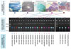
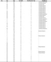
The conventional microbiological techniques indicated the presence of 23 pure cultures of Candida species that predominated by C. albicans, while any mixed culture was eliminated from further study (Table 1). However, those pure cultures were found to be distributed on five main species as it was shown by the presumptive identification by CHROMagar (Figure 1a). After being incubated for 72 hrs, C. albicans colonies exhibited distinctive apple-green colonies in CHROMagar, while pale pink colonies and large glossy pale pink to violet colonies were observed for C. parapsilosis and C. glabrata, respectively. Irrespective of these colonies, no concrete data were resolved in terms of the accurate detection of other Candida species due to high similarities found among their phenotypes. However, CHROMagar can identify few species of Candida on the basis of colonial morphology and color (21), and it has variable sensitivity for Candida detection (22). Moreover, the differentiation between closely related Candida species, such as C. albicans and C. dubliniensis was being difficult in CHROMagar (23). Therefore, each isolated pure colony was exposed to molecular analyses to confirm their species identity (Figure 1b).
Sequencing reactions of 23 subgingival Candida isolates were shown that these isolates had 99–100% homology with several deposited Candida databases in NCBI. Direct comparison of the assembled DNA sequences with highly related referring sequences subdivided these isolates into five highly related organisms belonged to Candida, including C. albicans, C. parapsilosis, C. glabrata, C. dubliniensis, and C. kefyr (Figure 1c). The latter organism was formerly known as C. pseudotropicalis (24). However, PCR sequencing results confirmed the prevalence of C. albicans and showed that the overall ratio of pure infection with Candida spp. was 46% (23/50) with a remarkable prevalence of C. albicans over other detected Candida species. The observed C. albicans showed prevalence of 26% (13/50) in the investigated patients, while the other ratio of infections exhibited lower prevalence of other species, including C. parapsilosis, C. glabrata, C. kefyr, and C. dubliniensis of 8% (4/50), 6% (3/50), 4% (2/50), and 2% (1/50), respectively.
Phylogenesis of Candida spp.
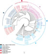
Phylogenetic analyses added another layer of confirmation with regard to the proven identity of the observed Candida species and indicated almost all entire divisions amongst the detected species. In addition to the analyzed Candida nucleic acid variations, the present 26S rRNA-based comprehensive tree was constructed by combining the accession numbers of 95 organisms of Candida. The comprehensive tree was generated to incorporate only species that belong to Candida, including C. albicans, C. glabrata, C. parapsilosis, C. dubliniensis, C. kefyr, C. lactis, and C. dobzhanskii (Figure 2). It was found that all five observed groups of the 26S rRNA variants were occupied distinctive positions within the tree. This data was clearly observed by the currently constructed phylogenetic tree which categorized each detected species in particular phylogenetic position. Thus, five groups were resolved from the present tree in five specified phylogenetic distances. Such informative speciation was provided from the utilization of 26S rRNA amplicons. The 26S rRNA-based differentiation had confirmed this ribosomal fragment ability to discriminate between five species of Candida. The latter discrimination has given new data about the pattern of distribution of these organisms in periodontitis patients.
DISCUSSION
This study indicated remarkable invasiveness of C. albicans distribution in the periodontitis patients. The present prevalence of this fungal organism over other detected species was not unusual. Several types of research indicated that the obvious superiority of C. albicans refers to a noticeable oral imbalance in the infected oral cavity which lead to overgrowth of this species over other species (25). Though no comparable study was taken place with regard to the distribution of Candida species in periodontitis patients, it is rational to account parallel outcomes for the higher percentage of infection of C. albicans than other species in other oral cavity infections. This observation is sustained by the reports that revealed a conversion of harmless fungal hyphae to invasive pathogenic forms once such imbalance is taken place in the mouth (26). However, such informative outcomes have been provided from the targeting of 26S rRNA locus that has not applied for such discriminating role yet.
The implementation of 26S rRNA-based amplification has proved the efficiency of this fragment in grouping Candida species into five different species showing sufficient data in the targeted large ribosomal sequences to discriminate Candida into five species. A clear distribution was provided from the currently constructed comprehensive tree in terms of different phylogenic localization of the investigated Candida species. Noteworthy, the detected species were closely related to each other due to their high ratio of homology (27). However, some strains of C. albicans, CP032012.1_410937; CP032012.1_398379, and C. dubliniensis, CR380958.2_1447277, were not found to be clustered within the same specified clade of their species. This observation may be due to the high variability witnessed in this genus which seems to be extremely distinctive as in such cases (2829).
Nevertheless, 26S rRNA-based amplification and phylogenesis has confirmed accurate speciation among those species despite these similarities. This observation leads to efficient detection and categorization of five species of Candida, while the utilization of large ribosomal subunit-based phylogenetic tree has not revealed such highly informative data as the closely related species were found to be interconnected with no specific clade identified (30). Noteworthy, the currently used 26S rRNA amplicons showed useful features in terms of its generalized ability to amplify variably different species within the oral cavity as well as with its specificity due to its ability in discriminating between those observed organisms into different species. This feature could be attributed to the specific characteristics that kept in ribosomal sequences (10). Moreover, the cost of undergoing such 26S rRNA-based speciation procedure is so low as compared with other costly techniques, such as those relied on DNA hybridization, or multiplex PCR detection (31). Therefore, the present manuscript suggests the implementation of 26S rRNA-based tool as a competent tool for low-cost detection of Candida spp., discrimination of its species in the oral cavity. This study could be utilized by dentists to evaluate the pattern of Candida infection and distribution in periodontitis patients.
In conclusion, data in the present study indicated a remarkable ability of 26S rRNA-based sequences to provide an easy guide to group Candida spp. into different species in the patients with periodontitis suggesting a direct, low-cost method for detection of Candida, discrimination of its species. The pattern of Candida distribution could be identified in chronic periodontitis without being complicated by other cost-effective diagnosis tools. Therefore, the 26S rRNA-based assessment of the status of invasiveness of the infected periodontitis cases is feasible to prescribe the proper antifungal therapy choice.
 XML Download