

 PDF
PDF ePub
ePub Citation
 Print
Print


Gleason grading is fundamental for making therapeutic decisions and predicting the prognoses of patients with prostate cancer, and is based on the architectural growth pattern of the tumor. This grading system was proposed by Donald F. Gleason in 1966, and has been continuously modified and revised since. In 2014, the International Society of Urological Pathology (ISUP) Consensus Conference introduced a new Gleason grading system; some of its principal components were that all cribriform glands should be labeled as Gleason grade 4, and that a Gleason score (GS) of 7 should be categorized as grade group 2 or 3 based on a primary Gleason grade of 3 or 4, respectively.1 Earlier, the 2005 ISUP Consensus Conference on Gleason Grading of Prostatic Carcinoma considered cribriform glands indicative of a Gleason pattern 3 or 4 depending on the glands' sizes, regularity of the contour, and morphology of the lumina. Cribriform glands were considered Gleason grade 3 when they were small with round lumina and regular contours; a GS of 7 was regarded as a single entity encompassing 3 + 4 or 4 + 3 patterns.2 At the 2014 ISUP Consensus Conference, there were important modifications to the Gleason grading system that highlighted the risk of Gleason grade 4 prostate cancers; these changes were largely attributed to milestone studies that demonstrated unfavorable oncological outcomes in Gleason grade 4 lesions, especially those that exhibited a cribriform pattern (CP). In 2011, Iczkowski et al. found that the presence of CP in prostate specimens after radical prostatectomy was significantly associated with biochemical recurrence (BCR) regardless of the size of the cribriform glands.3 Furthermore, Dong et al. suggested that CP was a predictive factor of biochemical failure and metastasis after radical prostatectomy,4 while Kweldam et al. demonstrated that CP was an adverse predictive factor for disease-specific survival as well as metastasis-free survival.56 In this study, we hypothesized that CP at the surgical margin is a strong risk factor for BCR after prostatectomy. The aim of our current study was to investigate the clinical significance of CP at the surgical margin in patients who underwent radical prostatectomy.
MATERIALS AND METHODS
Patient enrollment
We identified 817 consecutive patients who underwent radical prostatectomy between August 1999 and June 2016 at Pusan National University Hospital (PNUH), Busan, South Korea. Of these patients, 165 were found to have positive surgical margins after radical prostatectomy according to their pathological reports (radical retropubic prostatectomy in 11 cases, laparoscopic radical prostatectomy in 129, and robot-assisted laparoscopic radical prostatectomy in 25). The surgical margin CP statuses for 19 patients with positive margins were not clearly indicated in their pathological reports; these patients were therefore excluded from our analysis. The final cohort comprised the remaining 146 patients, 31 of whom had CP. This study was approved by the ethics committee of PNUH (Institutional review board number, H-1905-014-079).
Clinical characteristics
We obtained clinical data including age at the time of diagnosis (years), body mass index (BMI), preoperative prostate-specific antigen (PSA) level (ng/mL), nadir of PSA level (ng/mL), surgical method (radical retropubic prostatectomy, laparoscopic radical prostatectomy, or robot-assisted laparoscopic radical prostatectomy), BCR status (present or absent), and time to BCR (months) by reviewing the medical records. Preoperative PSA level was measured via laboratory testing performed during preoperative evaluation. Patients returned for follow-up visits 6 weeks and 3 months after radical prostatectomy, and then every 6 months thereafter. Laboratory tests including PSA levels were conducted serially during the follow-up visits. The PSA nadir was obtained from these office-based serial PSA level measurements. BCR was defined as 2 consecutive PSA level readings of 0.2 ng/mL or above. BCR-free survival was defined as the time to biochemical recurrence after radical prostatectomy.
Pathological data
Pathological parameters such as the tumor percentage involvement, preoperative GS, postoperative GS, pathologic T-stage, status of lymph node and perineural invasion, location and length of the positive surgical margin, and CP and GS at the positive margin were documented based on the pathological reports of preoperative biopsy samples as well as postoperative prostatic specimens. All prostate specimens were routinely evaluated at the Department of Pathology of PNUH after they were acquired. A well-experienced uropathologist with pathology board certification, and who was blinded to the patients' information, reviewed all prostatic histologic slides. Pathologic evaluations were performed according to the new Gleason grading system introduced at the 2014 ISUP Consensus Conference. Tumor mapping was used to calculate the tumor percentage involvement. The specimens obtained from radical prostatectomies were sliced and treated as histology slides. The tumor area was evaluated on every slide after being placed on a 1 mm2 background grid. All the tumor areas on these 2-dimensional slides were integrated to estimate the 3-dimensional volume of the tumor. All surgical margins of the prostatic specimens were examined to identify positive surgical margins. Inked edges in these prostatic samples were deemed to be positive surgical margins. The positive surgical margin area was defined according to the tumor location in the specimen: apex, periphery/radius, or base. Furthermore, the length and GS of the positive surgical margins were recorded. The assessment of CP in prostatic specimens is not routinely performed at our institute; therefore, we consulted the Department of Pathology regarding the presence of CP at the surgical margin in all the prostatic specimens derived from individuals in our study cohort. The presence of CP at the surgical margin was documented only when prostatic samples met the definition criteria as introduced by 2014 ISUP Consensus Conference.
Statistical analysis
We compared clinicopathological factors between specimens with and without CP at the surgical margin using the Mann-Whitney U-test for continuous variables and Pearson's chi-square test for categorical variables. We used Cox proportional hazard models to determine predictive factors for BCR. Survival probabilities were estimated using Kaplan-Meier analysis. The age, PSA level, PSA nadir, surgical method, BCR status, time to BCR, tumor percentage involvement, GS, pathologic T-stage, statuses of lymph node and perineural invasion, location and length of the positive margins, and GS and CP statuses at the surgical margin were subjected to multivariate analysis. All tests in this study were two sided with 5% significance level. The SPSS Statistics 20 software (IBM, Chicago, IL) was used for all statistical analysis.
RESULTS
Patient characteristics
The characteristics of the patients (n = 146) are listed in Table 1. The median follow-up period for 146 patients was 37.5 months (interquartile range [IQR] 18.93 – 62.47 months). The median follow-up periods for patients with CP at the surgical margin and for those without were 27.6 (IQR 20.10 – 55.65) and 38.3 (IQR 16.89 – 62.59) months, respectively (P = 0.58). The CP-present and CP-absent groups had similar distributions of age, BMI, preoperative PSA, tumor percentage involvement, positive margin length, preoperative GS, surgical method, postoperative GS, pathologic T-stage, lymphatic invasion, and location of the positive surgical margins (P > 0.05 for all).
Relationships between CPs and adverse outcomes
The presence of CP was evaluated according to 2014 ISUP guidelines. The typical CP present and absent prostate cancer specimen were presented in Figure 1. Small round cribriform glands were identified in the cancer specimen from left peripheral zone (Fig. 1A). The resection margin was involved by prostate cancer. The cancer specimen showing infiltrative growth pattern without CP were demonstrated (Fig. 1B). The Gleason scores were 7 (4 + 3) and 9 (5 + 4), respectively.
The presence of a CP at the surgical margin was associated with the nadir PSA level after radical prostatectomy. The median value of the PSA nadir in CP-present patients was higher than that in CP-absent patients (0.03 vs. 0.01 ng/mL, respectively, P = 0.031). In contrast to lymphatic invasion (P > 0.05), perineural invasion was more frequently observed in CP-present patients than in their CP-absent counterparts (93.5% vs 77.4%, respectively, P = 0.043).
The proportion of BCR was higher in CP-present patients (P = 0.045); 19 of 26 CP-present patients (73.1%) and 48 of 94 CP-absent patients (51.1%) experienced BCR. Furthermore, the median time to BCR in patients with CPs at the surgical margins was 14.9 months, which was shorter than that in CP-absent patients (35.0 months).
Risk factors for BCR in margin-positive patients
We performed multivariate analysis to identify BCR predictors. On multivariate survival analysis using Cox proportional hazard models, postoperative GS, tumor percentage involvement, presence of CP at the surgical margin, and location and length of the positive surgical margin were predictive factors for BCR in patients with positive surgical margins after radical prostatectomy (P = 0.022, < 0.001, 0.022, 0.015, and 0.001, respectively) (Table 2). Higher postoperative GS was associated with a greater risk of BCR; surgical margin-positive patients with GS 7 and GS 8 had approximately 9- and 14-fold higher risks of BCR compared to those with GS 6, respectively (hazard ratios: 8.68 [P = 0.037] and 14.08 [P = 0.019], respectively). Pathologic tumor percentage involvement was also a strong predictive factor for BCR (relative hazard ratio: 1.03, P < 0.001). The BCR risks differed according to the positive surgical margin locations in the prostatic specimens (P = 0.015). The length of the positive surgical margin was also a predictive factor for BCR (P = 0.001).
Relationship between CP and BCR
On multivariate analysis, the presence of CP at the surgical margin produced a 3-fold higher risk of BCR than CP absence (hazard ratio: 3.41, P = 0.022).
Moreover, the median times to BCR in the CP-present and CP-absent groups were 14.9 (IQR 4.5–40.2) and 35.0 (IQR 7.3–62.8) months, respectively (log rank P = 0.022) (Fig. 2). BCR occurred approximately 20 months earlier in CP-present patients than in their CP-absent counterparts.
DISCUSSION
CP is known to be associated with unfavorable oncological outcomes such as BCR, distant metastasis, and death.3467 In our study, we aimed to identify the predictors for BCR and evaluate BCR-free survival in surgical margin-positive patients.
The presence of CP was identified as a strong predictor of BCR on multivariate analysis using Cox proportional hazard models, which was consistent with previous CP-related studies. The presence of CP negatively impacted BCR-free survival. The median time to BCR in CP-present group was 14.9 months, which was significantly shorter than that in the CP-absent group (35.0 months). Kweldam et al. identified CP as an adverse predictor of metastasis-free and disease-specific survival.67 Our study cohort comprised surgical margin-positive patients who underwent radical prostatectomy, and we showed that the negative effects of CP on BCR persist in surgical margin-positive patients.
The PSA nadir has also been shown to be a strong predictor of BCR. Shen et al. suggested that the ultrasensitive nadir PSA predicted the risk of biochemical failure after radical prostatectomy;8 patients with a nadir PSA < 0.01 ng/mL were at low risk for BCR. In our study, the nadir PSA level was not a predictive factor for BCR on multivariate analysis; however, the median nadir PSA level in the CP-absent group was lower than that in the CP-present group. This might be attributed to differences in patient characteristics. In the study by Shen et al., the proportion of surgical margin-negative patients was substantial (359/423 with a nadir PSA < 0.01 ng/mL, 64/75 with a nadir PSA of 0.01 ng/mL, 12/19 with a nadir PSA of 0.02 ng/mL, and 17/28 with a nadir PSA of 0.04 ng/mL or greater). On the other hand, our cohort comprised only of patients with positive surgical margins.
Clinical parameters such as preoperative PSA, clinical tumor stage, GS, and pathological stage were found to be predictive factors for BCR. In other studies, biopsy GS, PSA level, clinical TNM stage, postoperative GS, positive margin length, and organ confinement were predictive of BCR.5910111213 In this study we found that postoperative GS was such a significant predictor of BCR as CP on multivariate analysis (P = 0.022). Postoperative GS 7 and 8 showed 9 and 14 times higher risk for BCR than postoperative GS 6.
The length and location of positive surgical margin are known risk factors for BCR after radical prostatectomy. In a study of site-specific surgical margins in 117 patients, Hsu et al. showed that the lengths of the positive surgical margins located in the anterior fibromuscular zone and apex were significantly associated with BCR.10 In our study, the proportion of pathologic tumor tissue and the length of positive margin were strong adverse predictors of BCR (relative hazard ratio = 1.03 and 1.1, respectively; all P < 0.001). And we also showed that the location of positive margin was predictive of BCR (P = 0.015). Though tumor volume and positive margin length were identified to be more significant for BCR, the relative hazard ratios were only 1.03 and 1.1, respectively, which were even lower than the relative hazard ratio of CP (relative hazard ratio = 3.41). So the presence of CP might be the more effective risk factor on BCR than tumor volume or positive margin length. We identified the positive surgical margin area as a risk factor of BCR, but failed to discover the relative degrees of risk between different positive margin locations.
Histologic features of prostate cancer were known to be important prognostic factor for treatment. The grade for prostate cancer has changed over time.14 Some recent studies on CP of prostate cancer elucidated the association of molecular alterations and adverse oncological outcomes.1516 In this study, we focused on the associations between BCR ratio and conditions of positive surgical margin, such as presence of CP, lengths and areas of the positive surgical margins and positive surgical margin Gleason grade. Among these variables, presence of CP, lengths and areas of the positive surgical margins were identified as BCR risk factors. And CP showed substantial hazard ratio of 3.41 and proved to be a strong predictor of BCR.
Our retrospective cohort study has some limitations. Patients with positive surgical margins were not randomly distributed into the CP-present and CP-absent groups, as this was not possible. Moreover, this was a single center study comprising only 31 patients in the CP-present group. And the number of CP-absent patients was 115, which was as much as four times of CP-present cases. The difference between numbers of CP-present and CP-absent patients, could deteriorate the reliability of this study. Lastly, only a single uropathologist with pathology board certification reviewed all the pathologic specimens. There was no chance to correct the potential errors on pathological results.
In conclusion, postoperative Gleason score, tumor percentage involvement, location and length of the positive margin and the presence of a CP at the surgical margin were identified as risk factors for BCR in patients with positive surgical margins after radical prostatectomy. The presence of CP showed higher risk for BCR than CP absent cases and proved to be a prominent predictor of BCR after radical prostatectomy in the patients with positive surgical margins.

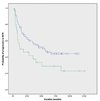
 : absence of cribriform pattern
 : presence of cribriform pattern
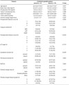

 XML Download