

 PDF
PDF ePub
ePub Citation
 Print
Print


Introduction
Acute toxic-encephalopathy (TME), which includes delirium and confusion, is an acute condition of global cerebral dysfunction in the absence of primary structural brain disease1). TME is common among critically ill patients and usually a consequence of systemic illness2). The electrolyte abnormalities that lead to encephalopathy include hypocalcemia or hypercalcemia, hypomagnesemia, and hypophosphatemia34). Possibly, hypophosphatemia occurs in 10-60% of patients receiving acute renal replacement therapy (RRT), especially continuous or prolonged therapy56). Moderate or severe hypophosphatemia has been recognized as a cause of respiratory muscle weakness7). Herein, we report the case of a 43-year-old woman who developed severe hypophosphatemia-induced acute TME during continuous RRT.
Case Report
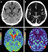
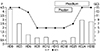
A 43-year-old woman visited the emergency room for oliguria. She was followed up in the hospital for type I diabetes mellitus, chronic kidney disease due to diabetic nephropathy, hypertension and heart failure with dilated cardiomyopathy. She had a history of volume depletion due to diarrhea and a decrease in urine volume. At the time of arrival, level of consciousness was drowsy and the Glasgow coma scale was 14. Physical examination revealed dehydration, including decrease in skin turgor, and tongue dehydration. The vital signs were blood pressure 110/60mmHg, pulse rate 104 beats/min, respiratory rate 20 breaths/min, and body temperature 36.2℃. In the chest auscultation the cardiac sound was normal without murmur and the breath sound was normal. Abdominal examination revealed no specific findings such as tenderness, and no pitting edema was observed in the lower extremities. Complete blood cell count examination revealed hemoglobin 8.3 g/dL; hematocrit 27.7%; WBC 12,070/mm3; and platelet count 195,000/mm3. The initial laboratory values were: serum sodium 134mEq/L; potassium 4.2mEq/L; calcium 8.71mg/dL; phosphorus 4.28mg/dL; magnesium 1.89mg/dL; serum urea nitrogen 46.7mg/dL; creatinine 6.86mg/dL; albumin 2.49 g/dL; total bilirubin 0.12mg/dL; AST/ALT 6.7/3.8 IU/L; glucose 235mg/dL; lactic acid 8.21mg/dL; alkaline phosphatase 68 IU/L; NGAL 739 ng/mL; ammonia 77 ug/dL; Pro-BNP >35,000 pg/mL; and CRP 3.95mg/dL. Arterial blood gas analysis revealed pH 7.14; pCO2 24mmHg; pO2 121mmHg, and tCO2 8.3mL/dL. The serum IgA was 109 mg/dL; ASO 32U; C3 92mg/dL; C4 35mg/dL; FANA negative; and ANCA negative. In the urine analysis, the specific gravity of urine was 1.007; pH 5.0; urine protein 2+; blood 3+; RBC 1–4/HPF; and WBC 5–9/HPF. Urine creatinine level was 14.9mg/dL, and urine protein was 6.78mg/dL. In the immune serum test, the HBs Ag/Ab, Anti-HCV Ab, Anti-HIV Ab, and VDRL results were all negative. Due to persistence of oliguria, continuous RRT was started. On the ninth hospital day, her mentation changed from alert to stupor mentation. The Glasgow coma scale (GCS) changed from 15 to 5. Brain images showed normal findings (Fig. 1). The following laboratory values were obtained: serum sodium 138mEq/L; potassium 4.5 mEq/L; calcium 8.26mg/dL; phosphorus <0.41mg/dL; magnesium 1.96 mg/dL; serum urea nitrogen 21.4mg/dL; creatinine 1.45 mg/dL; albumin 2.32 g/dL; glucose 209 mg/dL; and CRP 2.75mg/dL. After phosphate intravenous replacement, 0.25–0.5mmol/kg monobasic potassium phosphate (Phosten, JW, Korea) for 6 days, and administration of phosphate-containing replacement solution Phoxilium(containing phosphate at 1.2 mmol/L; Baxter Gambro, Deerfield, IL, USA) use, the phosphate level increased to 2.97mEq/L and mental state returned to alert (Fig. 2). On the 29th day of continuous RRT, normal urine volume was recovered with decrease in serum creatinine level. On the 33rd day, serum creatinine level decreased to 2.08mg/dL, subsequently RRT was stopped. At the time of discharge, serum creatinine and phosphorus level were 1.82mg/dL and 2.75mg/dL, respectively with alert mentation. The patient is still on outpatient follow-up.
Discussion
In the present case, severe hypophosphatemia-induced acute TME with oliguric acute kidney injury developed in a patient during continuous RRT. On the day of her hospital arrival, her mental status was alert with normal serum phosphate levels. On the ninth hospital day, her mentation changed from alert to stupor mentation with decrease in serum phosphate level during continuous CRRT. After phosphate intravenous replacement and administration of phosphate-containing replacement solution, her mental state returned to alert with normal serum phosphate level. Brain images and other studies showed normal findings. Based on these findings, severe hypophosphatemia-induced acute TME was diagnosed.
Continuous renal replacement therapy (CRRT) plays a crucial role in critically ill patients with acute kidney injury, particularity those with hemodynamic instability8). However, CRRT is associated with a risk of electrolyte imbalances, including hypophosphatemia, acid-base imbalances, hypotension, infection, bleeding and hypo- thermia910). The incidence of CRRT-related hypophosphatemia widely ranges from 27% to 78%5611). The CRRT-related hypophosphatemia can cause respiratory muscle failure, prolonged mechanical ventilation requirements, longer hospital length of stay, and mortality61112). It has been reported that mild hypophosphatemia is associated with poor outcome13). Lim et al., reported that the risk of prolonged mechanical ventilation increased even in patients with serum phosphorus <2.9mg/dL during CRRT12). In the present case, hypophosphatemia occurred 6 days after CRRT was initiated and her mentation was changed from alert to stupor on 9th day (Fig. 2).
There are several studies on phosphate supplementation for the prevention of hypophosphatemia during CRRT treatment. Troyanov et al. found that addition of 1.2mmol/L of phosphate to the dialysis fluid in adult CRRT patients14). Song et al. reported a dose of 2mmol/L of phosphate as appropriate for patients with CRRT-induced hypophosphatemia13). Recently, commercialized phosphate-containing dialysis solutions (e.g., Phoxilium) are used for CRRT patients15). In the present patient, after phosphate intravenous replacement and administration of phosphate-containing replacement solution, the phosphate level increased to 2.97mEq/L and mental state returned to alert (Fig. 2).
In the present case, severe hypophosphatemia-induced acute TME with oliguric acute kidney injury developed during continuous RRT. Consequently, a careful workup for hypophosphatemia and appropriate phosphate supplementation should be considered in addition to renal care in continuous RRT patient.
 XML Download