

 PDF
PDF Citation
 Print
Print


INTRODUCTION
The Herpesviridae family contains numerous important pathogens that have been classified into 3 subfamilies (Alpha-, Beta-, and Gammaherpesvirinae). Bovine gammaherpesvirus 4 (BoHV-4) belongs to the Gammaherpesvirinae subfamily and is a member of the Rhadinovirus genus [1]. Similar to its human counterparts, BoHV-4 is widespread in natural host populations, and BoHV-4 persists in most individuals resulting in lifelong, asymptomatic infections [2].
The BoHV-4 gene expression cascade is similar to that of other herpesviruses and comprises immediate early (IE), early (E), and late (L) gene expression. The IE gene products are expressed from 2 to 4 hours post-infection (hpi). The genes that encode these proteins are transcribed in the absence of de novo viral gene expression. Moreover, IE gene products activate the expression of E gene products. The E gene products are involved in viral DNA replication, after which the L genes are expressed. Activation of L gene expression requires DNA synthesis [3], and these genes give rise to the structural components of the virion.
The herpesvirus envelope contains various glycoproteins that are involved in virus attachment, penetration, budding, and spread among infected cells. Some of these proteins are highly conserved in both function and sequence, while others are typical of a specific virus genus or species [4].
While most enveloped viruses rely on a single fusogenic protein for entry, herpesviruses have a more complex entry mechanism. Indeed, they use a core fusion machinery that is conserved across the 3 subfamilies [5]. Most of the herpesviruses also employ one or more additional receptor-binding or -regulating proteins specific to subfamilies or genera. This complexity may indicate why herpesvirus entry, particularly its fusion mechanism, remains incompletely described.
The core machinery for herpesvirus entry comprises 3 highly conserved viral glycoproteins (g), gB, gH, and gL, along with one or more accessory glycoproteins necessary for binding to cell surface receptors [67]. In a number of beta- and gammaherpesviruses, including the human pathogens, 2 different gH/gL complexes have been observed on the virion envelope, and those complexes are necessary for the viruses to enter the full range of cell types that they infect in vivo.
The different proteins that compose the viral particles are therefore likely to have critical functions not only in viral structures and entry into target cells but also in evasion of the host's antiviral response [8]. The BoHV-4 mayor glycoproteins are gB, gH, gM, gL, and gp180, with gB, gH, and gp180 being the most glycosylated. These glycoproteins are often involved in cell binding, and some provide neutralization targets [9]. While gL is necessary for entry into the target cell and can form a heterodimer with gH, gB is responsible for adhesion to the host cell and is essential for the lytic replication of BoHV-4 [8]. Glycosylation of these envelope proteins could be involved in virion protection against neutralization by antibodies [9].
It has been suggested that neutralization of virus infectivity by antibodies, directly or indirectly, is a major immunological defense mechanism against virus invasion [1011]. However, during early infection events, these viruses manipulate pre-existing host cell signaling pathways to allow successful infection. In infected cattle, BoHV-4 induces an immune response characterized by low neutralizing antibody levels or an absence of such antibodies [12]. In countries where the virus has been detected, the prevalence is generally low: seropositivity varying from 16% to 30%, with low neutralizing antibody titers (1: 8 a1: 64) [13].
Based on published reports and our previous results, we infer that most BoHV-4 strains do not induce a neutralizing immune response in infected animals. Therefore, virus seroneutralization in vitro cannot always be easily demonstrated. The aim of the present study was to evaluate the neutralizing capacity of 2 Argentine BoHV-4 strains and to associate those findings with the gene expression profiles of the major envelope glycoproteins.
MATERIALS AND METHODS
Virus strains
BoHV-4 strains 07/435 and 10/154, which belong to the American and Argentine clades of BoHV-4 strains, respectively, were used in this study. They were originally isolated from vaginal discharges of adult aborted cows [14]. The strains were passaged twice in Madin-Darby Bovine Kidney (MDBK) cells. Viral stocks were propagated in MDBK cells in T-25 flasks (Greiner Bio-One, Germany) (1 × 10⁵ cells/mL) for 48 h. Supernatants were harvested and frozen at −80°C. Virus titers were determined by the endpoint titration method and expressed as tissue culture infective doses (TCID50), according to the method described by Reed and Müench [15].
Cell line
MDBK cells, cultured in minimum essential medium (MEM) supplemented with 10% fetal bovine serum (FBS) and antibiotic–antimycotic solution (Gibco, USA), were used for BoHV-4 propagation. MDBK cells were provided by the Argentinean Cell Bank (http://www.abac.org.ar/). The cells were free of BoHV-4 and certified as free of contaminating bacteria, mycoplasma, and adventitious viruses. The FBS was negative for anti-BoHV antibodies. The cells were incubated at 37°C in a 5% CO2 atmosphere.
Virus inoculation
MDBK cells were grown in 24-well plates (Greiner Bio-One) at a concentration of 2 × 10⁵ cells/mL and incubated at 37°C in a 5% CO2 atmosphere. Ninety percent confluent monolayers were infected with BoHV-4 strains at a multiplicity of infection (MOI) of 1. After 2 h of incubation, the supernatant was discarded and replaced by fresh medium. At different post-infection times (2, 4, 6, 8, 10, 24, 30, 48, 54, 60, 72, and 80 h) the infected cells and their respective uninfected controls were harvested and stored at −80°C until processing.
RNA extraction and reverse transcription polymerase chain reaction (RT-PCR)
Total RNA from infected and uninfected cells was isolated with TRIzol (Invitrogen, USA). The RNA concentration was determined using a spectrophotometer (Epoch Microplate Spectrophotometer) at absorbances of 260 and 280 nm. The extracted RNA was kept at −80°C until complementary DNA (cDNA) synthesis.
First-strand cDNA was prepared using random hexamer primers and M-MLV Reverse Transcriptase (Promega, USA), according to the manufacturer's protocols. The cDNA samples were stored at −20°C until further analyses. The cDNA products were amplified by polymerase chain reaction (PCR) using primers specific for ORF 50-encoding BoHV-4 IE transcript 2 (IE2), ORF 8-encoding gB, ORF 22-encoding gH, ORF 39-encoding gM, ORF 47-encoding gL, and Bo10-encoding gp180. In order to evaluate PCR specificity, a negative control (PCR mix with H2O) was reacted in parallel with the test samples. Strain 07/435 DNA was used as the positive control. Amplifications were carried out with the primers described in Table 1. The PCR was performed in a final volume of 50 μL, containing 5 mM deoxynucleotide triphosphates (Promega), 10 pmol primers, 2.5 mM MgCl2 (PB-L, Argentina), 10U DNA Taq polymerase (PB-L), 5X buffer (PB-L), 5% dimethylsulfoxide (Sigma Aldrich, Argentina) and water. For each reaction, 0.7 μg of DNA was added. Amplifications were performed in a Veriti Thermal Cycler (Applied Biosystems, USA). The PCR products were separated by electrophoresis in 2% agarose gel, stained with SYBR Safe for DNA gel stain (Thermofisher, Argentina) and visualized under ultraviolet light.
Table 1
Primers used in this study

| Gene | Reference | Orientation | Primer | Fragment (bp) |
|---|---|---|---|---|
| IE2 | Donofrio et al. [23], 2014 | Forward | 5′-ACA AAC ACA CAG ACC AGT CA-3′ | 386 |
| Reverse | 5′-GTT TCA CAA CAG ATT GAG CA-3′ | |||
| gH | Gillet et al., 2004 | Forward | 5′-CCG GGT GAA ACA AGT TCC TG-3′ | 570 |
| Reverse | 5′-GTC AGA GAA CAT ATG ATA ACA TC-3′ | |||
| gp180 | Tebaldi et al. [24], 2016 | Forward | 5′-CCA CCA ACC AGA CCA CAA AT-3′ | 372 |
| Reverse | 5′-ATC TGT CTA TAG CAA CAT CAA ATC C-3′ | |||
| gL | Lété et al. [8], 2012 | Forward | 5′-AAT CTG TCC AGG CCA CTG CTA T-3′ | 320 |
| Reverse | 5′-GCC CAT TCG GAA TAA CTT TAG A-3′ | |||
| gB | Wellenberg et al., 2001 | Forward | 5′-CCC TTC TTT ACC ACC ACC TAC A-3′ | 610 |
| Reverse | 5′-TGC CAT AGC AGA GAA ACA ATG A-3′ | |||
| gM | Lété et al. [8], 2012 | Forward | 5′-CAT CTG ATG ATA CGG GTT CCA-3′ | 960 |
| Reverse | 5′-TGG TTC CAA TCA CTG CCA CT-3′ |
![]()
Direct immunofluorescence (DIF) for detection of BoHV-4 strains
MDBK cells infected with BoHV-4 strains were tested for the BoHV-4 gB antigen, at different times (2, 4, 6, 8, 10, 24, 30, 48, 54, 60, 72, and 80 hpi), by using a direct fluorescent antibody procedure with a commercially available monoclonal antibody labeled with fluorescein isothiocyanate (anti-bovine BHV-4; Bio-X Diagnostics, Belgium). The slides were fixed at −20°C with acetone for 20 min, drained, and dried. The fixed preparations were kept at −20°C until processed following the manufacturer's instructions.
Extraction of viral DNA and restriction endonuclease analysis
To evaluate genotypic differences from the reference strain, field isolated BoHV-4 strains were analyzed with restriction enzymes. MDBK cells were grown in T-25 flasks (Greiner Bio-One) (0.5 × 10⁶ cells/mL). After 24 h of growth and with a 90% confluent monolayer, cells were infected with the indicated BoHV-4 strains at a MOI of 1.
DNA was extracted from infected cells using a commercially available kit (DNeasy Blood & Tissue Kit, Cat. 69504; Qiagen, Germany), according to the manufacturer's instructions. Viral DNA was digested by restriction endonucleases. An aliquot (5 mg) of extracted viral DNA was digested at 37°C with 20 U of EcoR1, BamH1, and HindIII (Promega). The digested product was subjected to electrophoresis at 15 V on 0.8% agarose gel using TAE (Tris–acetate–EDTA) electrophoresis buffer and was visualized with ethidium bromide. Lambda DNA HindIII fragments were used as molecular weight markers.
Virus neutralization (VN) assay
Since infection by BoHV-4 has been described in different animal species, particularly ruminants, we evaluated sera samples from different species by performing VN assays. Samples were submitted from different regions of Argentina for diagnostic purposes and the samples included sera from aborted and pregnant cows, bulls, aborted deer, sheep, and goats, as well as calves inoculated with 10/154 or 07/435 strains of BoVH-4. Experimental inoculations of the calves were carried out at INTA Balcarce (Argentina) in accordance with protocols approved by the Ethical Committee of Animal Welfare (CICUAE) of INTA Res 087/02. Fifteen calves aged 10 months were divided into 3 groups of 5 animals each. Group 1: calves were challenged with isolate 07/435; group 2: calves were challenged with isolate 10/154; and group 3 (mock-infected) calves were inoculated with MEM as a placebo. At the beginning of the experiment all animals were seronegative (SN) for bovine alphaherpesvirus type 1 (BoHV-1) and type 5 (BoHV-5) (checked by VN assay) and for BoHV-4 (checked by enzyme-linked immunosorbent assay [ELISA]; Bio-X Diagnostics, BIO K 263).
The VN assays were performed in 96-well plates (Greiner Bio-One) containing monolayers of MDBK cells; briefly, 60 μL of each viral suspension that contained 200 TCID of the virus were mixed with 60 μL of heat-inactivated (55°C for 30 min) diluted sera and incubated for 1.5 h. The mixtures were then seeded on 96-well culture plates and incubated at 37°C in 5% CO2 for 72 h, following the protocol previously described by Frazier et al. [13]. The cytopathic effect was evaluated and the inverse of the last dilution that completely inhibited viral replication was recorded as the titer value.
RESULTS
Gene expression of BoHV-4 glycoproteins by RT-PCR
BoHV-4 IE2 (RTA) is expressed during the entire virus replication cycle, a process whose initiation and progression has been shown to be strongly impaired in IE2 (ORF50/RTA) BoHV-4 mutants and to be rescued by expression of ORF50/RTA [16]. In keeping with the above functional features, the RT-PCR data (Fig. 1) showed that IE2 gene expression does indeed take place as early as 4 hpi (10/154 strain) or 6 hpi (07/435 strain). Moreover, gene expressions remained detectable until 10 and 24 hpi, respectively; a time that presumably coincides with the advanced stage of late gene expression.
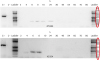 | Fig. 1Expression of the bovine gammaherpesvirus 4 immediate early transcript 2 gene, at different post-infection times, in Madin-Darby Bovine Kidney cells infected with 07/435 and 10/154 strains. C− (negative control): polymerase chain reaction mix with H20. C+ (positive control): 07/435 DNA.
|
Expression kinetics of BoHV-4 ORF 8, ORF 22, ORF 47, ORF 39 and Bo10 genes (coding gB, gH, gL, gM, and gp180, respectively) have not been previously described. Transcription from these genes was analyzed by RT-PCR at various times post-infection (input virus MOI = 1). Time course assays (Fig. 2) indicated that gH, gL, gM, gp180, and gB were expressed earlier in cells infected with strain 10/154 than in those infected with strain 07/435. In 10/154-infected cells, all genes were expressed by 2 hpi, except for gB for which expression was first observed at 10 hpi. On the other hand, in cells infected with strain 07/435, expression of genes coding for gB, gL, and gM was detected at 24 hpi, and expressions of those coding for gp180 and gH were evident at 10 hpi (Fig. 2).
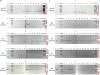 | Fig. 2Time course of BoHV-4 glycoprotein gene expressions. Expression of viral glycoprotein genes gp180, gB, gH, gL, and gM was investigated by reverse transcription polymerase chain reaction at various post-infection times. Input viruses were (A) 07/435 BoHV-4 strain and (B) 10/154 BoHV-4 strain on Madin-Darby Bovine Kidney cells (multiplicity of infection 1).BoHV-4, bovine gammaherpesvirus 4.
|
Time course detection of gB expression by DIF
DIF is used routinely in diagnostic services for virus identification after isolation in cell culture, followed by nested PCR for the BoHV-4-TK gene. Herein, DIF was carried out at different times post-infection of MDBK cells to evaluate whether gB expression is dependent on BoHV-4 strain. Furthermore, it was interesting to determine whether DIF results were in agreement with the gene expression results. DIF-positive cells were detected after 10 h of infection with strain 10/154, whereas in cells infected with strain 07/435, positive cells were visualized from 24 hpi. Thus, in both strains, the times at which positive immunofluorescence was detected coincided with the times from which gB gene expressions were evident. Examples of positive immunofluorescence for BoHV-4 gB and the negative control are presented in Fig. 3.

Enzyme restriction patterns of BoHV-4 strains
EcoR1, BamH1, and HindIII genomic restriction patterns of the 2 tested BoHV-4 strains are shown in Fig. 4. Variability was evident in the restriction enzyme analysis patterns for the strains studied. Variations in the number of restriction sites after digestion with the 3 enzymes were remarkable. The strain 07/435 pattern showed a higher number of fragments than strain 10/154, except for EcoR1 which yielded a number of fragments that was higher for strain 10/154.
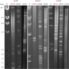 | Fig. 4Genomic restriction patterns of bovine gammaherpesvirus 4 strains 07/435 and 10/154 after digestion with EcoRI, BamHI, or HindIII.M, molecular weight marker (Lambda phage DNA digested with HindIII fragments); DN599, reference strain DN599-like group.
|
The patterns generated after EcoR1 showed some coincidences between the fragments obtained for the 2 BoHV-4 strains studied herein and the American prototype strain DN 599. BamH1 profiles showed remarkable differences in the number of fragments. Only 2 fragments were distinguished for the reference strain, while for strains 10/154 and 07/435, 6 and fourteen fragments, respectively, were obtained after enzymatic digestion. A similar degree of variation in the restriction patterns was observed between strains after HindIII digestion, for which strain 07/435 had the higher number of fragments.
It is important to highlight that the observed differences in restriction enzyme profiles were remarkable. This was previously observed in other studies comparing BoHV-4 strains [14], suggesting that BoHV-4 strains have a complex epidemiological background.
Detection of neutralizing antibodies against BoHV-4
The humoral immune response in bovines infected with BoHV-4 is characterized by low efficiency or low levels of neutralizing antibodies [17]. In experimental infections, high levels of positive serology have been detected by ELISA and immunoblotting. However, neutralizing antibodies were not detected by viral neutralization testing [18].
Serum neutralization assays were performed on 34 archived serum samples obtained from the Specialized Veterinary Diagnostic Service of INTA Balcarce. Samples were from dairy cows, aborted cows, and bulls, from different ruminant species such as deer, goats, and sheep, as well as from calves experimentally-infected with BoHV-4 (Tables 2, 3, 4). All samples evaluated in VN assays with strain10/154 were SN. On the contrary, the same samples were all seropositive when tested with the 07/435 strain, with sample titers ranging from 256 to >512 in dairy cows, aborted cows, and bulls (Table 2). Samples from all of the evaluated dairy cows had the highest titers. Only 3 of 5 sheep serum samples were positive (titer range, 64–128) in VN assay with strain 07/435. Three of seven serum samples from goats were positive. In both sheep and goats neutralizing antibodies were not observed for strain 10/154 (Table 4).
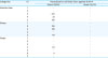
Table 2
BoHV-4 neutralization assay results using serum samples from different bovine categories
![]()
Table 3
VN titers using BoHV-4 isolates 07/435 and 10/154 with homologous and heterologous serum samples from infected and control calves

![]()
Table 4
Virus neutralization titers in serum samples from different ruminant species infected with 07/435 or 10/154 BoHV-4 isolates
![]()
The serologic response of the experimentally-infected calves was weak, regardless of the inoculated BoHV-4 isolate. The presence of neutralizing antibody titers (highest titer 1:64 at 25 days post-infection, dpi) was detected in VN tests performed using isolate 07/435 and in serum samples from calves in the Group 1 (07/435) and Group 2 (10/154) strains. Seroconversion was not detected in calves from any group. It is important to note that at day 0 and in mock-infected calves neutralizing antibodies were detectable when the test was performed with isolate 07/435. These results have also been observed when other Argentine BoHV-4 strains were used to perform VN assays (data not shown). When isolate 10/154 was used for VN assays, all serum samples (homologous and heterologous) were negative (Table 3). Calves in all groups at all times had no neutralizing antibody titers to BoHV-1 (data not shown). Neutralizing antibodies to the 2 strains (07/435 and 10/154) were not detected when sera from colostrum-deprived calves were evaluated.
DISCUSSION
Gammaherpesviruses are responsible for many important diseases in both animals and humans [19]. Some important aspects of their life cycle are still incompletely described, with viral transmission being a key element among those aspects. During co-evolution with their hosts, viruses have developed sophisticated mechanisms in order to hijack some important cellular functions to promote infection. Deciphering these interactions could not only allow a better understanding of virus biology but also could reveal unknown cellular pathways, as has been previously shown [20]. Polysaccharides play central roles in many important biological processes; therefore, it is not surprising that several viruses have exploited glycosylation to their benefit [21]. This study reports on the expression kinetics of genes coding glycoproteins of 2 BoHV-4 Argentine strains. An activator of virus replication and transcription, BoHV-4 IE2 (or BoHV-4 RTA) is conserved among gammaherpesviruses [22] and, in this study, was employed as a primary readout to ensure the infection status of the cells under study. In fact, in permissive cells, BoHV-4 IE2 is expressed during the entire virus replication cycle [23]. The results of this study confirm that IE2 gene expression effectively begins early after cell infection in both BoHV-4 strains tested, and its transcription continues until the advanced stage of late gene expression. Nevertheless, RT-PCR data showed the detection of gB expression when IE2 transcription was undetected in cells infected with the BoHV-4 strains. This is in agreement with the data reported by Tebaldi et al. [24], which demonstrated that the expression of IE2 takes place as early as 2 hpi and reaches a plateau between 10 and 12 hpi, when the expression of late genes, such as gB and IL 1.7, begins.
Regarding the expression of genes coding for envelope glycoproteins, it was evident that the expression of these genes occurred earlier in cells infected with the isolate 10/154 than in those infected with strain 07/435. Lété et al. [8] detected significant glycosylation in 3 of these envelope proteins: gB, gH, and gp180, and Machiels et al. [9] suggested that gp180 provides part of a glycan shield for otherwise vulnerable viral epitopes from gB, gH, and gL, thus contributing to evading the neutralizing antibodies. Considering these findings, it can be suggested that a modification in the glycosylation of gp180 might be responsible for the differences in the capacity of BoHV-4 strains to be neutralized.
Our results clearly demonstrate the differences between the 2 strains under study. The variations found in restriction enzyme analysis patterns are evidently due to the different genotypes of the 2 strains. However, the results obtained in the serological analysis can be attributed to differences in the expression of antigenic proteins or to post-translational modifications that mask neutralizing epitopes. Generally, BoHV-4 is recognized as a virus that does not induce an important humoral immune response, which is characterized by the production of low neutralizing antibody levels [12]. However, strain 07/435 was shown to significantly induce the production of high titers of neutralizing antibodies in different animal species, as well as in bovines.
Considering that antibodies have greater access to gB, gH, and gL, including neutralization epitopes on gp180-deficient virions [8] and the results obtained on gp180 expression, we assume that the delayed expression of this glycoprotein in strain 07/435 leaves unmasked viral antigenic epitopes, which are targets for neutralizing antibodies, resulting in blockage of viral activity. Nevertheless, it remains to be elucidated why neutralizing antibodies are detected in uninfected animals. It is also likely that non-specific neutralization may take place in certain circumstances.
Finally, the determination of the structure of the protein complexes of these strains and the mapping of their functional domains will allow a better understanding of why certain strains are able to induce neutralizing antibodies while others are not. This may lead to the development of more effective diagnostic tests for use in the detection of BoHV-4 infections.
In summary, we describe herein the results of the first comprehensive analysis of the expression kinetics of genes coding BoHV-4 glycoproteins in 2 BoHV-4 Argentine strains that were previously characterized as being of genotypes 1 and 2 [14]. While O-glycans contribute to BoHV-4 evasion of neutralizing antibodies, all together, our results suggest that BoHV-4 strain 07/435 has the ability to neutralize the virus in serum samples. The most relevant serological results were observed in adult animals. In the future, such findings could enhance our understanding of gammaherpesviruses (e.g., BoHV-4) life cycle, including elucidation of the processes of viral entry, assembly, and egress, and they may also help to clarify the genetic variability of the strains circulating in Argentina.
 XML Download