

 PDF
PDF ePub
ePub Citation
 Print
Print


I. Introduction
An inadequate vestibular depth (VD) in the flange region of a removable partial denture (RPD) can impede retention of the denture1. If the VD is insufficient, retention can be achieved using an implant-supported denture, but an RPD without implant support may be required for financial reasons. Vestibuloplasty is a procedure that mainly improves retention of prostheses and produces an environment where oral hygiene can be properly managed. The currently known vestibuloplasty techniques for obtaining VD suffer loss of obtained depth due to scar formation or continuous stimulation of the vestibule over time2.
Many studies on development of techniques to prevent relapse after vestibuloplasty have been carried out. Many modifications, such as Clark's technique, the Kazanjian technique, autogenous grafts (skin, palatal, or cheek), and allodermal and xenodermal grafts combined with autogenous grafts, have been introduced3. Skin grafts or mucosa grafts can reduce the relapse rate by 20% to 30%, but a free gingival graft (FGG) is technique sensitive, so careful attention is needed to achieve successful results4. Therefore, it is essential to develop less technique-sensitive methods to effectively perform vestibuloplasty with FGG.
Titanium is used in a variety of medical fields because of its high corrosion resistance, low toxicity, very low allergenic potency, and good biocompatibility5. In general, titanium mesh is used in the submucosal region, mainly for guided bone regeneration (GBR), because of its forming force, strength, osteocyte activity of the titanium oxide layer, and biocompatibility6. In addition, titanium is used widely as a material for dental prostheses because of its ability to adapt well to the oral environment invaded by bacteria and foods with multiple acidities7. Therefore, titanium mesh can also be used in situations where it is exposed to the oral environment.
This paper introduces a case of vestibuloplasty with a FGG performed easily and effectively in the anterior mandible titanium mesh to obtain the appropriate VD and keratinized gingiva (KG).
II. Technical Note
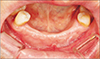
A female patient was admitted to the Emergency Department of Chonbuk National University Hospital for a #34 to #44 avulsion and lip penetration wound caused by a bicycle accident. A suture repair was performed. At 10 months after trauma, the patient visited the clinic for RPD fabrication. Vestibuloplasty was planned before fabrication of the RPD because of insufficient VD for retention of the RPD due to scar tissue constriction of the injured area.(Fig. 1)
Local anesthesia (Lidocaine HCl; Huons, Seongnam, Korea) was administered at the surgical site. An approximately 3 cm horizontal incision was created along the margin of the remaining KG with a No. 15 blade (Braun Manufacturing, Frankfurt, Germany), and a vertical incision was made from the distal aspect of the horizontal incision toward the vestibule. A curette was used to dissect and separate the mucous membrane and the mentalis muscle from the periosteum to create a recipient site.(Fig. 2. A)
Two rectangular FGGs (2 cm long and 1 cm wide) were harvested from the left and right palatal mucosa using a conventional technique. The incision depth was approximately 1.5 mm, and the graft and adipose tissues were well-separated during harvest to obtain an adequate amount of mucosal tissue. The interrupted suture for the harvested graft was performed using 5-0 Vicryl (Ethicon, Somerville, NJ, USA).(Fig. 2. B) The size of the Neo Titanium mesh (depth 0.085 mm, hole size diameter 0.4 mm; Neo Biotech, Seoul, Korea) was adjusted to that of the graft, and the mesh was fixed above the graft using mini-screws (diameter 1.5/2 mm, length 4 mm, bone screw system; Osung MND, Gimpo, Korea).(Fig. 2. C)
Surgicel (Johnson & Johnson, New Brunswick, NJ, USA) was applied to the palatal donor site to protect the wound, and a 0.5 mm Omnivec (3A MEDES, Goyang, Korea) obturator that was previously fabricated was placed on the site1.
Cephalosporin antibiotics and ibuprofen were prescribed for one week, and the patient was instructed to apply 0.1% chlorhexidine gluconate (Alpha-hexidine; Firson, Cheonan, Korea) twice a day for two weeks. The obturator was removed after five days, and the patient was provided instructions regarding a soft diet and conventional oral hygiene care. In this study, to prevent a relapse four weeks after surgery8, all the soft tissue that grew over the mesh was trimmed before removing the titanium mesh.(Fig. 3)
Four weeks after removing the mesh, normal recovery was observed, and sufficient KG and VD were obtained.(Fig. 4) At 10.3 months after surgery, the patient visited the hospital for fabrication of the RPD in the edentulous state without prosthetic treatment, and the VD was properly maintained. The average relapse rate was calculated by measuring the vertical depth from the scar margin of the gingival crest to the movable alveolar mucosa at the mesial, distal, and center of both operation sites. The relapse rate of VD was 27.38% and 24.57% on the right and left sides, respectively.
III. Discussion
An adequate VD is an important factor for proper oral hygiene care9. The sequelae of various intraoral surgical approaches, such as bone grafting, implant placement, and fracture surgery, may result in loss of KG and reduced VD. If the oral vestibule is shallow or KG is insufficient, it is necessary to produce an environment suitable for retention, stability, and function of the prostheses10. Vestibuloplasty is performed when the amount of alveolar bone remaining is appropriate, and there is a lack of covering mucosal tissue, or it is unsuitable for denture support because of pathology, such as scar formation. This is a procedure to secure the prosthesis support area by preventing muscle pull and producing a deep vestibule. Unfortunately, vestibuloplasty is widely known to have a tendency for shrinkage because of the action of the adjacent muscles or scar tissue formation2. An attempt was made to reduce the relapse rate using a range of surgical techniques, including the Clark method, the Kazanjian method, the Edlan-mejchar method, and a graft with autologous, xenoderm, and alloderm materials11. In 2016, Kumar et al.2 reported that an appropriate VD was obtained using cotton rolls and a lip-periosteum mattress suture using the modified Kazanjian vestibuloplasty technique in the area of the anterior region. On the other hand, complications, such as scar formation on the lip, occurred in some patients. Therefore, technological developments are still necessary2. On the other hand, vestibuloplasty with a graft is advantageous because it reduces the incidence of relapse when there is insufficient alveolar bone or a bone defect. The level of postoperative satisfaction of patients who have undergone vestibuloplasty with a graft has been reported to be high with reduced healing period and denture placement time after surgery12. Nevertheless, many patients refuse this treatment because of the pain and discomfort at the grafting site and the need for an additional surgical site. Although many alternatives for soft tissue grafting such as living cellular construct (LCC), extracellular matrix membrane (ECM), acellular dermal matrix (ADM), bilayer collagen membrane (BCM), and LCC have been introduced, a recent systematic review has shown that they do not result in the same amount of keratinized tissue obtained through FGG13. Therefore, in this study, vestibuloplasty combined with FGG was performed to secure the VD despite the patient's discomfort. Even if vestibuloplasty using grafting is performed, relapse can occur due to graft shrinkage. In general, shrinkage of the graft occurs most frequently after one to four weeks in cases of FGG to increase the attached gingiva8. Therefore, in this study, an attempt was made to minimize the effects of a reduction in VD due to graft shrinkage by maintaining the titanium mesh for four weeks, when the most shrinkage occurs. The obtained VD and KG were maintained at eight weeks after vestibuloplasty, and the tissue remaining after removing the titanium mesh appeared to have undergone remodeling with the recipient site in a desirable way.
To obtain successful outcomes of FGG in general, close attention is needed to prevent dead space, hematoma formation, and instability or movement of the graft that can cause tissue necrosis through pre-operative preparation of the recipient site, suitability of the graft, and rigid fixation of the graft4. Studies on reduction of scar tissue after grafting using the pressure of the temporary prosthesis have shown that application of uniform pressure contributes to rapid revascularization, which leads to successful grafting and minimal graft shrinkage14. On the other hand, issues such as necrosis of the graft due to excessive pressure have been reported15. In this study, stable fixation to minimize the sutures that could serve as a pathway of infection was obtained using a titanium mesh and miniscrews, while avoiding overpressure15. In addition, the surgery time could be drastically reduced by avoiding sutures for an apically positioned flap of a conventional vestibuloplasty2. Although additional surgery is necessary for mesh removal, it could provide an opportunity to separate and remove any undesired tissue from the graft site.
Titanium has many attractive properties such as corrosion resistance, low toxicity, very low allergenic potency, and good biocompatibility5. Therefore, it has been used frequently with a high rate of success in dentistry, in internal fixations and artificial joints in orthopedics, and in instruments such as pacemakers and stents in cardiovascular surgery5. Recently, it has been used widely in the form of dental implant fixtures, miniscrews, and mesh for bone grafts as a result of developments in implant dentistry. In general, titanium meshes with a thickness of 0.2 mm are used mainly in GBR because they are durable enough to retain the shape of grafted bones. The meshes are also flexible enough to minimize the risk of mucosal dehiscence16. In this study, because the aim of using a titanium mesh was to obtain close contact of the graft and rigid fixation, a thin mesh with a thickness of 0.085 mm was used to minimize the patient's discomfort. The titanium mesh located at the subgingiva for GBR does not block blood flow from the bone and mucosa because of its pores16. On the other hand, remodeling of the implanted bone is not achieved easily if the mesh is exposed to the gingiva, even if there is no inflammation in the exposed area due to titanium biocompatibility17. This results from the pores providing pathways for external saliva and bacteria18. Few studies using titanium meshes in the oral environment have been reported because oral exposure of the titanium mesh itself is considered a complication18. In this study, no complications were detected because a relatively thick FGG (1.2–1.3 mm) that is resistant to infections was applied with the titanium mesh. In addition, although patients who underwent vestibuloplasty using FGG generally suffer from pain due to exposure of the surgical site, difficulty in oral hygiene care, and difficulty in postoperative management due to delayed recovery19, the patient with mesh covering the surgical site experienced almost no discomfort or pain. Titanium can be dissolved in fluoride-containing solutions. Nevertheless, at the concentration of fluoride in commercially available toothpaste (at a fluoride concentration of approximately 1,000 ppm), titanium alloys do not corrode20; therefore, conventional oral self-care can be performed easily. These results suggest that the myth of exposure to the oral environment as a complication can be overcome by applying titanium mesh only to the submucosal area in GBR and applying a titanium mesh with effective biomechanical properties under various oral conditions.
In 1976, Ward9 recommended vestibuloplasty when the vestibule depth was 4 mm or less. There have been few studies on the criteria for VD suitable for retention of prostheses since. Recently, it was reported that a reduced VD is a risk factor for marginal bone loss around implants, loss of KG, and gingival recession10. Therefore, further studies on vestibuloplasty using a titanium mesh need to be conducted in implant patients with insufficient KG and VD.
In conclusion, this case demonstrates a successful outcome for vestibuloplasty on the anterior mandible. No complications following the vestibuloplasty using a titanium mesh were observed, and the patient did not complain of pain or discomfort. The VD produced by vestibuloplasty using titanium mesh was maintained well for eight weeks after surgery, even though no additional pressure had been applied using the prosthesis. These results suggest that a vestibuloplasty using a titanium mesh can be used effectively in clinical practice.


 XML Download