III. Results
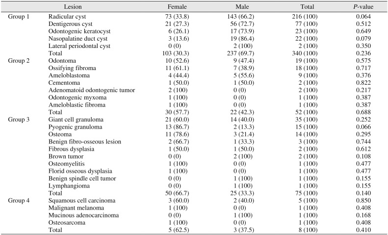
A total of 475 oral biopsies was located among the archived records. Of these, 287 cases were diagnosed in males (60.4%) and 188 cases were diagnosed in females (39.6%). The distribution of the groups by sex and the statistical significance are shown in
Table 1.
Table 1
Distributions of groups by sex

|
Lesion |
Female |
Male |
Total |
P-value |
|
Group 1 |
Radicular cyst |
73 (33.8) |
143 (66.2) |
216 (100) |
0.064 |
|
Dentigerous cyst |
21 (27.3) |
56 (72.7) |
77 (100) |
0.512 |
|
Odontogenic keratocyst |
6 (26.1) |
17 (73.9) |
23 (100) |
0.649 |
|
Nasopalatine duct cyst |
3 (13.6) |
19 (86.4) |
22 (100) |
0.079 |
|
Lateral periodontal cyst |
0 (0) |
2 (100) |
2 (100) |
0.350 |
|
Total |
103 (30.3) |
237 (69.7) |
340 (100) |
0.236 |
|
Group 2 |
Odontoma |
10 (52.6) |
9 (47.4) |
19 (100) |
0.575 |
|
Ossifying fibroma |
11 (61.1) |
7 (38.9) |
18 (100) |
0.717 |
|
Ameloblastoma |
4 (44.4) |
5 (55.6) |
9 (100) |
0.376 |
|
Cementoma |
1 (50.0) |
1 (50.0) |
2 (100) |
0.822 |
|
Adenomatoid odontogenic tumor |
2 (100) |
0 (0) |
2 (100) |
0.217 |
|
Odontogenic myxoma |
1 (100) |
0 (0) |
1 (100) |
0.387 |
|
Ameloblastic fibroma |
1 (100) |
0 (0) |
1 (100) |
0.387 |
|
Total |
30 (57.7) |
22 (42.3) |
52 (100) |
0.688 |
|
Group 3 |
Giant cell granuloma |
21 (60.0) |
14 (40.0) |
35 (100) |
0.252 |
|
Pyogenic granuloma |
13 (86.7) |
2 (13.3) |
15 (100) |
0.066 |
|
Osteoma |
11 (78.6) |
3 (21.4) |
14 (100) |
0.295 |
|
Benign fibro-osseous lesion |
2 (66.7) |
1 (33.3) |
3 (100) |
0.744 |
|
Fibrous dysplasia |
1 (50.0) |
1 (50.0) |
2 (100) |
0.612 |
|
Brown tumor |
0 (0) |
2 (100) |
2 (100) |
0.108 |
|
Osteomyelitis |
1 (100) |
0 (0) |
1 (100) |
0.477 |
|
Florid osseous dysplasia |
1 (100) |
0 (0) |
1 (100) |
0.477 |
|
Benign spindle cell tumor |
0 (0) |
1 (100) |
1 (100) |
0.155 |
|
Lymphangioma |
0 (0) |
1 (100) |
1 (100) |
0.155 |
|
Total |
50 (66.7) |
25 (33.3) |
75 (100) |
0.140 |
|
Group 4 |
Squamous cell carcinoma |
3 (60.0) |
2 (40.0) |
5 (100) |
0.850 |
|
Malignant melanoma |
1 (100) |
0 (0) |
1 (100) |
0.408 |
|
Mucinous adenocarcinoma |
0 (0) |
1 (100) |
1 (100) |
0.168 |
|
Osteosarcoma |
1 (100) |
0 (0) |
1 (100) |
0.408 |
|
Total |
5 (62.5) |
3 (37.5) |
8 (100) |
0.410 |

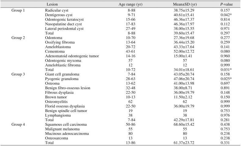
The mean age of all patients was 39.78±16.7 years (range, 7–88 years). The age distribution and statistical values of the groups are presented in
Table 2.
Table 2
Distributions of groups by age

|
Lesion |
Age range (yr) |
Mean±SD (yr) |
P-value |
|
Group 1 |
Radicular cyst |
8-88 |
38.75±15.29 |
0.157 |
|
Dentigerous cyst |
9-71 |
40.61±15.41 |
0.042*
|
|
Odontogenic keratocyst |
15-66 |
46.36±17.37 |
0.814 |
|
Nasopalatine duct cyst |
17-83 |
46.36±17.97 |
0.112 |
|
Lateral periodontal cyst |
27-49 |
38.00±15.55 |
0.971 |
|
Total |
8-88 |
39.60±15.47 |
0.297 |
|
Group 2 |
Odontoma |
10-70 |
27.36±19.68 |
0.277 |
|
Ossifying fibroma |
13-64 |
36.44±15.20 |
0.259 |
|
Ameloblastoma |
20-72 |
43.33±17.64 |
0.141 |
|
Cementoma |
43-61 |
52.00±12.72 |
0.080 |
|
Adenomatoid odontogenic tumor |
14-16 |
15.00±1.41 |
0.960 |
|
Odontogenic myxoma |
57 |
57 |
0.080 |
|
Ameloblastic fibroma |
12 |
12 |
0.999 |
|
Total |
10-72 |
34.01±18.61 |
0.031*
|
|
Group 3 |
Giant cell granuloma |
7-84 |
43.05±20.74 |
0.158 |
|
Pyogenic granuloma |
28-63 |
47.06±20.74 |
0.025*
|
|
Osteoma |
13-62 |
41.00±13.98 |
0.697 |
|
Benign fibro-osseous lesion |
32-48 |
38.00±8.71 |
0.891 |
|
Fibrous dysplasia |
22-50 |
36.00±19.79 |
0.148 |
|
Brown tumor |
10-13 |
11.50±2.12 |
0.150 |
|
Osteomyelitis |
62 |
62 |
0.999 |
|
Florid osseous dysplasia |
22-50 |
36.00±19.79 |
0.999 |
|
Benign spindle cell tumor |
19 |
19 |
0.753 |
|
Lymphangioma |
38 |
38 |
0.976 |
|
Total |
7-84 |
42.29±17.81 |
0.281 |
|
Group 4 |
Squamous cell carcinoma |
50-86 |
68.60±15.42 |
0.438 |
|
Malignant melanoma |
55 |
55 |
0.753 |
|
Mucinous adenocarcinoma |
80 |
80 |
0.238 |
|
Osteosarcoma |
13 |
13 |
0.238 |
|
Total |
13-86 |
61.37±23.72 |
0.331 |

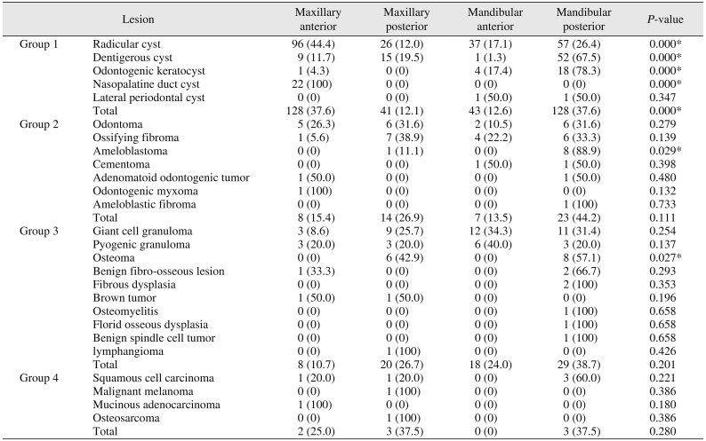
Of the 475 cases, 251 cases (52.8%) occurred in the mandible and 224 cases (47.2%) in the maxilla. The location distribution of pathologies according to group and associated statistical values are shown in
Table 3.
Table 3
Distributions of groups by anatomical location of the lesion

|
Lesion |
Maxillary anterior |
Maxillary posterior |
Mandibular anterior |
Mandibular posterior |
P-value |
|
Group 1 |
Squamous cell carcinoma |
96 (44.4) |
26 (12.0) |
37 (17.1) |
57 (26.4) |
0.000*
|
|
Dentigerous cyst |
9 (11.7) |
15 (19.5) |
1 (1.3) |
52 (67.5) |
0.000*
|
|
Odontogenic keratocyst |
1 (4.3) |
0 (0) |
4 (17.4) |
18 (78.3) |
0.000*
|
|
Nasopalatine duct cyst |
22 (100) |
0 (0) |
0 (0) |
0 (0) |
0.000*
|
|
Lateral periodontal cyst |
0 (0) |
0 (0) |
1 (50.0) |
1 (50.0) |
0.347 |
|
Total |
128 (37.6) |
41 (12.1) |
43 (12.6) |
128 (37.6) |
0.000*
|
|
Group 2 |
Odontoma |
5 (26.3) |
6 (31.6) |
2 (10.5) |
6 (31.6) |
0.279 |
|
Ossifying fibroma |
1 (5.6) |
7 (38.9) |
4 (22.2) |
6 (33.3) |
0.139 |
|
Ameloblastoma |
0 (0) |
1 (11.1) |
0 (0) |
8 (88.9) |
0.029*
|
|
Cementoma |
0 (0) |
0 (0) |
1 (50.0) |
1 (50.0) |
0.398 |
|
Adenomatoid odontogenic tumor |
1 (50.0) |
0 (0) |
0 (0) |
1 (50.0) |
0.480 |
|
Odontogenic myxoma |
1 (100) |
0 (0) |
0 (0) |
0 (0) |
0.132 |
|
Ameloblastic fibroma |
0 (0) |
0 (0) |
0 (0) |
1 (100) |
0.733 |
|
Total |
8 (15.4) |
14 (26.9) |
7 (13.5) |
23 (44.2) |
0.111 |
|
Group 3 |
Giant cell granuloma |
3 (8.6) |
9 (25.7) |
12 (34.3) |
11 (31.4) |
0.254 |
|
Pyogenic granuloma |
3 (20.0) |
3 (20.0) |
6 (40.0) |
3 (20.0) |
0.137 |
|
Osteoma |
0 (0) |
6 (42.9) |
0 (0) |
8 (57.1) |
0.027*
|
|
Benign fibro-osseous lesion |
1 (33.3) |
0 (0) |
0 (0) |
2 (66.7) |
0.293 |
|
Fibrous dysplasia |
0 (0) |
0 (0) |
0 (0) |
2 (100) |
0.353 |
|
Brown tumor |
1 (50.0) |
1 (50.0) |
0 (0) |
0 (0) |
0.196 |
|
Osteomyelitis |
0 (0) |
0 (0) |
0 (0) |
1 (100) |
0.658 |
|
Florid osseous dysplasia |
0 (0) |
0 (0) |
0 (0) |
1 (100) |
0.658 |
|
Benign spindle cell tumor |
0 (0) |
0 (0) |
0 (0) |
1 (100) |
0.658 |
|
lymphangioma |
0 (0) |
1 (100) |
0 (0) |
0 (0) |
0.426 |
|
Total |
8 (10.7) |
20 (26.7) |
18 (24.0) |
29 (38.7) |
0.201 |
|
Group 4 |
Squamous cell carcinoma |
1 (20.0) |
1 (20.0) |
0 (0) |
3 (60.0) |
0.221 |
|
Malignant melanoma |
0 (0) |
1 (100) |
0 (0) |
0 (0) |
0.386 |
|
Mucinous adenocarcinoma |
1 (100) |
0 (0) |
0 (0) |
0 (0) |
0.180 |
|
Osteosarcoma |
0 (0) |
1 (100) |
0 (0) |
0 (0) |
0.386 |
|
Total |
2 (25.0) |
3 (37.5) |
0 (0) |
3 (37.5) |
0.280 |

The group distributions were as follows: group 1 (n=340; 71.6%), group 2 (n=52; 10.9%), group 3 (n=75; 15.8%), and group 4 (n=8; 1.7%). Among all lesions, radicular cysts (n=216; 45.5%) were the most common biopsied lesion, followed by dentigerous cysts (n=77; 16.2%), giant cell granulomas (n=35; 7.4%), and odontogenic keratocysts (n=23; 4.8%).
Within group 1, 216 (63.5%) of the 340 cases were of radicular cyst, followed by 77 cases (22.6%) of dentigerous cyst, 23 cases (6.8%) of odontogenic keratocyst, 22 cases (6.5%) of nasopalatine duct cysts, and two cases (0.6%) of lateral periodontal cysts. One hundred sixty-nine of the cysts involved the maxilla (49.7%), while 171 were present in the mandible (50.3%). The most commonly affected sites were the mandibular posterior region (n=128; 37.6%) and maxillary anterior region (n=128; 37.6%), followed by the mandibular anterior region (n=43; 12.6%) and maxillary posterior region (n=41; 12.1%).(
Table 3) Overall, radicular cyst was the most frequent type observed in group 1. The mean age of patients in this group was 38.75±15.29 years (range, 8–88 years) and the female:male ratio was 1:2. Radicular cysts were most commonly observed in the maxillary anterior region and secondly in the mandibular posterior region.
In group 2, odontoma (n=19; 36.5%) was the most frequent odontogenic tumor of the 52 cases, followed by ossifying fibroma (n=18; 34.6%), ameloblastoma (n=9; 17.3%), cementoma (n=2; 3.8%), adenomatoid odontogenic tumor (n=2; 3.8%), odontogenic myxoma (n=1; 1.9%), and ameloblastic fibroma (n=1; 1.9%). Thirty cases (57.7%) of odontogenic tumors were in the mandible and 22 cases (42.3%) were in the maxilla. The most affected site was the mandibular posterior region (n=23; 44.2%), followed by the maxillary posterior region (n=14; 26.9%), maxillary anterior region (n=8; 15.4%), and mandibular anterior region (n=7; 13.5%).(
Table 3) The mean age among the odontoma cases, which was the most frequent odontogenic tumor, was 27.3±19.6 years (range, 10–70 years), and the female:male ratio was 10:9. Odontomas were seen equally in the mandibular posterior and maxillary posterior regions.
In group 3, 35 (46.7%) of the 75 cases were giant cell granuloma, followed by 15 cases (20.0%) of pyogenic granuloma; 14 cases (18.7%) of osteoma; and 11 cases (14.7%) of other lesions including benign fibro-osseous lesions (n=3; 4.0%), fibrous dysplasia (n=2; 2.7%), brown tumor (n=2; 2.7%), osteomyelitis (n=1; 1.3%), florid osseous dysplasia (n=1; 1.3%), benign spindle cell tumor (n=1; 1.3%), and lymphangioma (n=1; 1.3%). Bone tumor and related lesions were seen in 47 cases (62.7%) in the mandible and 28 cases (37.3%) in the maxilla. The most affected site was the mandibular posterior region (n=29; 38.7%), followed by the maxillary posterior region (n=20; 26.7%), mandibular anterior region (n=18; 24.0%), and maxillary anterior region (n=8; 10.7%).(
Table 3) The most frequent lesion in this group was giant cell granuloma, and the mean age of these patients was 43.05±20.74 years (range, 7–84 years), with a female:male ratio of 3:2. Giant cell granulomas were seen most commonly in the mandibular anterior region and secondly in the mandibular posterior region.
Finally, in group 4, five (62.5%) of the eight cases were squamous cell carcinoma (SCC), followed by one case (12.5%) of malign melanoma, one case (12.5%) of mucinous adenocarcinoma, and one case (12.5%) of osteosarcoma. Malignant tumors of the jaw were seen in five cases (62.5%) involving the maxilla and thee cases (37.5%) involving the mandible. The maxillary posterior region and the mandibular posterior region were affected equally. The most frequent type of lesion in this group was SCC. The mean age of patients was 68.6±15.42 years (range, 50–86 years), and the female:male ratio was 3:2. SCC was most commonly seen in the mandibular posterior region, while the maxillary anterior and maxillary posterior regions were affected equally.(
Table 3)
Go to :

IV. Discussion
OMF lesions can cause many pathological changes in the hard and soft tissues of affected regions. The prevalence and incidence of these lesions may vary according to age, sex, and location of the pathology; knowledge of these factors can aid in identifying the lesion type and informing the patients correctly. However, these factors alone are often not enough for clear determination of the lesion; clinical and radiological examination, biopsy, and histopathological examination are crucial for final diagnosis and treatment planning.
In the literature, OMF lesion names, groups, and subtypes have changed regularly since Broca
5 first described them in 1868. The first edition of histological classification of odontogenic tumors was published by the WHO in 1971 and has been revised three times to date
6. In particular, there were five major changes in the new classification released in 2017 compared with the third version from 2005
1: odontogenic cysts, which were not included in the previous classifications, are classified mainly based on their true nature
2; odontogenic keratocyst and calcifying odontogenic cyst were removed from classification of odontogenic tumors and were included instead in classification of odontogenic cysts
3; new entities such as sclerosing odontogenic carcinoma and primordial odontogenic tumor were identified
4; ameloblastic fibrodentinoma, ameloblastic fibro-odontoma, and odontoameloblastoma were removed from the classification scheme entirely
5; and osseous dysplasia and ossifying fibroma were renamed as cemento-osseous dysplasia and cemento-ossifying fibroma
7.
In the present study, the most common group of lesions was odontogenic cysts (71.6%), followed by bone tumors and related lesions (15.8%), odontogenic tumors (10.9%), and malignant tumors of the jaw (1.7%). According to a study by Jaafari-Ashkavandi and Akbari
8 conducted in Iran, the odontogenic cyst group (63%) was the most common, followed by groups of benign bone pathologies (15.9%), odontogenic tumors (11.9%), and malignant tumors of the jaw (2.8%) and these results were very similar to those in our study. Separately, in a prior retrospective study conducted in Turkey, cystic lesions were found to compose 17.13%, benign tumor and related lesions composed 8.25%, malignant tumors composed 1.72%, inflammatory and reactive lesions composed 35.69% and other lesions composed 37.2% of the total findings
9.
Tekkesin et al.
10 evaluated 5,088 odontogenic and nonodontogenic cyst cases and reported that the most common cyst present was the radicular cyst, followed by the odontogenic keratocyst and dentigerous cyst, and these authors claimed that Turkish populations have high risk of aggressive cysts because of the more frequent appearance of the odontogenic keratocyst than the dentigerous cyst. Another study conducted in the southeast region of Turkey showed that 63% of the cysts were reported as radicular cysts, 26.9% as dentigerous cysts, 6.1% as odontogenic keratocysts, 3.4% as residual radicular cysts, and 0.6% as nasopalatine cysts
11. The most frequent cyst observed was the radicular cyst (63.5%), followed by the dentigerous cyst (22.6%) and the odontogenic keratocyst (6.7%) in the present study. de Souza et al.
12 found that the most frequent odontogenic cysts in their investigation were radicular cysts (61.4%), followed by dentigerous cysts (20.1%) and odontogenic keratocysts (6.4%), showing similar results to those of our research.
Age is one of the major factors for the distribution of odontogenic cysts and the distribution pattern of odontogenic cysts can change with age. In our study, the mean age of patients was 39.6±15.47 years. In the study of Tekkesin et al.
10, also conducted in Turkey, the mean age was 36.33 years, while, in another study
12 performed in the Brazilian population, the mean age of patients was 31 years.
In the present study, cysts were found in men (69.7%) more frequently than in women (30.3%). Similar results were found among studies conducted in our country but to a rate that was less than 69.7%, even though the incidence remained higher in males. In the study by Tekkesin et al.
10, cysts were more common in male patients (57.7%) according to 5,088 biopsies, and Açıkgöz et al.
13 also reported a male predominance (53.8%) in their study involving a Turkish population.
In this study, 169 cysts were located in the maxilla (49.7%) and 171 were located in the mandible (50.3%), with the most affected sites being the mandibular posterior region (n=128; 37.6%) and maxillary anterior region (n=128; 37.6%), followed by mandibular anterior region (n=43; 12.6%), and maxillary posterior region (n=41; 12.1%). Radicular cysts more often affected the maxillary anterior region, while dentigerous cysts and odontogenic keratocyst were more commonly observed in the mandibular posterior region, similar to in other reports in the literature
1011121314.
Members of the odontogenic tumor group composed 10.9% of cases among all biopsies in the present study. In the literature, it was reported that odontogenic tumor rates range from 1.2% to 2.5% of all biopsies across studies, a ratio that is less than that in our research
151617. The main reason for this difference may be exclusion of soft tissue reactive lesions and inflammatory processes in this study.
The most frequent odontogenic tumors were, in order, odontoma (36.5%), cemento-ossifying fibroma (34.6%), ameloblastoma (17.3%), adenomatoid odontogenic tumor (3.8%), cementoma (3.8%), odontogenic myxoma (1.9%), and ameloblastic fibroma (1.9%) in the present study. As previously mentioned, odontogenic keratocyst was considered a cyst according to the second edition of the WHO classification, while it was regarded as a tumor in the third edition. In the fourth edition published in 2017, odontogenic keratocyst was described as a cyst again, and this modification likely influences the rankings of odontogenic tumors in the literature. According to Jaeger et al.
18, odontoma (39.67%) was the most common odontogenic tumor, similar to in our research, when odontogenic keratocyst was defined as a cyst. However, when it was described as an odontogenic tumor, the most common tumor was the keratocystic odontogenic tumor (41.07%); in this situation, the ratio of odontogenic tumors to all pathologies increased from 2.04 to 11.51% in their study. In a multicentric retrospective study of 2,000 cases in the Turkish population, the keratocystic odontogenic tumor (classified as a tumor at the time) was the most frequent finding (57.2%), followed by odontoma (16.7%) and ameloblastoma (15.08%)
19. In another study
20 conducted in Turkey, ameloblastoma (36.5%) was reported as the most common tumor, followed by odontoma (18%) and cementoma (15%). However, the incidence of aggressive tumors such as ameloblastoma is much higher in African countries. According to Akinmoladun et al.
21, whose study was conducted in Nigeria, 1,754 ameloblastomas were reported among 3,075 cases (57.04%), while, in the study by Adebayo et al.
22, cases of ameloblastoma composed 73% of all tumors. Thus, in studies conducted in Africa
2122, the frequency of ameloblastoma is significantly higher in comparison with other regions of the world, including Turkey. This may be due to limited health care services in countries with populations of lower socioeconomic levels, for which people can only receive health care after emergence of severe symptoms, and routine treatment is not often available to catch signs and symptoms early
23.
According to Jing et al.
24, the age of patients with odontogenic tumors varied from three to 84 years with a mean age of 32.1 years, while, in Turkey, Sekerci et al.
25 reported that the age of their patients ranged from 10 to 84 years, with a mean age of 34.52 years. These results are very similar to those in our study; we reported a mean age of 34.01±18.61 years in patients with odontogenic tumor.
In the present study, odontogenic tumors were reported in women (57.7%) more frequently than in men (42.3%). Olgac et al.
26 reported that odontogenic tumors were seen in female patients (53%) more so than in male patients (47%) in Turkey, which is similar to in our study. According to another study
25 conducted in our country, a slight predominance of afflicted males (50.5%) was reported, but this result was not statistically significant.
Odontogenic tumors in the study of Jing et al.
24 were observed in 75.5% of all mandibular jaw cases in China, while another study in Turkey
25 reported 77.9% of such cases in the mandible. In the present study, the most typically affected site was the mandibular posterior region (44.2%), followed by the maxillary posterior region (26.9%), maxillary anterior region (15.4%), and mandibular anterior region (13.5%), similar to other studies.
The bone tumors and related lesions group included 15.8% of all pathologies in the present study. Of the 75 cases, 46.7% were giant cell granuloma; 20% were pyogenic granuloma; 18.7% were osteoma; and 14.6% were other lesions including fibrous dysplasia, osteomyelitis, florid osseous dysplasia, brown tumor, lymphangioma, and benign spindle cell tumor. According to Jaafari-Ashkavandi and Akbari
8, benign bone pathologies were the second most common lesions (15.9% of all lesions) after odontogenic cysts, and central giant cell granuloma was the most common lesion of all benign bone pathologies. However, some researchers reported odontogenic tumors more frequently than benign bone pathologies in their study populations
2728. In the study of Jaafari-Ashkavandi and Akbari
8, ossifying fibroma was accepted as a benign bone lesion, while we included it in the odontogenic tumor group as cemento-ossifying fibroma, based on the fourth edition of the WHO classification
7. In our study, if ossifying fibroma had been included in the bone tumors and related lesions group, the incidence among all pathologies would have increased from 15.7% to 19.5%.
As indicated above, the most frequent type of lesion among bone tumors and related lesions was giant cell granuloma, with a mean age of affected patients of 43.05 years (range, 7–84 years). Of this subpopulation of 35 cases, 21 (60.0%) were female and 14 (40.0%) were male. In the Boffano et al.'s study
29, the mean age of patients with peripheral giant cell granuloma was 48.8 years (range, 8–89 years), and the lesions showed female preponderance (62.3%). Although the sex distribution of such lesions between their study and ours was fairly equal, the lesion appeared more often in older patients in our study. In the literature, peripheral giant cell granuloma has been seen more commonly in the mandible than the maxilla
30. However, the maxilla was involved in 50.6%, and the mandible affected 49.4% of about 874 cases in the Boffano et al.'s study
29. In that study, giant cell granuloma was reported at 65.7% (n=23 cases) in the mandible and 34.3% (n=12 cases) in the maxilla; the most affected region was the mandibular anterior region (n=12 cases), followed by the mandibular posterior region (n=11 cases). However, in our study, we did not describe giant cell granulomas as either central or peripheral giant cell granulomas, so it is not possible to make a precise comparison.
In the present study, cases of malignant tumors of the jaw were observed as 1.7% (in eight cases) of all pathologies, with the most frequent lesion being SCC (n=5 cases), while osteosarcoma, mucinous adenocarcinoma, and malign melanoma were reported in one case each. In a multicenter study in Turkey, 0.37% of the tumors were malignant
19. Malignant tumors of the jaw were reported to range from 2.1% to 5.4% in other countries
83132. Surprisingly, the incidence of malignant tumors here was low compared with other studies, although Kocaeli is a region with a high rate of cancer. Although the cancer population is high in Kocaeli, the reason for the lower incidence of malignant tumors could be explained as the presentation of patients firstly to ear, nose, and throat clinics. Our patients in this group were encountered during the initial examination.
SCC was the most frequent malignant lesion in this study, reported as 62.5% of all malignant tumors, and the mean age of patients was 68.6 years. In the study of Tay
32, SCC constituted 67% of all malignant lesions, similar to our research. However, in the study of Jaafari-Ashkavandi and Akbari
8, osteosarcoma (28.1%) was the most frequent malignant tumor. Oral SCC is seen mostly in the older patient population; in our study, the mean age of the SCC population was 68.6 years. However, the incidence of oral malignant tumors has been increasing among the young population in recent years. Almoznino et al.
31 reported five SCC patients under the age of 30 years. In the present study, malignant tumors affected mostly the mandibular posterior region
33, and SCC was reported in three of five cases with posterior mandibular involvement.
Go to :


