

 PDF
PDF ePub
ePub Citation
 Print
Print


Introduction
A recent review [1] revealed that the frequency of multiple renal arteries is about 30% in normal subjects and varies with ethnicity ranging from 4% in Malaysians to 61.5% in Indians. It tends to be less common in East Asians, reported to be 13.0% in Korean, 14.5% in Chinese, and 19.0% in Japanese. The number of additional renal arteries has been reported to range from 1 to 4 with a prevalence of 19.81%, 2.88%, 0.44%, and 0.05%, respectively [2] or from 1 to 6 with a prevalence of 9%, 7%, 1.6%, 0.3%, 0.2%, and 0.1%, respectively [3].These additional renal arteries can originate from anywhere between the aorta and the iliac arteries, but rarely from the external iliac, lumbar, gonadal, inferior mesenteric, superior suprarenal, inferior phrenic, right colic, subcostal, contralateral renal, splenic or thoracic aorta [4]. Although multiple renal arteries can be observed unilaterally or bilaterally [5], a case of bilateral multiple renal arteries with an extra-aortic origin is extremely rare. Herein, we report a rare case of asymmetric bilateral multiple renal arteries originating, not only from the aorta but also from the testicular artery in which the accessory renal arteries from the left testicular artery were accompanied by a pair of veins arising from the four testicular veins.
Case Report
During routine dissection, bilateral multiple renal arteries and quadruple testicular veins were observed in a 75-year-old Korean male cadaver, whose cause of death was pneumonia.
On the right side, the main artery arose from the abdominal aorta, in which a saccular aneurysm (about 5 mm in size) was found at the main bifurcation. The first and second additional renal arteries arose from the anterolateral aspect of the abdominal aorta with a precaval course (Fig. 1A). The first additional renal artery entered the renal hilum, but the second showed early division and the separated branches entered the inferior pole of the right kidney (Fig. 1B).
On the left side, the main renal artery arose from the abdominal aorta with an early division. The second, the third, and the fourth additional renal arteries arose from the lateral aspect of the abdominal aorta (Fig. 1). In addition, two additional renal arteries arose from the testicular artery and entered the anteroinferior aspect of the left kidney (Fig. 1B, rectangle). The left testicular veins subdivided into four veins; two of them drained into the left renal vein and the other two were accompanied by additional renal arteries originating from the left testicular artery. The microscopic section clearly showed these vessels penetrating the renal parenchyma (Fig. 1C, D).
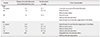
We analyzed the distance from the bifurcation of the abdominal aorta, the vertebral level at the origin, and other characteristics of each renal artery (Table 1), based on a previous classification [6]. On each side, the first renal artery was the main renal artery and the additional arteries were located below the main renal artery.
Discussion
The multiple renal arteries in the present case consisted of one accessory hilar and one inferior polar artery on the right side and two accessory hilar and one inferior polar artery on the left side (Figs. 1, 2), which coincided with a previous report that showed 61% of hilar artery and 29.6% of inferior polar artery out of the additional renal arteries [5]. However, the asymmetric bilateral multiplicity of this report is very rare because the frequency of these findings has been reported to be below 0.1% [23] or 1.1% [5]. Moreover, another rare variation, two accessory renal parenchymal branches originating from the testicular artery entering the anteroinferior aspect of the left kidney, was found. Thus, the frequency of accessory renal arteries originating from the testicular artery would be extremely rare, because only 1.1% of multiple renal arteries were found to have another urovascular variation [5]. Inferior polar and renal parenchymal branches originating from the gonadal artery have rarely been reported. The inferior polar and renal parenchymal branches arising from the testicular artery in the present case were consistent with a previous report that most accessory gonadal arteries supplied the inferior pole of the kidney as inferior capsular arteries [7]. Thus, the gonadal artery may serve as a source of collateral blood supply to the kidney through the gonadal-renal capsular artery since the gonadal artery and the inferior capsular artery have a common origin [8].
A recent study [9] suggested that the mesonephric arteries were obliterated and eliminated before the development of glomeruli in the metanephros. Therefore, the metanephros is unlikely to use mesonephric arteries as feeding arteries during renal ascent. Felix's theory [10], however, is widely acknowledged because it can explain variations associated with the renal arteries originating from the aorta. The developing mesonephros, metanephros, suprarenal glands, and gonads are supplied by nine pairs of lateral mesonephric arteries arising from the dorsal aorta. These mesonephric arteries are divided into the cranial, middle, and caudal groups. The middle and caudal arteries give rise to the renal and gonadal artery, respectively, and form the rete arteriosum urogenitale. At the early stage of kidney development, the metanephros is situated within the pelvis. While the metanephros develops definitive renal arteries after the ascent, some mesonephric arteries which did not regress remain as accessory renal arteries. Accordingly, the finding of multiple renal arteries from the aorta in the present case cannot exclude the possibility that the middle and caudal groups of the lateral mesonephric arteries persisted and caused an associated variation in the renal and testicular arteries.
In addition to multiple renal arteries, we found quadruple testicular veins on the left side, which were further subdivided into two pairs and then drained into the left renal vein or were accompanied by the renal parenchymal branches originating from the left testicular artery, respectively. The testicular veins are anatomically asymmetrical. These testicular veins originated from the pampiniform plexus that condenses to form four veins at the superficial inguinal ring, two veins at the deep inguinal ring, and one vein at variable levels [11]. Variations in the number, location of drainage, and termination angle of the testicular vein were summarized in previous studies [112]. Although quadruple gonadal veins have been reported in only two studies to date [1113], to the best of our knowledge, the renal parenchymal drainage seen in the present case has never been reported.
The development of the gonadal vein and the renal vein is closely related to the development of the inferior vena cava (IVC). The embryogenesis of these veins involves the development, regression, anastomosis, and replacement of three pairs of venous channels: posterior cardinal, sub-cardinal, and supra-cardinal. Anastomosis between the supra-cardinal and the sub-cardinal veins, which occur bilaterally, forms the renal segment of the IVC [14]. The gonadal vein develops from the caudal part of the sub-cardinal vein and drains into the supra-sub cardinal anastomosis. On the right side, this suprasub cardinal anastomosis and a small portion of the subcardinal vein are incorporated into the formation of the IVC. Therefore, the right gonadal vein usually drains into the IVC. On the left side, this supra-sub cardinal anastomosis forms part of the left renal vein where the left gonadal vein drains [15]. Failure or abnormal involution of the intersubcardinal anastomosis leads to multiple testicular veins and abnormal drainage, such as that seen in the present case.
In summary, we herein report an extremely rare case of asymmetric multiple bilateral renal arteries with aortic and extra-aortic origin and quadruple testicular veins, in which two inferior polar, renal parenchymal branches from the testicular artery were accompanied by a pair of testicular veins. The renal parenchymal branches from the testicular vessels could be regarded as components of the perinephric arterial arch or venous anastomoses, which usually involved in the formation of minor branches from the testicular vessels. However, histopathological examination confirmed that the renal parenchymal branches penetrated the renal parenchyma. Although variations in the urogenital vessels are relatively common, anatomists, clinicians, and especially nephrologists should be aware of combined urogenital vessel variations.


 XML Download