Journal List > Anat Cell Biol > v.52(4) > 1140528

Salman, Shabana, El-ghazouly, and Maha: Protective effect of glucosamine and risedronate (alone or in combination) against osteoarthritic changes in rat experimental model of immobilized knee

Abstract

This study is aiming to investigate the protective effect of glucosamine, risedronate (alone or in combination) on articular cartilage in experimental model of immobilized rat knee. Twenty-five adult male albino rats were divided into five groups (five rats each): control group, immobilized group, glucosamine-treated group, risedronate-treated group, and group treated by a combination of glucosamine and risedronate. The articular cartilage was obtained for histological, immunohistochemical and morphometric studies. The immobilized group showed manifestations of osteoarthritis in the form of significant decrease of articular cartilage thickness with surface erosions, shrunken chondrocytes with pyknotic nuclei and marked manifested fall of chondrocyte number. There was manifested reduction of collagen contents of the articular cartilage using Masson trichrome stain. Safranin O–Fast Green revealed low proteoglycan contents. The collagen type II was also declined. The manikin score was 7.8. Risedronate improved this manifestation slightly more than glucosamine, but combination of booth drugs caused significant improvement of the damaged articular cartilage caused by immobilization. Oral administration of glucosamine and risedronate improved the degenerative changes of rat knee articular cartilage that follow immobilization. This improvement was more remarkable when both drugs were used in combination.

Introduction

The articular cartilage is a load-bearing resistant in joints. It gets its nourishment by diffusion through the matrix. It is composed of chondrocytes, which form 1%–5% of its volume of the articular cartilage, a framework formed of collagen-rich fibrils and a hydrated substance contains cartilage-specific proteoglycan (PG) aggrecan [1]. The collagen found in adult articular cartilage is mainly composed of type II collagen [2].
Injured articular cartilage is less capable of healing than other tissues in the body due to lack of a vascular system, the chondrocytes immobility and the restricted ability of mature chondrocytes to proliferate and rejuvenate new cartilage [34].
Immobilization of joint is usually used for the management of joint injuries as ligament injuries and periarticular fractures. Plaster cast is used to immobilize the knee for management of femoral, patellar, or tibial fractures. Moreover, it is used to stabilize the knee in case of cruciate, collateral, meniscus ligament injuries, after knee surgery or management of the quadriceps tendon rupture [5].
It is stated that immobilization induces degeneration of articular cartilage due to a reduction in chondrocytes activity [67].
In another experiment on an immobilized dog knee with a splint, softening of the femoral and tibial cartilages and joint stiffness were noticed [8].
Furthermore, immobilization of rat knee with a plate and screws, Hagiwara et al. (2009) [9] observed hypertrophy of chondrocytes in the transitional area and reduced number of chondrocytes in the contact area. Besides, it was noticed that immobilization caused rapid reduction of bone mass, osteoporosis and increases risk of bone fracture [10].
It was stated that glucosamine is synthesized by chondrocytes from glucose and its precursors, which share to form the non-cellular part of the connective tissue. This component is mainly responsible for the mechanical function of cartilage [11].
The efficacy of glucosamine in healing the articular cartilage has been demonstrated in animal models [1213] and many clinical trials [1415]. In postmenopausal women, glucosamine sulfate reduced the progression of knee osteoarthritis and diminished the symptoms [16].
Glucosamine has a mild anti-inflammatory activity [12] and aids to retrieve the PG matrix of the articular cartilage, to guard injured cartilage from metabolic impairment [17].
Risedronic acid is one of the most potent Bisphosphonates. It revealed numerous serviceable effects on osteoarthritis treatment. This effect has been reported by several studies on animals and human [1819].
In rabbit models, risedronic acid demonstrated a protective effect on mechanical properties of the ligaments and periarticular bone and reduced the mineral loss at the bony attachment of the medial cruciate ligament [20]. Moreover, it reduced joint cartilage lesion in guinea pig models [21]. In the early stages of osteoarthritis of a rat model, using non-steroidal anti-inflammatory drug and risedronate, they decreased the impact of osteophyte bony adaptations and preserve trabecular bone mass [22].
In clinical trials of osteoarthritis, treatment with risedronate, improved both symptoms and joint structure in patients with primary knee osteoarthritis [18].
The current study was carried out to detect changes that occur in the rat knee joint following immobilization and to evaluate whether the oral administration of a combination of risedronate and glucosamine is capable to improve these changes compared to using each drug separately.

Materials and Methods

Drugs

The rats were given glucosamine sulfate (EVA Pharma Company, Cairo, Egypt) 40 mg/kg/day orally diluted in saline solution (NaCl) 0.9% [23]. Risedronate (actonel 35 mg, Sanofi Aventis Pharmaceutical Company, Cairo, Egypt) was given orally to rats in a dose of 0.2 mg/kg/day. Gypsona was obtained from International Medical Company (Cairo, Egypt) under license of BSN Medical Limited (London, UK).

Animals

Twenty-five adult male albino rats weighing 200±20 g were used in this study after approval of the protocol by the Ethical Committee of faculty of medicine Ain Shams University. Animals were obtained from Ain Shams animal house Egypt. They were housed under standard conditions of temperature (23℃±2℃) and lighting (12-hour light/dark cycles) and were allowed free access to food and drinking water. All rats received care in accordance with the rules and regulations of the Medical Research Ethics Committee of Faculty of Medicine Ain Shams University.

Groups

The animals were randomly divided into five groups (five rats each) as follows: group I, sacrificed and served as control; group II (immobilized group), immobilized by casting their right hindlimb for 6 weeks; group III, immobilized and received oral glucosamine; group IV, immobilized and received oral risedronate; and group V, immobilized and received a combination of oral risedronate and glucosamine.

Method of immobilization

The knee joints of the right hindlimb of rats were immobilized in full extension, using a plaster cast for 6 weeks. The plaster cast was wrapped from above knee to above ankle. The plaster cast was replaced at least every 3 days to prevent loosening and edema in the hind limb. The rats were able to move freely in the cage by using the three limbs that were not immobilized [2425].
At the end of the experiment, the rats were anesthetized by diethyl ether. Animals were sacrificed by cervical dislocation. The skin above knee joint was removed and the knee joint was exposed, Then the knee joint was cut in sagittal plane. The specimen contained tibia, femur with articular cartilages and menisci. The specimen was fixed in 10% formaldehyde for 48 hours. The specimen was decalcified using ethylenediaminetetraacetic acid. After processing for making paraffin blocks, 7-µm sections were cut and stained with hematoxylin and eosin (H&E) stain for routine histological examination, Masson trichrome stain for detection of collagen fibers, and Safranin O–Fast Green for detection of PG content of the cartilage matrix.

Immunohistochemical

Tissue sections were de-waxed, followed by treatment with hyaluronidase and trypsin (0.1% hyaluronidase and 0.2% trypsin 1 hour 37℃ for wax sections; Sigma-Aldrich, St. Louis, MO, USA) to unmask the collagen antigens. Sections were then incubated for 1 hour at room temperature with primary antibodies against type II collagen (Collagen, Type II, Bovine Joint Cartilage, Sigma-Aldrich). Endogenous peroxidase was blocked with 0.3% hydrogen peroxide in methanol before sections were incubated with secondary antibody, anti-mouse for collagen types II primary antibody, then incubate sections in ABC-peroxidase solution for 30 minutes at room temperature followed by incubation with diaminobenzidine chromogen to detect immunoreactivity. Mayer's hematoxylin was used for counterstaining [26].

Histomorphometric and statistical studies

Articular cartilage thickness

Thickness (mm) of total articular cartilage was defined as the distance between the cartilage surface and the osteochondral junction at the mid-portion of any area. To determine the cartilage thickness, histological sections stained with H&E were analyzed using a digital image analysis system (ImageJ software open source, UK, contributors worldwide) for quantitative histomorphometry. Each microscopic image was projected to a monitor and the thickness of the articular cartilage was measured at contact area of the articular surface. The mean thickness of each experimental group was calculated.

The number of chondrocytes

As the thickness of articular cartilage was different from area to area, we set a certain range of interest (rectangles 100 µm deep and 400 µm long) in the articular cartilage and superimposed it over histological sections stained with H&E to count the number of chondrocytes, using a digital image analysis program. The chondrocytes were counted and the means were calculated.

Histological scoring

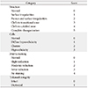
The histological appearance of the articular cartilage of the knee joints was evaluated using a modified Mankin scoring system (Table 1) [27], examining the surface, cellularity, matrix staining, and tidemark integrity. This scoring method consists of four different parameters; each parameter has scores, with higher scores reflected worse degenerative change. The highest possible score was 13. Thus, 13 points represented the worse degenerative change of cartilage and zero signified no change. The hematoxylin and eosin–stained sections were used to assess the structures, surface, cells, and tidemark integrity. Loss of PG staining was assessed from Safranin O sections.
All data (data for articular cartilage thickness, number of the chondrocytes and histological scores) were analyzed statistically using GraphPad prism version 4. Data were expressed as mean±SD and analyzed by using One-way analysis of variance followed by Bonferroni's multiple comparison post-hoc tests for comparison between all groups. Differences were regarded as non-significant if P-values were >0.05, and significant if P-values were <0.05.

Results

Light microscopic results

Histological study

H&E staining

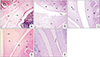
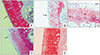
The examination of H&E-stained sections of knee joint of control group revealed the normal histological architecture of the articular cartilage and meniscus with no apparent degeneration in all of the specimens of the control group. The articular cartilage showed regular smooth intact surface and normal chondrocytes with normal organization. The chondrocytes appeared in non-calcified and calcified regions of cartilage which were separated by a clear intact tidemark appearing as a basophilic line in between the two regions. The chondrocytes in the non-calcified region were arranged in three zones: superficial (tangential), transitional (intermediate), and radial (deep) zone. The superficial zone had small flat chondrocytes arranged parallel to the articular surface. The transitional and radial zones had rounded, oval or triangular chondrocytes arranged in columns perpendicular to surface. The chondrocytes appeared to have pale basophilic cytoplasm with central rounded nuclei and are located inside their lacunae either singly or in groups forming cell nests. The calcified region had scattered rounded chondrocyte located in their lacunae. The subchondral bone appeared intact. The meniscus was composed of homogenous eosinophilic staining well-organized collagen fibers with fibrochondrocytes in between them. The fibrochondrocytes were located singly in their lacunae and appeared rounded to oval with vesicular nucleus. The meniscus showed smooth surface with no fraying or undulation (Figs. 1A, 2A, 3A). On the other hand, sections from immobilized group revealed many histological changes as compared to control group. These changes were variable in severity. The changes of articular cartilage were as follows: irregular notched surface, apparent reduction in thickness of cartilage, chondrocytes appeared shrunken with pyknotic nuclei, disorganized and few in number, loss of chondrocytes in some areas, tidemark was not clearly visible and degenerative changes in subchondral bone. The meniscus showed severe fraying and tears with unorganized disrupted collagen fibers and markedly shrunken darkly stained fibrochondrocytes (Figs. 1B, 2B, 3B). Interestingly, sections from the immobilized treated groups revealed better histological appearance as compared to the immobilized group. The immobilized group treated with both glucosamine and risedronate showed least degenerative changes in the which appeared nearly normal except for few shrunken chondrocytes, few empty lacunae and slight erosion in the surface of the meniscus (Fig. 1C–E, 2C–E, 3C–E).

Masson trichrome staining

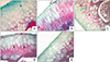
Masson trichrome staining was used for the evaluation of the collagen of the cartilage matrix. Masson trichrome commonly stains the cartilage matrix green, the nuclei dark blue, and the zone of calcifying cartilage red. In the control group, the matrix of articular cartilage was well stained with Masson trichrome (green color) reflecting normal content of collagen fibers (Fig. 4A). On the other hand, articular cartilage of the immobilized group showed marked reduction of Masson trichrome–stained area for collagen with appearance of an extensive red color reflecting marked reduction of collagen fibers in the matrix. Also, minimal erosion of the surface of articular cartilage was observed (Fig. 4B). Interestingly, the matrix of the three treated immobilized group revealed increase in the Masson trichrome–stained area for collagen with a reduction in the red color compared with the immobilized group especially in the immobilized group treated with both glucosamine and risedronate which showed a picture nearly similar to the control group (Fig. 4C–E).

Histochemical study

Safranin O–Fast Green staining

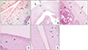
The articular cartilage in the control group was well stained with Safranin O (stains PG red) with no apparent loss of staining intensity reflecting normal PG content of the matrix (Fig. 5A). On the other hand, articular cartilage of the immobilized group showed marked reduction of Safranin O staining intensity in the entire non-calcified region of the articular cartilage and slight reduction in its calcified region reflecting marked decrease of the PG content of the matrix. In addition, the surface of articular cartilage showed fibrillation (Fig. 5B). However, the reduction in Safranin O staining intensity was less pronounced in the three treated immobilized group as compared to the immobilized group with better conservation of Safranin O staining intensity in the immobilized group treated with both glucosamine and risedronate reflecting nearly normal PG content of the matrix (Fig. 5C–E).

Immunohistochemical study

Immunohistochemical staining for collagen type II

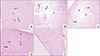
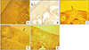
Immunohistochemical staining for expression of collagen type II fibers in the articular cartilage of the control group revealed very strong immunostaining intensity (brown color) reflecting dense and uniform distribution of collagen type II fibers in the matrix (Fig. 6A). On the other hand, immunohistochemical staining of collagen type II fibers of articular cartilage of the immobilized group revealed weak immunostaining intensity reflecting marked decrease of collagen type II fibers in the matrix of the articular cartilage (Fig. 6B). However, collagen type II fibers expression was stronger in the three treated immobilized group as compared to the immobilized group especially in the immobilized group treated with both glucosamine and risedonate which revealed strong immunostaining intensity for collagen type II (Fig. 6C–E).

Histomorphometric and statistical results

Articular cartilage thickness

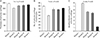
The mean thickness of the articular cartilage (at the contact area) in the immobilized group (group II) showed a highly significant decrease (P<0.01) as compared to control. The mean thickness of the articular cartilage in both the immobilized group treated with glucosamine (group III) and the immobilized group treated with risedronate (group IV) showed a significant decrease (P<0.05) as compared to control. The mean thickness of the articular cartilage in the immobilized group treated with both glucosamine and risedronate (group V) showed non-significant decrease (P>0.05) as compared to control (Fig. 7A).

The number of chondrocytes

The mean number of the chondrocytes in the articular cartilage in the immobilized group (group II) showed a highly significant decrease (P<0.01) as compared to control. The mean number of the chondrocytes in the articular cartilage in both the immobilized group treated with glucosamine (group III) and the immobilized group treated with risedronate (group IV) showed a highly significant decrease (P<0.01) as compared to control. The mean number of the chondrocytes in the articular cartilage in the immobilized group treated with both glucosamine and risedronate (group V) showed non-significant decrease (P>0.05) as compared to control (Fig. 7B).

Histological scoring

The Mankin score of the control group (I) was 0. The examination of the stained sections of the immobilized group (II) showed highly significant articular cartilage damage (irregular notched surface, hypocellularity, severe reduction in the matrix staining intensity and invisible tidemark) as compared to the control group, with a score of 7.80±0.27 (P<0.01). The examination of the stained sections of the treated immobilized group revealed highly significant less degenerative changes in the articular cartilage as compared to the immobilized group, with the least degenerative changes in the immobilized group treated with both glucosamine and risedronate (group V) with a score of 1.20±0.27 (P<0.01) indicating that the treatment of the immobilized group with both glucosamine and risedronate was associated with better preservation of articular cartilage (Fig. 7C).

Discussion

It has been proven that immobilization of joints decreases patient pain, stop additional damage, and encourage healing of injured structures [28]. However, this may cause articular cartilage degeneration. Many authors have proved the occurrence of the articular cartilage damage due to joint immobilization [29303132333435], while others reported that the joint mobility guards the cartilage from biochemical changes caused by immobilization. These changes were in the form of reduction of PG content [3637], upswing of hydration, which causes swelling and softening of the cartilage [38], reduction of cartilage thickness [839], decrease collagen II content [40] and downgrading histological scoring system [41]. These changes were classified as features of osteoarthritis. Moreover, osteoarthritis due to immobilization may be explained by loss of weight-bearing force, shortening and thickening of joint capsule [4243], and contraction of the muscles [44] and cartilage swelling which leads to increase tension inside the joint that compresses the articular cartilage and causes its degeneration.
The present study revealed the presence of morphological changes in the rats' knee articular cartilages following immobilization for 6 weeks. The chondrocytes appeared shrunken and pyknotic [45] reported the same finding. Our results have shown a significant thinning and softening of articular cartilage [373946] were also in concordance with these findings.
On the contrary, other studies reported an increase in the cartilage thickness following immobilization [93747] while others reported no alteration in cartilage thickness [324849].
According to some authors [3950], these contradictory results may be explained by a lack of standard measurement sites, the difference in animals age, or the use of contralateral knees as controls.
The thinning of the cartilage and its layers in immobilized rats which were found in this study could be explained by the decline in chondrocytic activity during immobilization, which influenced the structure of the extracellular matrix and led to a gradual decrease in cartilage thickness [3748]. It is also possible that the reduction in cartilage thickness occurred by decreasing synovial fluid production and the nutrient supply to the cartilage, as determined by the lack of motion and load, and thus produced deficits in the diffusion of liquids and pumping these elements into the cartilage [48].
In our study, the number of chondrocytes was markedly decreased in the immobilized rats, which corresponded well with Trudel et al.'s study [32], who explained this reduction by chondrocyte death due to necrosis or apoptosis. On the other side, others explained this reduction by alterations in chondrocyte biosynthesis [385152].
In the present experiment, a reduction of collagen and PG content of the articular cartilage in the immobilized group was detected, by using Masson trichrome and Safranin O Green stains, which could affect cartilage elasticity.
In agreement with these findings, a reduction in the PG content in the immobilized knee was also detected by other studies [938535455], while Trudel et al. [32] reported no change in PG content of the deep part of the cartilage after immobilization.
Moreover, a decrease in collagen content was also reported by Haapala et al. [39]. On the other hand, Saamanen et al. [29] and Muller et al. [53] reported no change of the same parameter. The reduction of PG and collagen cartilaginous contents were clarified by Jortikka et al. [55] who reported that there was an imbalance between synthesis and degeneration. This imbalance was due to the activity of matrix metalloproteinase aggrecanase II (ADAMTSs) which has a role in debasing the collagen and PG constituents of articular cartilage [5657]. Echtermeyer et al. [58] have suggested that one of the members of matrix metalloproteinase in the cartilage is matrix metalloproteinase-3 (MMP-3) that digests many components of extracellular cartilaginous matrix and activates aggrecanase II (ADAMTS-5). Many studies detected an elevation of MMP-3 in acute injury or osteoarthritis [5960].
Furthermore, Grumbles et al. [61] detected an elevation of matrix metalloproteinase in immobilized canine knee. This could be explained by elevation of the MMP-3 during 6 hours after immobilization and persistence uprising though 21 days of immobilization [62].
Our results have shown changes in chondrocytes and cartilage extracellular matrix due to immobilization. This also led to irregularity in cartilage surface which was also reported by Trudel et al. [32] and Helminen et al. [63].
Glucosamine is an amino-monosaccharide and one of the main constituents of the disaccharide parts of articular cartilage glycosaminoglycans. Besides, it is considered a symptomatic slow-acting drug for osteoarthritis treatment. Muller et al. [53] have confirmed the higher efficacy of glucosamine in treating knee osteoarthritis compared to non-steroidal anti-inflammatory drugs. On the other hand, immobilization induced marked reduction of glycosaminoglycans in canine articular cartilage [7546465]. The precise mechanism of action of glucosamine has not been fully elucidated yet [66].
The protective effect of glucosamine may be explained by the in vitro study of Vidal y Plana et al. [66] which stated that the synthesis of cartilage glycosaminoglycans was increased by adding glucosamine to cartilage culture. Moreover, glucosamine balanced between PG production and degeneration as it stimulated chondrocytes to synthesis PG core protein [6768].
The use of glucosamine in osteoarthritis is a matter of controversy. The benefits of the use of glucosamine for osteoarthritis have long been agreed with skepticism due to the lack of reliable information regarding their absorption, pharmacokinetics, and mechanism of action. Pharmacokinetic studies on glucosamine in dogs using 14C-glucosamine and 35S-labeled chondroitin sulfate found that 87% of an orally administered dose of radiolabelled glucosamine and 70% of the labeled chondroitin sulfate were absorbed [69]. Other studies reported that glucosamine was bioavailable after oral dosing and had a tropism for articular cartilage [70].
Our results confirmed the efficacy of glucosamine in protection of articular cartilage from osteoarthritis caused by knee immobilization. These results were in consistence with the clinical studies of Naito et al. [70] and Richy et al. [71] who proved the modifying effect of glucosamine in knee osteoarthritis.
Risedronate was considered as osteoarthritis modifying drug due to its anti-inflammatory effect. It diminished swelling of the articular cartilage [72] and caused marked reduction of CTX-II (marker of cartilage degeneration) [1973]. Moreover, it was considered as bone antiresorptive drug which protected periarticular bone characteristic [7475].
It was found that patients with osteoarthritis had high level of bone turnover markers [76]. Furthermore, treatment of Paget's disease patients with risedronate improved bony pathology and decreased biochemical bone markers [77].
In our study, we observed an improvement of cartilage pathology in the risedronate-treated group, the same finding was obtained by Permuy et al. [78] who detected an improvement in resedronate treated animals in a rabbit model of osteoarthritis using Safranin O–Fast Green. In another study, resedronate improved bone metabolism in subchondral level which alleviated osteoarthritis symptoms [21]. On the other hand, Thomsen et al. [79], in his study on Dunkin Hartley guinea pigs, reported no significant differences between control animals and risedronate-treated one.
In conclusion, our findings have suggested that the use of risedronate and glucosamine combination improves the damage to the knee articular cartilage in an immobilized rat model compared to the use of each drug separately.

Figures and Tables

Fig. 1

Hematoxylin and eosin (H&E)–stained sections of the knee joint of different groups. (A) Control group showing articular cartilage with regular smooth intact surface and well-organized chondrocytes which appeared in non-calcified (NCC) and calcified (CC) regions of cartilage with a clear intact tidemark (arrows) in between. The subchondral bone (SC) appears intact. The meniscus shows regular smooth surface with no fraying or undulation. (B) Immobilized group showing reduction in thickness of articular cartilage, shrunken chondrocytes, absence of chondrocytes in some areas (*) and invisible tidemark. The SC appears intact. Meniscus shows severe fraying and tears and erosion of surface with necrotic cells shedding from it (arrow). (C) The immobilized group treated with glucosamine; the articular cartilage shows smooth surface, shrunken chondrocytes and visible tidemark (*). The SC appears intact. The meniscus shows smooth surface with some cracks (arrow). (D) The immobilized group treated with risedronate; the articular cartilage shows smooth surface, shrunken chondrocytes, some empty lacunae, and hardly visible tidemark. The SC appears intact. The meniscus shows smooth surface with some cracks (arrow). (E) immobilized group treated with glucosamine and risedronate; the articular cartilage shows smooth surface, few shrunken chondrocytes, some empty lacunae, and visible tidemark. The SC appears intact. The meniscus shows minimal erosion of its surface (arrow) (H&E, ×200). ar, articular cartilage; L, lacunae; m, meniscus; t, tears.

acb-52-498-g001
Fig. 2

Hematoxylin and eosin (H&E)–stained sections of articular cartilage of knee joint of different groups. (A) Control group showing the chondrocytes in non-calcified region (NCC) of the articular cartilage arranged in three zones: superficial (S), transitional (T), and radial (R) zone. The superficial zone contains small flat chondrocytes. The transitional and radial zone contain columns of rounded, oval, or triangular chondrocytes (*). The chondrocytes are located inside their lacunae forming cell nests. The calcified region (CC) is separated from radial zone by a tidemark (arrows). The subchondral bone (SC) appears intact. (B) Immobilized group; articular cartilage shows irregular notched surface (arrows) and its chondrocytes appear shrunken with pyknotic nuclei, disorganized and few in number. Tidemark is invisible. The subchondral bone shows degenerative changes (*). (C) The immobilized group treated with glucosamine; articular cartilage shows irregular degenerated surface (arrow) and shrunken chondrocytes which appear disorganized and few in number. Tidemark is visible. The SC appears intact. (D) The immobilized group treated with risedronate; articular cartilage shows smooth surface, shrunken chondrocytes which appear disorganized and few in number. Tidemark is not clearly visible. (E) The immobilized group treated with glucosamine and risedronate; articular cartilage shows smooth surface, few shrunken chondrocytes, few empty lacunae, and visible tidemark (arrow) (H&E, ×400). ch, chondrocyte; f, flat chondrocyte; L, lacunae; ne, cell nests.

acb-52-498-g002
Fig. 3

Hematoxylin and eosin–stained sections of meniscus of the knee joint of different groups. (A) Control group showing the meniscus composed of homogenous eosinophilic staining well-organized collagen fibers (arrows) with fibrochondrocytes in their lacunae. The meniscus surface is smooth with no fraying or undulation (*). (B) Immobilized group showing many meniscal tears with unorganized disrupted collagen fibers (arrows) and markedly shrunken darkly stained fibrochondrocytes. (C) The immobilized group treated with glucosamine showing some tears and cracks (arrows) and moderately shrunken darkly stained fibrochondrocytes. (D) The immobilized group treated with risedronate showing few tears and cracks (arrows) and slightly shrunken fibrochondrocytes. (E) The immobilized group treated with glucosamine and risedronate showing nearly normal meniscus except for minimal erosion of its surface (arrow) (H&E, ×400). fc, fibrochondrocytes.

acb-52-498-g003
Fig. 4

A photomicrograph of Masson trichrome–stained sections of articular cartilage of knee joint of different groups. (A) Control group showing the articular cartilage which is well stained with Masson trichrome for collagen (green color) (*). (B) Immobilized group showing marked reduction of Masson trichrome–stained area for collagen with appearance of an extensive red color (*). Minimal erosion of the articular cartilage surface is observed (arrow). (C) The immobilized group treated with glucosamine showing marked reduction of Masson trichrome–stained area for collagen with appearance of a red color (*). (D) The immobilized group treated with risedronate showing moderate reduction of Masson trichrome–stained area for collagen with appearance of a red color (*). (E) The immobilized group treated with glucosamine and risedronate showing slight reduction of Masson trichrome–stained area for collagen with appearance of a slight red color (*) (Masson trichrome, ×400).

acb-52-498-g004
Fig. 5

A photomicrograph of Safranin O–Fast Green (SO)–stained sections of articular cartilage of knee joint of different groups. (A) Control group showing the articular cartilage which appears well stained with SO. The smooth surface of the articular cartilage (arrow) with normal cellular distribution is observed. (B) Immobilized group showing marked reduction in SO staining intensity in the non-calcified region of the articular cartilage (*) and slight reduction in its calcified region. The surface of articular cartilage shows fibrillation (arrow). (C) The immobilized group treated with glucosamine showing marked reduction in SO staining intensity in the superficial part (arrow) of the articular cartilage and moderate reduction in its deeper part (*). The smooth surface of the articular cartilage is observed. (D) The immobilized group treated with risedronate showing moderate reduction in SO staining intensity in the calcified region of the articular cartilage (*) with disruption in the superficial part of the articular cartilage (arrow). (E) The immobilized group treated with glucosamine and risedronate showing slight reduction in SO staining intensity in the superficial part of the articular cartilage (*). The smooth surface of the articular cartilage is observed (SO staining, ×400).

acb-52-498-g005
Fig. 6

Immunohistochemical stained sections for collagen type II of the articular cartilage of the knee joint of different groups. (A) Control group showing very strong immunostaining intensity for collagen type II in articular cartilage (brown color). The smooth surface of the articular cartilage is observed. (B) Immobilized group showing weak immunostaining intensity for collagen type II in articular cartilage with disruption in the superficial part of cartilage (arrow). (C) The immobilized group treated with glucosamine showing moderate immunostaining intensity for collagen type II in articular cartilage (*). Irregularity of the surface (arrow) and presence of a space in superficial part are observed. (D) The immobilized group treated with risedronate showing moderate immunostaining intensity for collagen type II in articular cartilage (*) with necrosis of superficial part of the articular cartilage (arrow). (E) The immobilized group treated with glucosamine and risedronate showing strong immunostaining intensity for collagen type II in the articular cartilage. The smooth surface of the articular cartilage is observed (immunohistochemical staining for collagen type II, ×400). s, space.

acb-52-498-g006
Fig. 7

(A) The thickness of the articular cartilage (µm) of different experimental groups. Pairwise significant differences were detected between: group I and group II (P<0.001); group II and group III (P<0.05), group IV (P<0.01) and group V (P<0.001); group III and groups IV and V (P>0.05); group IV and group V (P>0.05). (B) The number of the chondrocytes in the articular cartilage of different experimental groups. Pairwise significant differences were detected between: group I and group II (P<0.001); group II and groups III, IV and V (P<0.001); group III and groups IV and V (P>0.05); group IV and group V (P>0.05). (C) The number of the chondrocytes in the articular cartilage of different experimental groups. Pairwise significant differences were detected between: group I and group II (P<0.001); group II and groups III, IV, and V (P<0.001); group III and group IV (P>0.05), V (P<0.001); group IV and group V (P<0.001). Group I, control; group II, immobilized knee; group III, immobilized group treated with glucosamine; group IV, immobilized group treated with risedronate; group V, immobilized group treated with glucosamine and risedronate. Analysis was done using GraphPad Prism version 4. Data were analyzed by using one-way analysis of variance test followed by Bonferroni's multiple comparison post-hoc test for comparison between all groups. P>0.05, nonsignificant; P<0.05, significant.

acb-52-498-g007
Table 1

Modified Mankin histological scores

acb-52-498-i001

Notes

AUTHOR CONTRIBUTIONS

  • Conceptualization: Atef Shabana.

  • Data acquisition: Ahmed Salman, Atef Shabana.

  • Data analysis or interpretation: DEE.

  • Drafting of the manuscript: Ahmed Salman, ME.

  • Critical revision of the manuscript: ME.

  • Approval of the final version of the manuscript: all authors.

Conflicts of Interest No potential conflict of interest relevant to this article was reported.

References

1. Iqbal K, Khan Y, Minhas LA. Effects of immobilization on thickness of superficial zone of articular cartilage of patella in rats. Indian J Orthop. 2012; 46:391–394.
2. Eyre D. Collagen of articular cartilage. Arthritis Res. 2002; 4:30–35.
3. Hunter W. Of the structure and disease of articulating cartilages. 1743. Clin Orthop Relat Res. 1995; (317):3–6.
4. Newman AP. Articular cartilage repair. Am J Sports Med. 1998; 26:309–324.
5. Sprouse RA, McLaughlin AM, Harris GD. Braces and splints for common musculoskeletal conditions. Am Fam Physician. 2018; 98:570–576.
6. Buckwalter JA. Osteoarthritis and articular cartilage use, disuse, and abuse: experimental studies. J Rheumatol Suppl. 1995; 43:13–15.
7. Setton LA, Mow VC, Muller FJ, Pita JC, Howell DS. Mechanical behavior and biochemical composition of canine knee cartilage following periods of joint disuse and disuse with remobilization. Osteoarthritis Cartilage. 1997; 5:1–16.
8. Jurvelin J, Kiviranta I, Tammi M, Helminen JH. Softening of canine articular cartilage after immobilization of the knee joint. Clin Orthop Relat Res. 1986; (207):246–252.
9. Hagiwara Y, Ando A, Chimoto E, Saijo Y, Ohmori-Matsuda K, Itoi E. Changes of articular cartilage after immobilization in a rat knee contracture model. J Orthop Res. 2009; 27:236–242.
10. Mosekilde L, Thomsen JS, Mackey MS, Phipps RJ. Treatment with risedronate or alendronate prevents hind-limb immobilization-induced loss of bone density and strength in adult female rats. Bone. 2000; 27:639–645.
11. Towheed TE, Maxwell L, Anastassiades TP, Shea B, Houpt J, Robinson V, Hochberg MC, Wells G. Glucosamine therapy for treating osteoarthritis. Cochrane Database Syst Rev. 2005; (2):CD002946.
12. Shikhman AR, Amiel D, D'Lima D, Hwang SB, Hu C, Xu A, Hashimoto S, Kobayashi K, Sasho T, Lotz MK. Chondroprotective activity of N-acetylglucosamine in rabbits with experimental osteoarthritis. Ann Rheum Dis. 2005; 64:89–94.
13. Tiraloche G, Girard C, Chouinard L, Sampalis J, Moquin L, Ionescu M, Reiner A, Poole AR, Laverty S. Effect of oral glucosamine on cartilage degradation in a rabbit model of osteoarthritis. Arthritis Rheum. 2005; 52:1118–1128.
14. Pavelká K, Gatterová J, Olejarová M, Machacek S, Giacovelli G, Rovati LC. Glucosamine sulfate use and delay of progression of knee osteoarthritis: a 3-year, randomized, placebo-controlled, double-blind study. Arch Intern Med. 2002; 162:2113–2123.
15. Herrero-Beaumont G, Ivorra JA, Del Carmen, Blanco FJ, Benito P, Martin-Mola E, Paulino J, Marenco JL, Porto A, Laffon A, Araújo D, Figueroa M, Branco J. Glucosamine sulfate in the treatment of knee osteoarthritis symptoms: a randomized, double-blind, placebo-controlled study using acetaminophen as a side comparator. Arthritis Rheum. 2007; 56:555–567.
16. Bruyere O, Pavelka K, Rovati LC, Deroisy R, Olejarova M, Gatterova J, Giacovelli G, Reginster JY. Glucosamine sulfate reduces osteoarthritis progression in postmenopausal women with knee osteoarthritis: evidence from two 3-year studies. Menopause. 2004; 11:138–143.
17. da Camara CC, Dowless GV. Glucosamine sulfate for osteoarthritis. Ann Pharmacother. 1998; 32:580–587.
18. Spector TD, Conaghan PG, Buckland-Wright JC, Garnero P, Cline GA, Beary JF, Valent DJ, Meyer JM. Effect of risedronate on joint structure and symptoms of knee osteoarthritis: results of the BRISK randomized, controlled trial [ISRCTN01928173]. Arthritis Res Ther. 2005; 7:R625–R633.
19. Garnero P, Aronstein WS, Cohen SB, Conaghan PG, Cline GA, Christiansen C, Beary JF, Meyer JM, Bingham CO 3rd. Relationships between biochemical markers of bone and cartilage degradation with radiological progression in patients with knee osteoarthritis receiving risedronate: the Knee Osteoarthritis Structural Arthritis randomized clinical trial. Osteoarthritis Cartilage. 2008; 16:660–666.
20. Doschak MR, Wohl GR, Hanley DA, Bray RC, Zernicke RF. Antiresorptive therapy conserves some periarticular bone and ligament mechanical properties after anterior cruciate ligament disruption in the rabbit knee. J Orthop Res. 2004; 22:942–948.
21. Spector TD. Bisphosphonates: potential therapeutic agents for disease modification in osteoarthritis. Aging Clin Exp Res. 2003; 15:413–418.
22. Jones MD, Tran CW, Li G, Maksymowych WP, Zernicke RF, Doschak MR. In vivo microfocal computed tomography and micro-magnetic resonance imaging evaluation of antiresorptive and antiinflammatory drugs as preventive treatments of osteoarthritis in the rat. Arthritis Rheum. 2010; 62:2726–2735.
23. Aghazadeh-Habashi A, Kohan MH, Asghar W, Jamali F. Glucosamine dose/concentration-effect correlation in the rat with adjuvant arthritis. J Pharm Sci. 2014; 103:760–767.
24. Nakabayashi K, Sakamoto J, Kataoka H, Kondo Y, Hamaue Y, Honda Y, Nakano J, Okita M. Effect of continuous passive motion initiated after the onset of arthritis on inflammation and secondary hyperalgesia in rats. Physiol Res. 2016; 65:683–691.
25. Tuukkanen J, Peng Z, Väänänen HK. The effect of training on the recovery from immobilization-induced bone loss in rats. Acta Physiol Scand. 1992; 145:407–411.
26. Roberts S, McCall IW, Darby AJ, Menage J, Evans H, Harrison PE, Richardson JB. Autologous chondrocyte implantation for cartilage repair: monitoring its success by magnetic resonance imaging and histology. Arthritis Res Ther. 2003; 5:R60–R73.
27. Mankin HJ, Dorfman H, Lippiello L, Zarins A. Biochemical and metabolic abnormalities in articular cartilage from osteo-arthritic human hips. II. Correlation of morphology with biochemical and metabolic data. J Bone Joint Surg Am. 1971; 53:523–537.
28. Gault SJ, Spyker MJ. Beneficial effect of immobilization of joints in rheumatoid and related arthritides: a splint study using sequential analysis. Arthritis Rheum. 1969; 12:34–44.
29. Säämänen AM, Tammi M, Kiviranta I, Jurvelin J, Helminen HJ. Maturation of proteoglycan matrix in articular cartilage under increased and decreased joint loading: a study in young rabbits. Connect Tissue Res. 1987; 16:163–175.
30. Evans EB, Eggers GW, Butler JK, Blumel J. Experimental immobilization and remobilization of rat knee joints. J Bone Joint Surg. 1960; 42:737–758.
31. Hall MC. Cartilage changes after experimental immobilization of the knee joint of the young rat. J Bone Joint Surg. 1963; 45:36–44.
32. Trudel G, Himori K, Uhthoff HK. Contrasting alterations of apposed and unapposed articular cartilage during joint contracture formation. Arch Phys Med Rehabil. 2005; 86:90–97.
33. Akeson WH, Amiel D, Woo SL. Immobility effects on synovial joints the pathomechanics of joint contracture. Biorheology. 1980; 17:95–110.
34. Videman T. Experimental osteoarthritis in the rabbit: comparison of different periods of repeated immobilization. Acta Orthop Scand. 1982; 53:339–347.
35. Iqbal K, Khan MY, Minhas LA. Effects of immobilisation and remobilisation on superficial zone of articular cartilage of patella in rats. J Pak Med Assoc. 2012; 62:531–535.
36. Tammi M, Saamanen AM, Jauhiainen A, Malminen O, Kiviranta I, Helminen H. Proteoglycan alterations in rabbit knee articular cartilage following physical exercise and immobilization. Connect Tissue Res. 1983; 11:45–55.
37. Palmoski MJ, Brandt KD. Running inhibits the reversal of atrophic changes in canine knee cartilage after removal of a leg cast. Arthritis Rheum. 1981; 24:1329–1337.
38. Behrens F, Kraft EL, Oegema TR Jr. Biochemical changes in articular cartilage after joint immobilization by casting or external fixation. J Orthop Res. 1989; 7:335–343.
39. Haapala J, Arokoski JP, Hyttinen MM, Lammi M, Tammi M, Kovanen V, Helminen HJ, Kiviranta I. Remobilization does not fully restore immobilization induced articular cartilage atrophy. Clin Orthop Relat Res. 1999; (362):218–229.
40. Aldahmash AM, El Fouhil AF, Mohamed RA, Ahmed AM, Atteya M, Al Sharawy SA, Qureshi RA. Collagen types I and II distribution: a relevant indicator for the functional properties of articular cartilage in immobilised and remobilized rabbit knee joints. Folia Morphol (Warsz). 2015; 74:169–175.
41. Narmoneva DA, Cheung HS, Wang JY, Howell DS, Setton LA. Altered swelling behavior of femoral cartilage following joint immobilization in a canine model. J Orthop Res. 2002; 20:83–91.
42. Videman T, Michelsson JE, Langenskiold A. The development of radiographic changes in experimental osteoarthritis provoked by immobilization of the knee in rabbits. Int Res Commun Syst Med Sci. 1977; 5:62.
43. Videman T, Eronen I, Friman C, Langenskiöld A. Glycosaminoglycan metabolism of the medial meniscus, the medial collateral ligament and the hip joint capsule in experimental osteoarthritis caused by immobilization of the rabbit knee. Acta Orthop Scand. 1979; 50:465–470.
44. Thaxter TH, Mann RA, Anderson CE. Degeneration of immobilized knee joints in rats: histological and autoradiographic study. J Bone Joint Surg Am. 1965; 47:567–585.
45. Maldonado DC, Silva MC, Neto Sel-R, de Souza MR, de Souza RR. The effects of joint immobilization on articular cartilage of the knee in previously exercised rats. J Anat. 2013; 222:518–525.
46. Kiviranta I, Tammi M, Jurvelin J, Arokoski J, Säämänen AM, Helminen HJ. Articular cartilage thickness and glycosaminoglycan distribution in the young canine knee joint after remobilization of the immobilized limb. J Orthop Res. 1994; 12:161–167.
47. Sood SC. A study of the effects of experimental immobilisation on rabbit articular cartilage. J Anat. 1971; 108(Pt 3):497–507.
48. O'Connor KM. Unweighting accelerates tidemark advancement in articular cartilage at the knee joint of rats. J Bone Miner Res. 1997; 12:580–589.
49. Leroux MA, Cheung HS, Bau JL, Wang JY, Howell DS, Setton LA. Altered mechanics and histomorphometry of canine tibial cartilage following joint immobilization. Osteoarthritis Cartilage. 2001; 9:633–640.
50. Gyarmati J, Foldes I, Kern M, Kiss I. Morphological studies on the articular cartilage of old rats. Acta Morphol Hung. 1987; 35:111–124.
51. Tammi M, Kiviranta I, Peltonen L, Jurvelin J, Helminen HJ. Effects of joint loading on articular cartilage collagen metabolism: assay of procollagen prolyl 4-hydroxylase and galactosylhydroxylysyl glucosyltransferase. Connect Tissue Res. 1988; 17:199–206.
52. Videman T, Eronen I, Candolin T. [3H]proline incorporation and hydroxyproline concentration in articular cartilage during the development of osteoarthritis caused by immobilization. A study in vivo with rabbits. Biochem J. 1981; 200:435–440.
53. Muller FJ, Setton LA, Manicourt DH, Mow VC, Howell DS, Pita JC. Centrifugal and biochemical comparison of proteoglycan aggregates from articular cartilage in experimental joint disuse and joint instability. J Orthop Res. 1994; 12:498–508.
54. Haapala J, Arokoski J, Pirttimaki J, Lyyra T, Jurvelin J, Tammi M, Helminen HJ, Kiviranta I. Incomplete restoration of immobilization induced softening of young beagle knee articular cartilage after 50-week remobilization. Int J Sports Med. 2000; 21:76–81.
55. Jortikka MO, Inkinen RI, Tammi MI, Parkkinen JJ, Haapala J, Kiviranta I, Helminen HJ, Lammi MJ. Immobilisation causes longlasting matrix changes both in the immobilised and contralateral joint cartilage. Ann Rheum Dis. 1997; 56:255–261.
56. Mow VC, Proctor CS, Kelly MA. Biomechanics of articular cartilage. In : Nordin M, Frankel VH, editors. Basic Biomechanics of the Musculoskeletal System. 2nd ed. London: Lea & Febiger;1989. p. 31–58.
57. Cawston TE, Wilson AJ. Understanding the roleof tissue degrading enzymes and their inhibitors in development and disease. Best Pract Res Clin Rheumatol. 2006; 20:983–1002.
58. Echtermeyer F, Bertrand J, Dreier R, Meinecke I, Neugebauer K, Fuerst M, Lee YJ, Song YW, Herzog C, Theilmeier G, Pap T. Syndecan-4 regulates ADAMTS-5 activation and cartilage breakdown in osteoarthritis. Nat Med. 2009; 15:1072–1076.
59. Lohmander LS, Hoerrner LA, Dahlberg L, Roos H, Björnsson S, Lark MW. Stromelysin, tissue inhibitor of metalloproteinases and proteoglycan fragments in human knee joint fluid after injury. J Rheumatol. 1993; 20:1362–1368.
60. Walakovits LA, Moore VL, Bhardwaj N, Gallick GS, Lark MW. Detection of stromelysin and collagenase in synovial fluid from patients with rheumatoid arthritis and posttraumatic knee injury. Arthritis Rheum. 1992; 35:35–42.
61. Grumbles RM, Howell DS, Howard GA, Roos BA, Setton LA, Mow VC, Ratcliffe A, Muller FJ, Altman RD. Cartilage metalloproteases in disuse atrophy. J Rheumatol Suppl. 1995; 43:146–148.
62. Leong DJ, Gu XI, Li Y, Lee JY, Laudier DM, Majeska RJ, Schaffler MB, Cardoso L, Sun HB. Matrix metalloproteinase-3 in articular cartilage is upregulated by joint immobilization and suppressed by passive joint motion. Matrix Biol. 2010; 29:420–426.
63. Helminen HJ, Jurvelin J, Kuusela T, Heikkilä R, Kiviranta I, Tammi M. Effects of immobilization for 6 weeks on rabbit knee articular surfaces as assessed by the semiquantitative stereomicroscopic method. Acta Anat (Basel). 1983; 115:327–335.
64. Kiviranta I, Jurvelin J, Tammi M, Säämänen AM, Helminen HJ. Weight bearing controls glycosaminoglycan concentration and articular cartilage thickness in the knee joints of young beagle dogs. Arthritis Rheum. 1987; 30:801–809.
65. Reginster JY, Deroisy R, Rovati LC, Lee RL, Lejeune E, Bruyere O, Giacovelli G, HenrotinY , Dacre JE, Gossett C. Long-term effects of glucosamine sulphate on osteoarthritis progression: a randomised, placebo-controlled clinical trial. Lancet. 2001; 357:251–256.
66. Vidal y Plana RR, Bizzarri D, Rovati AL. Articular cartilage pharmacology: I. In vitro studies on glucosamine and non steroidal antiinflammatory drugs. Pharmacol Res Commun. 1978; 10:557–569.
67. Bassleer C, Rovati L, Franchimont P. Stimulation of proteoglycan production by glucosamine sulfate in chondrocytes isolated from human osteoarthritic articular cartilage in vitro. Osteoarthritis Cartilage. 1998; 6:427–434.
68. Fenton JI, Chlebek-Brown KA, Peters TL, Caron JP, Orth MW. Glucosamine HCl reduces equine articular cartilage degradation in explant culture. Osteoarthritis Cartilage. 2000; 8:258–265.
69. McCarthy G, O'Donovan J, Jones B, McAllister H, Seed M, Mooney C. Randomised double-blind, positive-controlled trial to assess the efficacy of glucosamine/chondroitin sulfate for the treatment of dogs with osteoarthritis. Vet J. 2007; 174:54–61.
70. Naito K, Watari T, Furuhata A, Yomogida S, Sakamoto K, Kurosawa H, Kaneko K, Nagaoka I. Evaluation of the effect of glucosamine on an experimental rat osteoarthritis model. Life Sci. 2010; 86:538–543.
71. Richy F, Bruyere O, Ethgen O, Cucherat M, Henrotin Y, Reginster JY. Structural and symptomatic efficacy of glucosamine and chondroitin in knee osteoarthritis: a comprehensive metaanalysis. Arch Intern Med. 2003; 163:1514–1522.
72. Corrado A, Santoro N, Cantatore FP. Extra-skeletal effects of bisphosphonates. Joint Bone Spine. 2007; 74:32–38.
73. Iwamoto J, Takeda T, Sato Y, Matsumoto H. Effects of risedronate on osteoarthritis of the knee. Yonsei Med J. 2010; 51:164–170.
74. MacNeil JA, Doschak MR, Zernicke RF, Boyd SK. Preservation of periarticular cancellous morphology and mechanical stiffness in post-traumatic experimental osteoarthritis by antiresorptive therapy. Clin Biomech (Bristol, Avon). 2008; 23:365–371.
75. Walker K, Medhurst SJ, Kidd BL, Glatt M, Bowes M, Patel S, McNair K, Kesingland A, Green J, Chan O, Fox AJ, Urban LA. Disease modifying and anti-nociceptive effects of the bisphosphonate, zoledronic acid in a model of bone cancer pain. Pain. 2002; 100:219–229.
76. Bettica P, Cline G, Hart DJ, Meyer J, Spector TD. Evidence for increased bone resorption in patients with progressive knee osteoarthritis: longitudinal results from the Chingford study. Arthritis Rheum. 2002; 46:3178–3184.
77. Brown JP, Chines AA, Myers WR, Eusebio RA, Ritter-Hrncirik C, Hayes CW. Improvement of pagetic bone lesions with risedronate treatment: a radiologic study. Bone. 2000; 26:263–267.
78. Permuy M, Guede D, López-Peña M, Muñoz F, González-Cantalapiedra A, Caeiro JR. Effects of glucosamine and risedronate alone or in combination in an experimental rabbit model of osteoarthritis. BMC Vet Res. 2014; 10:97.
79. Thomsen JS, Straarup TS, Danielsen CC, Oxlund H, Brüel A. No effect of risedronate on articular cartilage damage in the Dunkin Hartley guinea pig model of osteoarthritis. Scand J Rheumatol. 2013; 42:408–416.
TOOLS
ORCID iDs

Ahmed Salman
https://orcid.org/0000-0002-4382-3125

Atef Ibrahim Shabana
https://orcid.org/0000-0002-7341-9377

Dalia El-sayed El-ghazouly
https://orcid.org/0000-0001-6532-9104

Elbeltagy Maha
https://orcid.org/0000-0002-4777-2388

Similar articles