

 PDF
PDF ePub
ePub Citation
 Print
Print


Introduction
The median atlanto-axial joint is a pivot joint between the dens (odontoid process of the axis) and a ring formed by the anterior atlantal arch and transverse ligament of the atlas [1, 2]. A vertically ovoid facet on the anterior dens articulates with another on the posterior aspect of the anterior arch. The fibrous capsule, which is lined by the synovial membrane, is relatively weak and loose, especially superiorly. We have previously reported fetal development of the transverse atlantis and alar ligaments at the median atlanto-occipital joint [3]. We found that the joint cavity in the anterior compartment appears from the inferior part at 7–8 weeks of gestation and extends superiorly to reach a future apical ligament containing the notochord remnant. Because of a tight connection between the occipital and dens apex by the notochord, the growing dens temporarily rides over the occipital. Thus, the anterior joint cavity faces the apical ligament in the roof. In previous studies and later ones, we incidentally found a “rim” or disk-like structure in the anterior compartment of the median atlanto-axial joint. In the present study, we aimed to clarify the development and morphology of the disk-like structure in the fetus. Because no previous report has described the disk in the adult median joint, the structure is considered likely to disappear in the late-stage fetus or after birth.
Materials and Methods
The study was performed in accordance with the provisions of the Declaration of Helsinki 1995 (as revised in Edinburgh 2000). We observed sagittal or horizontal sections of 44 human fetuses: 30 mid-term specimens (crown-rump length [CRL], 43–117 mm; approximately 9–15 weeks of gestational age [GA]) and 14 late-stage specimens (CRL, 250–310 mm; GA, 30–37 weeks). All specimens were part of the large collection kept at the Department of Anatomy and Embryology of the Universidad Complutense, Madrid, and had originated from miscarriages and ectopic pregnancies at the Department of Obstetrics of the University. The present mid-term specimens were sectioned serially, while the late-stage specimens were sectioned at intervals of 100 µm. Most sections were stained with hematoxylin and eosin, while a minor proportion were stained with Masson trichrome, azan, orange G or silver. The sectional plane was horizontal (25 mid-term, 4 late-stage) or sagittal (5 mid-term, 10 late-stage). The use of these specimens was approved by Complutense University ethics committee (B08/374). Most photographs were taken with a Nikon Eclipse 80 (Tokyo, Japan), whereas photographs at ultra-low magnification (objective lens less than ×1) were obtained using a high-grade flat scanner with translucent illumination (Epson scanner GTX970, Tokyo, Japan).
Ethical statement
The authors declare that all procedures contributing to this work complied with the ethical standards of the relevant national guidelines on human experimentation and with the Helsinki Declaration of 1995, as revised in 2000, and that the study was approved by the relevant institutional committees (No. 1428).
Results
In the present specimens, we did not find anomalies at the craniocervical junction such as Chiari malformations and neurocentral synchondrosis. The transverse atlantis and alar ligaments appeared to develop normally in comparison with our previous observations [3]. Sagittal sections allowed better visualization of the topographical anatomy of the joint cavity, whereas horizontal sections readily demonstrated the presence or absence of the disk-like structure along the superoinferior axis. Indeed, sagittal sections demonstrated the entire extent of the structure along the long axis, but we needed to carefully identify whether the site was marginal or central in the joint.
Thirty mid-term specimens
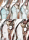
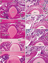
In the anterior component of the median atlanto-axial joint, i.e., between the anterior aspect of the dens and the anterior atlantal arch, we found vascularized synovial tissue projecting from the superior and/or lateral margins to the joint cavity. Thus, the disk-like structure was not a complete septum but appeared to be a superior or superolateral marginal fold. The smallest specimen containing the disk-like structure was CRL 52 mm (approximately GA, 10 weeks) and the structure was consistently present in specimens larger than CRL 82 mm (GA, 12 weeks) (Figs. 1, 2). Although it was absent or interrupted in the central or median area, the structure appeared to represent a complete septum depending on the sections chosen (such as horizontal sections across the superior part of the joint) (Fig. 1A, D).
The maximum thickness was 0.1–0.15 mm, being relatively thick and corresponding to more than 10% of the superoinferior diameter of the anterior atlantal arch (0.8–1.3 mm). Notably, in four specimens (CRL, 90 mm, 96 mm, 100 mm and 120 mm), we found evidence suggesting that a mesenchymal tissue plate was separated from a roof of the joint cavity, providing the disk-like structure. The roof was located close to the apex of the dens. As shown in Fig. 2A and B, the disk-like structure was continuous with mesenchymal tissue at the irregular superior margin of the joint cavity. As shown in Fig. 2F and G, the roof of the joint cavity was separated from the atlantal arch. The fovea dentis of the atlas had not yet not developed in any of the specimens. The transverse ligament provided a deep notch on the dens.
Fourteen late-stage specimens
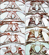
Instead of a disk, we found a short membranous structure in all specimens. In the majority (10 specimens) (Figs. 3, 4, 5), it was a short, thin synovial fold projecting from the roof of the joint cavity in association with tissue fragments scattered within the cavity. Usually, the short fold was highly flexed and pushed to the roof of the joint cavity by the convex surface of the anterior atlantal arch (Fig. 3E, F). In a minority of specimens (4 in all), the membranous structure appeared to provide an incomplete septum (Fig. 4E) interrupted in the central or median area (Fig. 4F). In two of the four specimens showing the septum, the dens did not reach the level of the upper margin of the atlantal arch (Fig. 4C). Thus, in the two specimens, the joint cavity extended above the apex of the dens and the transverse ligament was attached to the apex, rather than appearing as a neck-like depression. The fovea dentis of the atlas had not yet developed in any of the specimens. The fold or septum was vascularized, at least at the base.
In late-stage specimens, the maximum thickness of the fold or septum was less than 0.15 mm: it appeared to be very thin compared with the size of the surrounding bony structures. The atlas and axis were 2–3 times larger than at mid-term (e.g., the anterior atlantal arch, 2.4–2.9 mm), and the maximum thickness of the joint cavity increased from 0.2 mm to 0.8 mm.
Discussion
The present disk-like structure at the anterior component of the median atlanto-axial joint was similar to the so-called fibroadipose meniscoid present in adult lumbar zygapophysial joints [4]. Like the zygapophysial meniscoid, the present disk-like structure did not form a complete septum in the joint cavity. The zygapophysial meniscoid is considered to be a result of aging, and it develops to increase joint congruity. According to our unpublished observations, fetal lumbar zygapophysial joints sometimes carry a very short fold or rim. However, in contrast to the zygapophysial meniscoid, the present atlanto-axial disk appeared to degenerate in late-stage fetuses and possibly also in childhood. Other than real growth, the present measurements suggested that the disk became stretched and fragmented between the mid-term to late stages (approximately 16–29 weeks). We found evidence suggesting that a tissue plate was separated from the roof of the joint cavity at mid-term. In adults, the roof of the joint is characterized by a weak and loose capsule [1, 2].
The elbow (humeroradial) joint always carries synovial folds or rims in late-stage fetuses as well as in adults [5]. In the human fetal elbow joint at 9–10 weeks, Mérida-Velasco et al. [6] found that the rim or fold develops from interzone mesenchymal tissue filling the future joint cavity before cavitation. Likewise, in the fetal knee joint, both the synovial fold and meniscus are sculpted from interzone tissue of the future joint cavity [7]. In the median atlanto-axial joint at mid-term, when the cavitation reached the roof, the mesenchymal tissue plate appeared to become separated from the superior margin of the cavity to provide a disk-like structure. The roof of the joint cavity seemed to correspond to the initial sheath of the notochord or the base of the future apical ligament [3]. Therefore, there seemed to be traction stress against the roof of the joint cavity between the occipital and the dens during their bony growth at and after mid-term, resulting in easy separation of the roof. Consequently, rather than creating increased joint congruity, the present disk-like structure appeared to be a temporary structure during the process of cavitation and later growth.
We found that a relatively lower or shorter dens accompanied a thicker and larger disk in the median atlanto-axial joint. Without compression from the dens, incomplete degeneration might occur. Currently there is no information on the association of the disk with anomalies of the craniocervical junction, such as Chiari malformations and neurocentral synchondrosis [89101112]. Therefore, further detailed examinations of the relationship between bone anomalies and the persistent disk-like structure would be warranted.
 XML Download