

 PDF
PDF ePub
ePub Citation
 Print
Print


Introduction
Tongue muscles change in shape and position during speaking and swallowing [1]. Reductions in tongue muscle strengths trigger problems in both the pharyngeal and oral phases, particularly in terms of poor bolus formation during chewing and swallowing [2]. Tongue strength and accuracy must be improved if such problems develop; the TPS-100 device measures the maximum tongue isometric strength [34]. The buccinator muscle presses the cheek against the teeth, sending food to tooth occlusal surfaces to aid mastication [5]. Muscle strength and co-ordination may improve with training; normal functioning may resume. The strengths and activities of the tongue and buccinator muscles have been studied. However, the relationships between the muscles remain unclear.
Postural techniques improve swallowing safety by controlling food and liquid flow and reducing the aspiration risk. For example, in patients who have suffered cerebrovascular accidents, the head should be turned toward the more-involved side to close off the weaker pharyngeal wall, which makes swallowing safer [6]. In patients with pharyngeal disorders, head rotation toward the paralyzed side reduces the volume of the pyriform sinus, after which the bolus descends down the non-affected side [7], reducing pressure on the upper esophageal sphincter and pressurizing the thyroid cartilage. This in turn promotes the closure of the vocal cords [8]. Oral cancer patients who have undergone partial tongue resection can move a bolus from the mouth to the pharynx by extending the head [6]. Most stroke patients are at risk of food aspiration because the swallowing reflex is delayed. Head flexion reduces this risk by holding the bolus temporarily in the vallecula [9]. Thus, head rotations and tilts are often prescribed by occupational therapists. However, most prior studies have not explored individual swallowing physiologies. Few Korean studies have biomechanically analyzed oral pressures or suprahyoid/buccinator muscle activities.
Electromyography (EMG) is commonly used to explore movements of the upper and lower limbs. Commencing in 2017, oral pressure-measuring equipment has been increasingly used in clinics. However, no study has yet explored how head position affects oral pressure and associated muscle activities. Preliminary work with healthy adults is essential prior to studying patients with weak tongue or buccinator muscles. We thus gathered basic data on how head positioning might aid such patients, exploring how head rotation and tilt affect oral pressure and muscle activities.
Materials and Methods
Oral pressure measurement
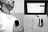
The TPS-100 device (Cybermedic, Iksan, Korea) is used to analyze and strengthen tongue (front and rear) movements in patients with swallowing disorders. Tongue coordination enhances bolus movement and oral pressurization during deglutition. The device features an air bulb, a tube, a sensor, and a pressure-measuring device. We measured tongue and cheek pressures three times, and calculated averages in hPa (Fig. 1).
Surface EMG
We used a surface EMG device (2EM 4D-MT, Relive, Gimhae, Korea) to measure suprahyoid and buccinator muscle activities. The signals were bandpass-filtered, preserving only those of 25–300 Hz. Skin resistance was minimized by removing hair and wiping the skin with an ethanol swab. The interelectrode distance was 1 cm. To measure suprahyoid activity, two electrodes were attached to the skin over the midline of the submental triangle; the ground electrode was placed on the right mastoid process. To assess right buccinator muscle activity, the first electrode was attached lateral to the mouth and the second just lateral to the first (Fig. 1) [1011]. To measure the percentage reference voluntary contraction (%RVC), each subject was asked to swallow saliva three times at intervals of 3 minutes.
Subjects
We briefed recruited subjects on oral anatomical structures, our experimental plan, and the clinical significance of the study. We enrolled only volunteers. We excluded those with surgical injuries to the tongue and cheek. The subjects included 30 Korean adults (15 males, 15 females; mean age, 23 years; range, 20–30 years). The study was approved by the Dongseo University Institutional Review Board (No. 1041493-A-2019-004) and we obtained informed consent from the subjects.
Experimental procedures
Head rotation and tilt served as independent variables. The tongue pressure applied to the front of the palate when the tongue was elevated was measured, and the activities of the suprahyoid and buccinator muscles were recorded. Next, the air bulb was positioned between the right upper and lower first molars and the right buccal mucosa, and cheek pressure and suprahyoid and buccinator muscle activities were recorded during cheek contraction. All tests were run three times at 3-minute intervals. The order of the head positions tested was randomized.
Statistical analysis
The SPSS software ver. 24 (IBM Corp., Armonk, NY, USA) was used to compare all data. The level of statistical significance (α value) was set to 0.05. The following items were analyzed via one-way analysis of variance accompanied by post-hoc (linear contrast) testing: (1) oral pressures of the tongue tip and cheek in the neutral position, and after left and right head rotation; (2) oral pressures of the tongue tip and cheek in the neutral position, and after head flexion and extension; (3) buccinator and suprahyoid muscle activities in the neutral position, and after left and right head rotation; (4) buccinator and suprahyoid muscle activities in the neutral position, and after head flexion and extension.
Results
Oral pressure in relation to head position and tongue elevation
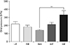
The tongue pressure after tongue elevation did not differ when the head was in the neutral position or rotated to the left or right (F=2.95, P>0.05). When the tongue was elevated, the pressure differed significantly when the head was in the neutral position versus when it was flexed or extended (F=8.25, P<0.05). On post-hoc analysis, the difference when the head was extended compared to the neutral position remained significant (P<0.01), but the difference when the head was flexed did not (P>0.05) (Table 1, Fig. 2).
Oral pressure in relation to head position and cheek constriction
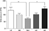
In terms of head rotation with cheek constriction, the cheek pressures differed significantly between the neutral position and upon left or right rotation (F=3.48, P<0.05). On post-hoc analysis, the difference between the neutral position and left rotation was maintained (P<0.01), but the effect of right rotation (compared to no or left rotation) was not (both P>0.05). The cheek pressure differed significantly when the head was in the neutral position versus flexed or extended (F=10.34, P<0.01). Post-hoc analysis showed that the difference upon extension (compared to neutral/flexed) (P<0.05) was maintained, but the difference upon flexion was not (both P>0.05) (Table 1, Fig. 3).
Buccinator muscle activity in relation to head position and tongue elevation
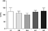
When the head was rotated with the tongue elevated, no significant difference in buccinator muscle activity between the neutral and left- or right-rotated positions was evident (F=0.33, P>0.05). In terms head flexion or extension with the tongue elevated, the buccinator muscle activity did not vary (F=0.74, P>0.05) (Table 2, Fig. 4).
Buccinator muscle activity in relation to head position and cheek constriction
No significant difference in buccinator muscle activity was noted when the head was in the neutral position versus rotated left or right (F=3.02, P>0.05). No significant difference in buccinator muscle activity was noted when the head was in the neutral position versus extended or flexed (F=1.27, P>0.05) (Table 2, Fig. 5).
Suprahyoid muscle activity in relation to head position and tongue elevation
In terms head rotation with the tongue elevated, the suprahyoid muscle activity varied significantly (F=255.65, P<0.01) when the head was in the neutral position versus rotated to the left or right (post-hoc analysis; both P<0.01), but the activities upon left and right rotation were equivalent (P>0.05). In terms of head flexion or extension with the tongue elevated, the suprahyoid muscle activity varied significantly (F=100.49, P<0.01) when the head was in the neutral position versus flexed or extended (post-hoc analysis; both P<0.01); the effects of flexion and extension differed (P<0.01) (Table 3, Fig. 6).
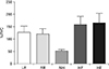
Suprahyoid muscle activity in relation to head position and cheek constriction
The suprahyoid muscle activity differed significantly (F=4.29, P<0.05) when the head was in the neutral position versus rotated left or right (P<0.05). The suprahyoid muscle activity differed significantly (F=4.30, P<0.05) when the head was in the neutral position versus flexed or extended (P<0.05) (Table 3, Fig. 7).
Discussion
The tongue is critical in terms of food movement, and the buccinator muscles of the cheek serve as lateral retainers that prevent food particles from falling into the sulcus between the jaw and cheek [12]. Postural interventions for those exhibiting swallowing impairments have traditionally sought to functionally modify the tongue and cheek biomechanics [13]. EMG has been used to aid patients with chronic dysphagia, affording useful biofeedback on oral muscle tone, thus improving swallowing [14]. We found that the tongue and cheek pressures increased significantly when the head was extended; specifically, the cheek pressure increased on contralateral (left) head rotation. When the muscle fibers and soft tissues are stretched, the tongue and cheek pressures change; passive pressure develops when the muscle and elastic components are stretched beyond their resting lengths [15]. If a patient exhibits low tongue or cheek tension, head extension or contralateral rotation is helpful. Passive tension induced by stretching increases internal pressure, and active tensioning on contraction enhances EMG activity. Suprahyoid muscle activity increases on ipsilateral (right) rotation because the pyriform sinus volume is reduced and the bolus descends on the opposite side; the suprahyoid muscles contract [16]. These muscles elevate the hyoid either anteriorly or posteriorly. When the swallowing response is triggered, the tongue base rises to direct the bolus into the pharynx and the hyoid becomes elevated and moves anteriorly [17]. We found that the suprahyoid muscle activity was higher when the head was flexed or extended, rather than neutral. Head flexion directly moves the hyoid upward and forward because of the length-tension relationship; the force produced by the contractile elements (lying parallel to the elastic components) of the suprahyoid tendons increases [18]. Head extension widens the laryngeal vestibule and narrows the vallecular space; physiological difficulties may follow [17]. However, if oral transfer is impaired in patients who have undergone supraglottic resection, head extension is a useful postural remedy [19]. In dysphagic patients, head flexion expands the vallecular space, pushes the tongue base toward the pharynx, and protects the epiglottis. Therefore, head flexion is frequently used during deglutition training [20].
We found that the suprahyoid muscle activity increased upon contralateral (left) head rotation. Such rotation would be less favored than the neutral position in healthy adults, who use the suprahyoid muscles symmetrically. However, many stroke patients use the perioral muscles asymmetrically [21]. In left hemiplegic patients, left rotation would increase right-side muscle activity and decrease left-side muscle activity. Thus, head rotation would compensate for the suprahyoid muscle weakness of the involved side. Turning the head toward that side eliminates that region of the pharynx from muscle activation, rendering the non-impaired side more active [22].
The buccinator muscle is used to position food for chewing and to control the bolus. We found that the cheek pressure increased significantly on contralateral head rotation and extension, compensating for buccinator weakness. Cheek pressure is influenced by cheek space between fixed part (teeth, alveolar bone) and unfixed part (buccinator muscle). Head position (contralateral rotation, extension) reduced the space and pressure was increased consequently. However, there were no significant changes in the buccinator muscle activity. We assumed the origin and insertion of the muscle was hardly affected by head position [5]. The change in head position significantly increases the pressure in the cheek area even though it does not affect the buccinator muscle activity, which may be helpful for patients with buccinator weakness.
We measured oral pressure and suprahyoid and buccinator muscle activities related to head position. Both the tongue and cheek pressure increased on head extension or contralateral rotation. The suprahyoid muscle activity increased upon head flexion and extension, and contralateral and ipsilateral rotation. In conclusion, head extension or contralateral rotation would aid those with poor tongue or cheek pressure. Head flexion/extension, or contralateral/ipsilateral rotation, increase suprahyoid muscle activity; the optimal head position will vary individually in patients with functional dysphagia or who have undergone supraglottic resection.


 XML Download