

 PDF
PDF ePub
ePub Citation
 Print
Print


Introduction
The practice of anatomical sciences has an elaborate legacy of more than two thousand years. However available literature suggests that modern human anatomy (the form that we are familiar with) was born in Europe during the 16th century [1]. Andreas Vesalius (1514–1564) the famous Paduan anatomist is widely regarded as the ‘father of modern human anatomy’ by virtue of his anatomical exploits which were based on self conducted human cadaveric dissections. The most remarkable contribution of this extraordinary anatomist was the publication of his textbook De humani corporis fabrica in 1543. The publication of De humani during the Renaissance period was a cascading event that trigerred a scientific revolution within the realm of anatomical sciences [2].
Prior to De humani, the most popular textbook in human anatomy was De anatomicis administrationibus which was authored by the ancient anatomist, Claudius Galenus (129–216 AD) who is also known as Galen of Pergamon [3]. The text was marred with erroneous descriptions as Galen's observations were based on animal dissection (human dissection was prohibited by religious authorities and societal norms) and did not have any illustrations [4]. Although Mondino de Liuzzi (1275–1326) supervised the first officially sanctioned human dissection, however he too blindly followed Galen's text as a Sector (surgeon/barber) conducted the dissection [5]. It was Vesalius who first held the dissection knife in his own hand, conducted the dissections all by himself and through his observations initiated the rectification of erroneous details prevalent from the time of Galen. His documentation of anatomical details and complementing them with illustrations in De humani (both were based on dissection based findings) changed the course of human anatomy [1]. The popularity of his published text triggered a new trend and encouraged future anatomists to undertake their own experiments and document their valuable insights through their own signature texts.
Since the Vesalian era, a number of noted anatomists published their own textbooks where they have detailed their observations and contributed towards the advancement of human anatomy. Although these anatomists belonged to different generations, however there is a common link in their published work as a progressive stepwise improvement in the quality of anatomical texts was witnessed with passage of time. This ongoing process of refining the practice of human anatomy through publication of signature texts eventually led to the emanation of the most popular anatomical treatise of all time, Gray's Anatomy authored by the English anatomist Henry Gray in 1858 [6]. In a way publication of Gray's Anatomy, which is the most widely followed anatomical text till date, was the culmination of a long process initiated by Vesalius more than three centuries ago. In this focussed review article we have tried to revisit the interesting journey from pathbreaking De humani to Henry Gray's masterpiece. We have detailed on other popular anatomical texts published during this timeline which formed vital cogs in the turning wheel of evolution of anatomical sciences to the form that we are familiar with in present times.
Methods
An extensive literature search was undertaken for this study and standard search engines such as PubMed, Scopus, Google search, Google Scholar, and Wikipedia were referred to for relevant literature. While searching for published texts in anatomy we limited our timeline between publication of first edition of De humani corporis fabrica (1543) and publication of first edition of Gray's Anatomy (1858). We started with De humani as it heralds the onset of modern human anatomical practice and we did not go beyond Gray's Anatomy as it is the most well known and popular text in Anatomy till date and very much in use with recent updated editions in present time. Within the selected timeline, we have included only those anatomical texts which were pertaining to the whole of human anatomy as such (popular texts focussed on a particular region of the human body were excluded from the study) and were in use within the academic domain over a considerable period of time. We also excluded anatomical atlases (primarily depicting pictorial representation of human anatomy) that were published during this timeline. Copies of the anatomical texts reviewed in the present study and their English translations were consulted from online libraries and wherever applicable have been referenced appropriately. The images used in the study were procured from the internet and it was ensured that all figures included in the present study are in the public domain.
De Humani Coporis Fabrica
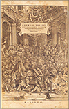
De humani corporis fabrica (on the fabric of human anatomy) is a treatise on human anatomy by the great Paduan anatomist, Andreas Vesalius and was first published in 1543 [7]. The text comprised of seven volumes and was compiled on a system based approach to human anatomy (unlike the present trend of region based study) (Fig. 1). The first two volumes details the anatomy of the connective tissue (Vol. I on bones and cartilages and Vol. II on ligaments and muscles). Vols. III and IV provides description of the vascular system and nervous system respectively. Vol. V was based on the anatomy of the gastro-intestinal system and genito-urinary system. Vol. VI details the anatomy of cardio-vascular system and vol. VII describes the anatomy of brain [8]. His work was a breakaway from the anatomical texts available during that period as Vesalius deviated from the anatomical knowledge documented in Galen's text which was based on animal dissections as human dissection was forbidden at that time [9]. The publication of De humani is a seminal event in the progress of anatomical sciences as the descriptions were for the first time very close to reality. This could be attributed to the fact that Vesalius was a pioneer among his contemporaries as he took the knife in his own hand and himself conducted human dissections (the prevalent trend until then was the barber surgeon would dissect the human body). Vesalius was driven by his conviction that truthful knowledge of anatomy can only be gained through dissection of human corpses and not by following authoritative texts [10]. His luminary footsteps proved to be a gamechanger in removing shackles of mythic speculations of authoritative texts thereby paving the way for the emergence of anatomy as the empirical science and torch bearer of medical sciences during the Renaissance.
Among the many errors of Galen rectified by Vesalius, the most notable one relates to the circulatory system. Regarding this system, the Galenic theory had two essential components: liver was the source of venous blood with the inferior vena cava originating from the liver itself and that the venous blood and arterial blood were two separate entities independent of each other with the arterial system being controlled by the heart. Vesalius through his observations during human cadaveric dissection could rectify the first component of Galenic theory as he documented that inferior vena cava was not originating from the liver and also established that it was emptying into right atrium of heart. However Vesalius continued with the understanding that arterial and venous blood were two separate systems as he was of the opinion that the right ventricle was regulating the venous and left ventricle the arterial circulation [1112]. Thus Vesalius was able to rectify the error in Galen's anatomical theory however he did not dispute Galenic principles of physiology. This was eventually rectified by William Harvey when he detailed the unified theory of circulation in his text Anatomica de motu cordis which was published in 1628 [13]. Vesalius also did not deny the patency of interventricular septum (another long standing Galenic theory) however he harboured doubts in his mind [14]. Subsequent dissection based observations enabled him to present a rectified version in the second edition of De humani (published in 1555) where Vesalius clearly denied the existance of any communication across the interventricular septum. However it also occurred to Vesalius that if blood could not pass from right to left ventricle through interventricular septum, then it was necessary to conceive an alternate pathway [15]. This query was eventually solved by one of his students Realdo Colombo, when he documented the pulmonary circulation in his text De re anatomica in 1559 [1617]. Vesalius also rectified Galen's anatomical errors with regards to the structure of human carotids. Galen's model represented left and right carotids emerging from a common trunk (truncus communis), which is actually the case in simians (dissected by Galen) but not in humans, where left carotid emerges separately from aortic arch [1819].
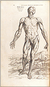
De humani was a trendsetter as it catalysed the use of illustrations alongside the written materials in anatomical texts. Although these illustrations were prepared by Stefan van Calcar (1499–1546) a renowned Italian painter from the Renaissance period, nevertheless these were relevant enough to convey the essential details [7]. The woodcut illustrations included in De humani underlined a seamless combination of artistic precision, refinement and anatomical systhesis [20]. Although prepared by an artist, these illustrations were complimented with minute details based on the inputs provided by Vesalius, gained through cumulative observations from human cadaveric dissections [21]. De humani initiated a paradigm shift in the use of illustrations in anatomy from serving a visual record to compensate for limited access to cadavers in teaching to becoming an indispensable tool to accurately convey detailed anatomical structure through medium of printing [22]. Nevertheless a notable drawback of these illustrations were the overwhelming presence of artistic flavours characterised by theatrical attitudes and ornate landscapes (Fig. 2). In fact De humani started the Vesalian tradition of anatomical illustrations with the typical presence of grandiose human figures, which emerged as a signature component of anatomical texts during this period [23].
De humani is a milestone in the long history of anatomical texts as it was an honest attempt to look beyond the boundaries established by age long Galenic thoughts and principles (etched in the minds of anatomists over centuries). In true sense, it was a flag bearer of the scientific revolution in the domain of medical sciences that took place during the Renaissance period. Undoubtedly, it played a crucial role in advancement of anatomical sciences and was integral to the rise in popularity of the subject among the general population. The crucial aspect of this text which enables it to stand out from all previously published anatomical texts were that the findings were based on direct observation of human dissection and use of quality illustrations with anatomical details (albeit with artistic influence). Vesalius rectified some of the Galenic errors and raised doubts based on logically derived thoughts on certain other theories that encouraged future anatomists to come up with further insights. De humani not only enriched anatomical sciences but also opened floodgates for many discoveries and developments that eventually contributed to the evolution of medical sciences to the state that we are familiar with in present times.
Syntagma Anatomicum
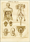
Syntagma anatomicum publicis dissectionibus was authored by German anatomist, Johann Vesling (1598–1649) who served as the Professor of Anatomy and Surgery at the University of Padua, Italy [24]. It was the most popular anatomical text of the seventeenth century and was first published in 1641 with two copper plate illustrations [25]. However, it was the second edition of the text which was published in 1647 with an elaborate 24 copper plate illustrations, became a much sought after treatise among anatomists in Europe [26]. Syntagma favoured practical knowledge which human dissection offers to the anatomist over theoretical conundrums. In Syntagma, Vesling described anatomical structures exactly as they are seen at dissection. The text is detailed on region based anatomy and guides the reader through a textual cum visual dissection from head to foot, proceeding according to the body parts an anatomist would encounter from the moment of first incision, beginning with skin and body fat. Gradually the reader is familiarised with the bones underlying the soft parts which are being dissected and eventually acquainted with the complexities of the nervous system, the blood vessels and the lymphatic system [25]. Such detailed and stepwise detailing of anatomical structures were among the earliest in medical science. Moreover the copper plate illustrations accompanying the text were of high quality and were original engravings that represented organs of the human body more accurately than their predecessors. These illustrations significantly contributed to the success and popularity of Syntagma and were frequently reused by contemporaries [19].
Syntagma was published almost hundred years after De humani and understandably there were considerable progress in prevalent anatomical knowledge. This is evident as Vesling documented rectified version of anatomical details with regards to hepatic portal vein and pulmonary veins. Vesling reported that the portal vein underwent bifurcation as it entered the liver. This was in contrast to the accepted theory until then that the portal vein divided into five branches on arriving at the gate of the liver as reported by Vesalius [27]. Vesling was the first to document the existence of four pulmonary veins emptying into the left atrium of heart [28]. It may be mentioned here that Vesalius had only thought about the existence of pulmonary circulation and Realdo Colombo had documented the same without details [16]. Syntagma was enriched with descriptions of new anatomical structures as well. Vesling gave an outline of the cerebral vasculature at the base of the brain in the second edition of Syntagma [26]. Later on Thomas Willis gave a detailed account of the arterial circle of brain in 1664 and it was eponymously linked to his name [29]. Vesling also detailed the existence of rete mirabile (wonderful net) representing the fine branches of the internal carotid artery spread over the surface of brain. The structure was mentioned previously by Herophilus and described by Galen in animals. However, it was Vesling who established its presence in humans [30]. Vesling was the first to describe the soleus muscle and additional ear ossicles which he observed as small structures attached to the side of stapes. He coined the terms ossiculum quartum (fourth ossicles) and ossiculum parvum (small one) in reference to these entities [3132].
Syntagma was the most popular anatomical text in Europe from second half of seventeenth and first half of eighteenth centuries [33]. The success of the text can be assessed from the fact that it was republished many times with as many as sixteen editions in different languages such as Latin, German, Dutch, and English [22]. In 1741, a Dutch version of the Syntagma became the first illustrated western anatomical text to reach Japan and pioneered the development of European medicine in Japan [34]. The widespread popularity of the Syntagma could be attributed to the simplicity and the diagrammatic nature of the illustration used in the text. Vesling avoided the theatrical attitudes and ornate landscapes that were prominent in anatomical figures as a part of tradition established by Vesalius. The illustrations used in Syntagma primarily focussed on anatomical details thus giving them a realistic outlook which was more useful for medical science (Fig. 3). The figures used in Syntagma often served as models for illustrating anatomical textbooks published later on in Europe [35].
In Syntagma, Vesling continued with documenting the dissection based anatomical exploits, a trend started by Vesalius. In a way Syntagma carried forward the light of discoveries and insights in the domain of anatomical sciences from De humani and even made it look brighter in glow. The text owes its success to the diligence of its author Vesling, who in the Vesalian mould was an ardent believer in unravelling anatomical details through resolute human cadaveric dissection. Vesling thrived in a period when anatomists were gradually shifting towards the Vesalian principles and acknowledging errors in Galenic theories. This possibly contributed in Vesling having the courage to document radical discoveries in Syntagma and those being readily embraced by his contemporaries. The illustrations in Syntagma ushered a new era in anatomical literature whereby realistic figures replaced the over indulgence of artistic flavours which became a trend from Vesalian era.
Adversaria Anatomica
Adversaria anatomica was the anatomical text written by the most influential anatomist of the 18th century, Giovanni Battista Morgagni (1682–1771). Morgagni was an Italian anatomist who served as the Professor of Anatomy at the University of Padua for more than 50 years [36]. The publication of Adversaria proved to be a watershed event in anatomical literature as it started the trend of detailing clinically oriented anatomy. In a way Adversaria provided the much-needed link between the advances in the theory and knowledge of anatomy with clinical practice [37]. This was a major shift from the trend prevalent until the seventeenth century, when anatomists were more focussed in detailing dissection based observations with little or negligible emphasis on their clinical implications. Morgagni pioneered the anatomo-clinical concept in medicine and established anatomy as the instrument to identify the seat and etiology of any disease [38]. His extraordinary ability to integrate and synthesize information drawn from anatomical dissections, set him apart from his contemporaries. He seamlessly correlated anatomical observations with clinical cases (anatomo-clinical method) and his standalone approach was a major breakthrough in medical science as it guided physicians to diagnose a disease, analyse its prognosis and prepare a management protocol for the same [39].
Adversaria was based on a series of research findings from immaculate anatomical dissections. The text was published in three volumes: the first volume Adversaria anatomica prima was published in 1706, followed by the second volume Adversaria anatomica altera in 1717 and the final volume Adversaria anatomica omnia in 1719 [404142]. Throughout the text, Morgagni followed a signature pattern. At first anatomical details of a particular organ was documented based on dissection based observation. This was followed by a comparative analysis of the findings with the details reported by previous authors which was based on extensive reading of available literature [40]. A characteristic feature of Adversaria, which separates the text from contemporaries is the reflection of modesty, open mindedness and due acknowledgement of noted anatomists by the author. Although Morgagni was held in high esteem by anatomists across Europe (evident from the fact that his colleagues referred to him as ‘his anatomic majesty’) he maintained his reticence in the documented details [43]. In Adversaria, whenever applicable the efforts of predecessors such as Harvey, Willis and many others have been acknowledged. He also advised his readers not to accept his views blindly and insisted them to undertake their own experiments for clarification of any doubts and never believe that the last words in science have ever been spoken [40].
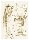
Morgagni continued with the tradition of his predecessors and rectified several errors in anatomical details documented by previous anatomists. However, Adversaria anatomica became popular due to the novel and valuable information on several aspects of human anatomy. Adversaria throws new light on the anatomy of trachea, glottis, laryngeal glands, lacrimal apparatus, paranasal air sinuses, heart with great vessels, kidneys and male as well as female genital organs [44]. The high point of Adversaria were the first time description of some structures which have been eponymized by the name of Morgagni. Morgagni's ventricle which happens to be the pouch located on the lateral aspect of the vestibule of larynx, Sinus of Morgagni which is the space between the base of skull and superior constrictor of pharynx and Morgagni's concha the eponym for superior nasal concha are few examples [40]. Adversaria was also enriched by the first-time description of the fissure between the sternal components and costal components on the right side of the diaphragm and this entity which is better known as trigonum sternocostale dextrum is referred to as Morgagni's foramen. Moreover, the incidence of congenital hernia through this foramen was described in Adversaria and is now known as Morgagni-Larry hernia [45]. Adversaria familiarizes the reader with previously unknown anatomical structures such as appendix of testis (Morgagni's hydatid) and fluid filled cysts often found attached to the fimbriae of the fallopian tubes (hydatids of Morgagni) [46]. In accordance with the prevalent trend, Adversaria was complimented with illustrations which were precise and dedicated to the display of anatomical details (Fig. 4). Morgagni thus followed the trend established by Vesling in keeping the illustrations simple yet detailed but more significantly was free from unnecessary artistic influences [36].
Morgagni pioneered the technique of correlating ante mortem symptoms (of his patients) with the post mortem anatomical details observed while dissecting the bodies of his deceased patients. Thus he gave birth to a new scientific approach within the realm of anatomy which became popular as Pathologic Anatomy. The detailing of pathologic anatomy in Adversaria which was based on anatomo-clinical approach, stands apart from the texts authored by Morgagni's predecessors in the precision of reasoning he applied to the subject. Morgagni gave vivid descriptions of anatomo-clinical details related to lesions of heart (angina pectoris, pericardial effusion, endocarditis, rupture cordis and many more), pathology of respiratory system (pulmonary tuberculosis, pneumonia, bronchitis) and abnormal anatomy of the kidneys (asymmetrical kidney, solitary kidney, horse shoe-shaped kidney) [464748]. Morgagni also detailed the anatomo-clinical changes in malignant lesions like osteosarcoma, carcinoma of stomach, cervical cancer, breast carcinoma, testicular carcinoma among others [49]. His monumental work which was the result of years of dedication to the practice of anatomical sciences eventually proved to be a great service to the subject and was decisive for the advancement of medical sciences.
Anatomy of the Human Body
Anatomy of the human body was co-authored by John Bell (1763–1820) who was a Scottish anatomist and surgeon and his younger brother Charles Bell (1774–1842) who was also an eminent surgeon, famous as an anatomy teacher and a prolific author [50]. Anatomy of the human body was published in four volumes, with Vols. 1 and 2 authored by John Bell and were published in 1797. However for Vols. 3 and 4, Charles Bell assisted his brother in authoring the text and prepared the parts on nerves, the sensory organs and the viscera (published in 1803 and 1804, respectively) [21]. In this well compiled text, John Bell introduced the hitherto unexplored domain of Surgical Anatomy. Before the publication of Anatomy of the human body, the concept of anatomical details was well known and even the trend of clinically oriented anatomy was spreading roots. However, there was no effort to detail anatomy exclusively from the surgeon's point of view. As surgery was fast emerging as a prominent science, Anatomy of the human body was able to gain massive popularity among surgeons and also served the core of anatomical sciences [50]. As a student of medicine, John Bell realized that anatomists were teaching the subject without any first-hand knowledge of the problems encountered by surgeons. He endeavoured to minimalize this lacunae by teaching anatomy from a surgeon's viewpoint to his students while practising surgery. During this period, he started preparing his notes on anatomy as well as surgical anatomy which were primarily based on his lectures. Later on these notes were used as the platform for writing his anatomical text [51]. As an anatomist he took the entire human body as his province and in his writings the details of the topographical anatomy manifest themselves as a source of unfailing strength to the surgeon. Before his time the anatomy of the human body was well known, however Bell's text provided a new prism and a fresh outlook to the subject [52]. Anatomical facts that were long established were given a new significance and those that were nebulous or equivocal were now presented with new clarity. His contributions were well recognized by the fact that he was referred to as the ‘father of surgical anatomy' by his contemporaries [50].
A noteworthy feature of Anatomy of the human body were the extraordinary illustrations plates which were prepared by John Bell himself, unlike most of his predecessors who had relied on eminent artists to prepare illustrations for their work [21]. As an artist John Bell was one of the few eminent medical men who illustrated their own work and one to whom drawing, sketching or engraving came equally at ease [53]. The four volumes of Anatomy of the human body were complimented with a total of 125 illustration plates which were prepared as neatly detailed engravings. John Bell took the liberty in demonstrating anatomical details in his own individual manner in most of his illustrations thereby emphasizing on the directness of representation to the reader [54]. His drawings were never intended to be anything other than handmade pieces and there were no attempts to make the figures appear as if they were alive. This lack of a recognizable style in the engravings of Anatomy of the human body emerged as a new style and became popular as ‘pliable naturalism’ a trend which was embraced by contemporary and future anatomists [20].
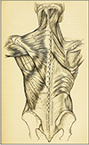
The illustration plates in volumes 3 and 4 were mostly prepared by Charles Bell, the younger brother of John. Prior to assisting John in preparing the draft of Anatomy of the human body, Charles had published his own text titled as the System of dissection (in two volumes) for which he had prepared his own illustrations [51]. Charles was himself a gifted artist and in a way carried forward the legacy of John Bell in the final two volumes of the text. Charles prepared the engravings of nerves, the sensory organs and the viscera, which are a testimony of his incredible artistic skills [52]. Charles' work was driven by his conviction that preparing the illustrations would actually discipline a surgeon's hand and the study of anatomy would discipline the physician's eye [55]. This ideology formed the very soul of Anatomy of the human body as the text was complemented with accurate and simply rendered illustrations depicting anatomical details which were used by students of anatomy as well as a preparatory text for surgical study and practice (Fig. 5).
John and Charles Bell believed that true understanding of anatomy was aided in pairing accurate drawing with thorough description and this was reflected in the core of their text [20]. For the description of anatomical structures, a number of variations observed in different bodies were considered but the emphasis was always on the most typical anatomical details [54]. Any deviation from usual forms were noted and explained in the text. The key feature of the text was to devote sustained attention to how anatomy could best be communicated visually to aspiring surgeons. After the publication of Anatomy of the human body written text and accompanying illustrations were given equal importance in published literature related to anatomical science [21]. Through their anatomical treatise, the Bell brothers brought the printed text and the illustrations into such harmony that they gave a holistic overview to the reader thus leading to double impact of single information. The degree to which quality illustrations were prioritised can be assessed from Charles Bell's view that anatomy without plates is ‘no better than a book of geography without maps’ [53]. In a way Anatomy of the human body as a text put an end to two practices prevalent until then in the published materials: the ongoing conflict between the anatomist (insisting for details) and the artist (striving for individual styles) and also the inclination towards the use of full-page real size anatomical illustrations [51]. The text was able to convince the contemporaries that an effective representation need not be of the same size as the object itself [20]. Published at the very beginning of the nineteenth century, Anatomy of the human body proved to be an eye opener for generations of anatomists and possibly paved the way for better things to happen in the domain of anatomical sciences.
Anatomy: Descriptive and Surgical
The legendary work of Henry Gray titled Anatomy: Descriptive and Surgical, popularly known as Gray's Anatomy was published in 1858 and the voluminous text had 750 pages which were complimented with 363 illustrations (Fig. 6) [56]. Gray was an English anatomist & surgeon and he was a Fellow of the Royal College of Surgeons at the time his work was first published. As the title of the text as well as the background of its author would suggest, Gray's text provided deep insights into the application of the knowledge of anatomy to surgical practice [6]. In fact, the primary objective of the author was to induce the reader to apply the anatomical details to hone one's skills as a surgeon. In a way Gray's Anatomy followed the trend established by John and Charles Bell in their anatomical text, however Gray was able to present the details in a more evolved, elaborate and precise manner based on his long dissection sessions as well as through more number of illustrations used in the text [56]. Moreover, Gray's Anatomy also introduced the concept of sectional anatomy by the use of illustrative depictions of methodically prepared sections of the human body [57]. Although the concept presented in the text was in a rudimentary state, nevertheless it laid the foundation of a major advancement within the domain of anatomical sciences. In order to propel his readers forward Gray himself travelled the reverse path when as a painstaking and methodical worker, Gray (although he was a trained surgeon) advanced his anatomical knowledge by the slow but invaluable method of making dissections for himself [58]. The key component of the success story of Gray's Anatomy were the observations noted during the course of these detailed human dissections as well as examination of surgical specimens with substantial focus on the study of normal anatomy and possible variations thereof [59]. The monumental exercise that Gray undertook over a considerable period of time is evident across the pages of the text that served as the very basis of medical science for generations to come.
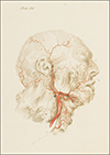
The phenomenal success of Gray's text can be attributed to a large extent to the excellence of its illustrations [21]. The drawings used in the text were prepared by Henry Vandyke Carter (1831–1897). When Gray began work on his text, Carter was working as a demonstrator of anatomy at St. George's Hospital Medical school where Gray was his senior and serving as a lecturer. Both Carter and Gray had similar interest and soon became good friends. Gray relied to a great extent on Carter to assist him with the dissections also to prepare the large number of illustrations required for the text [6]. Continuing with the ‘lack of style’ approach introduced by John and Charles Bell, Carter restrained himself from any attempt to place the figures in graceful poses or against unrealistic/whimsical backgrounds. The use of life size views of the human was consciously avoided and even a complete skeleton was not depicted anywhere in the text [20]. Carter's illustrations were aimed at scientific description of the anatomical structures and the core value of these well-prepared wood engravings was the illustrative clarity which was remarkably atypical in relation to that period (Fig. 7) [60]. In those days, illustrations in anatomy texts were typically proxy labelled, whereby the reader had to refer to the key which was usually provided in the footnote. The illustrations used in Gray's Anatomy contrastingly represented unified nomenclature and structures which enabled the reader to assimilate both simultaneously [61]. Carter's work ensured that throughout the text the focus remain on the anatomical details and this was in accordance to Gray's dream to provide visual descriptions in anatomy that would be useful for aspiring surgeons and clinicians.
The completed manuscript and illustrations for Gray's Anatomy were forwarded to the publishers John W Parker & Sons towards the end of 1857. When the book was eventually published a year later, Gray and Carter were not able to celebrate together as Carter had already left for India, where he was appointed as Professor of Anatomy & Physiology at Grant Medical College in Bombay [62]. Within one year of its publication in England, Gray's Anatomy was made available in the United States and the resulting surge in popularity of the text reflected in the sales. Within a short timespan Gray's Anatomy emerged as the most sought-after textbook of anatomy across the English-speaking world [63]. Encouraged by the success of his work, Gray began working on a second edition being assisted by his colleague Timothy Holmes who was also a surgeon. Unfortunately, Gray passed away at a very young age in 1861 due to small pox and could not oversee the completion of his work. This responsibility was ably tendered by Timothy Holmes, who prepared the subsequent seven editions of this masterpiece in anatomy [64].
Henry Gray authored his text keeping in mind an audience of medical students and physicians, particularly the surgeons. Across all these years, successive editors have been entrusted with the responsibility to maintain the relevance of this wonderful text. The untiring efforts of luminaries (past and present) in the subject of anatomy have ensured that the legacy of Henry Gray continues till date. Emphasis on preparing each edition in a manner that the text emerges as the most comprehensive account of the anatomical descriptions (keeping in view the clinicians' perspective) available at the time of publication, possibly played a key role in maintaining the popularity of the text over all these years. The authenticity and authority of Gray's Anatomy even in present times is evident from the fact that it is widely referred to by scholars for undertaking research activities pertaining to anatomical sciences [65]. From the 39th edition onwards of this landmark textbook a major change was incorporated as anatomical details were documented according to regional anatomy as all editions previous to this were organized by systemic anatomy [66]. The 40th edition of this legendary text was published in 2008, to celebrate the 150th anniversary of the publication of the first edition [67]. The most recent 41st edition was published in 2015 as a meticulously revised and thoroughly updated edition, reflecting the very recent understanding of clinical anatomy from leaders in respective fields around the globe. In keeping with changing times, in addition to the regular printed version, the latest edition of Gray's Anatomy is also available in an e-book version [68]. Over all these years and across several editions, Gray's Anatomy has remained true to the vision of Henry Gray and Henry Carter in that it continues to detail the anatomical basis of clinical practice, from the perspective of leading clinicians and anatomists. Till present day Gray's Anatomy remains the most updated, definitive and comprehensive reference on anatomical details, offering ready access to information essential to ensure safe medical practice.
Conclusion
In this review article, we revisited the journey from De Humani Corporis Fabrica to Gray's Anatomy as we felt it is necessary to familiarize the present generation of anatomists with the legacy of past stalwarts whose visionary efforts across centuries have ensured an ongoing process of refinement of texts pertaining to practice of modern anatomy. As we have observed the road was not an easy one and it required remarkable courage from Vesalius to rectify errors in Galenic principles and exemplary foresight to compliment the text with illustrations. Vesling continued the tradition and made the illustrations more scientific. Morgagni introduced the concept of clinical anatomy and made anatomical sciences more relevant towards clinical practice. John and Charles Bell emphasized the relevance of surgically oriented anatomy, a concept which was further enriched by Henry Gray in his anatomical masterpiece. Moreover, Gray's Anatomy presented a new style of illustrations with unified nomenclature which is followed till date. Henry Gray also introduced the concept (albeit in a rudimentary fashion) of sectional anatomy in his text, which rose to prominence in the years to come. Each of the anatomical texts detailed in this study played a significant role in overall development of anatomical sciences and the gradual process of refinement brought about by these legendary anatomists provided the key component towards the evolved outlook of anatomical texts in present times.
 XML Download