

 PDF
PDF ePub
ePub Citation
 Print
Print


INTRODUCTION
Biliary adenofibroma is an extremely rare biliary epithelial tumor that is characterized by tubulocystic bile duct proliferation lined with non-mucin secreting biliary epithelium and supported by abundant fibroblastic stroma.1 The MRI findings of biliary adenofibroma are not well established owing to its rarity. This paper presents histopathologically confirmed cases of biliary adenofibroma found in two patients who underwent a preoperative MRI, along with a review of the relevant literature focusing on the MRI findings.
CASE REPORT
Case 1
A 63-year-old man with chronic hepatitis B and hepatocellular carcinoma treated previously with locoregional therapy was referred to the authors' medical institution for an evaluation of a solitary hepatic lesion increasing in size, as shown on the regular follow-up CT. The patient denied abdominal pain, weight loss, nausea, fever, or jaundice. The laboratory tests revealed a normal liver function. The tumor markers, including alpha-fetoprotein, protein induced by vitamin K absence or antagonist-II, CA 19-9, and CEA, were not elevated.
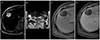
The CT scans obtained at an outside hospital revealed a tumor size of 1.1×0.7 cm, 2.3×1.4 cm, and 3.6×3.0 cm 5, 3, and 1 year ago, respectively, presenting a slowly growing nature. MRI demonstrated a well-circumscribed multiseptated mass within segments IV and VIII of the liver, measuring 4.7×4.5 cm. The T2-weighted image revealed a multicystic tumor with bright signal intensity (Fig. 1A, B). The precontrast T1-weighted image of the tumor showed low signal intensity (Fig. 1C). After gadolinium administration, thin septa within the tumor were enhanced (Fig. 1D). Communication with the intrahepatic bile ducts was not evident on MRI. No lymphadenopathy was observed.
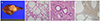
The patient underwent a central bisegmentectomy. The gross pathological examination showed a well-circumscribed multicystic tumor, measuring 4.5×4.3 cm with thin fibrous septa (Fig. 2A). The tumor was comprised of tubulo-glandular and microcystic structures embedded in the fibrous stroma (Fig. 2B). The tubules and cysts were lined with cuboidal or low columnar epithelial cells. The epithelial component did not contain atypia. The background stroma contained scattered spindle cells. The stroma was fibrous, but ovarian-like stroma was not detected (Fig. 2C). No communication was observed between the lesion and intrahepatic bile ducts, either grossly or microscopically. Immunohistochemically, the epithelial component was positive for cytokeratins 7 and 19, suggesting a bile duct origin. P53 was focally positive. Ki-67/index proliferation was low (<2%). The stromal cells were stained with α-smooth muscle actin (Fig. 2D). The background liver showed septal fibrosis and features of chronic hepatitis consistent with a HBV infection. The patient has been well with no recurrence after a 41-month follow-up period.
Case 2
A 38-year-old man was referred to the authors' medical institution for an evaluation of a hepatic tumor detected incidentally on a CT scan. A physical examination and laboratory, data including tumor markers, were within the normal range. He denied any personal or family history of hepatobiliary disease.
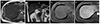
Abdominal CT revealed a well-circumscribed low attenuation mass within the left lateral section of the liver, measuring 2.7×2.4 cm. MRI revealed a multiseptated multicystic mass with hypointensity on the T1-weighted image and bright signal intensity on the T2-weighted image (Fig. 3A–C). After gadolinium administration, septa and wall enhancement were noted (Fig. 3D). No communication was observed between the lesion and the intrahepatic bile ducts seen on MRI. An enlarged lymph node was not demonstrated.
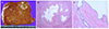
The patient underwent a left lateral sectionectomy. The gross examination revealed an unencapsulated tumor, measuring 2.5×2.2 cm (Fig. 4A). Microscopically, the tubules were lined with cuboidal or low columnar non-mucin secreting epithelial cells, embedded in a fibrous stroma (Fig. 4B, C). No epithelial atypia was observed. Pathology analysis did not reveal evident communication with the bile ducts. The background liver showed macrovesicular and microvesicular fatty changes. The patient did not develop any recurrence during the 39-month follow-up.
DISCUSSION
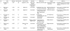
Biliary adenofibroma is a rare primary intrahepatic tumor with an unknown etiology. Tsui et al.1 first described the disease in 1993, and it is now recognized as a distinct tumor of a bile duct origin according to the 2010 World Health Organization (WHO) classification.2 Biliary adenofibroma is characterized by a complex tubulocystic biliary epithelial tumor without mucin production and with abundant fibroblastic stromal components. To the best of the authors' knowledge, only 21 cases of biliary adenofibroma have been reported thus far in the medical literature.1345678910111213141516171819 Previously reported cases focused on the clinical and pathological features of biliary adenofibroma, while the imaging findings were recorded only sporadically. Currently, MRI is the imaging modality of choice for evaluating a range of intrahepatic tumors. MRI has advantages regarding the characterization and diagnosis of these lesions owing to its high contrast resolution. Consequently, the literature was reviewed to characterize the MRI features of biliary adenofibroma.
Table 1 provides brief clinical information and lists the MRI features of the tumor. Biliary adenofibroma occurs in both men and women. The MRI features usually reveal a well-circumscribed multiseptated multicystic tumor that varies in diameter from 2.7 cm to 16 cm. The tumor exhibits hypointensity on the precontrast T1-weighted image and bright signal intensity on the T2-weighted image. After contrast administration, septa and wall enhancement are noted. The present cases also showed no communication with the bile ducts on MRI.
The differential diagnosis for biliary adenofibroma includes intraductal papillary neoplasms of the bile duct (IPNB) and mucinous cystic neoplasm (MCN) of the liver. The tumors grew in a papillary architecture and may be histopathologically similar to biliary adenofibroma.16 On the other hand, mucin secretion was absent in biliary adenofibroma, whereas IPNB and MCN form mucin-containing cysts. The presence of communication between the lesion and the bile duct system is essential for a diagnosis of IPNB. In contrast to IPNB, biliary adenofibroma has no bile duct communication. MCN requires the presence of ovarian-like stroma and occurs almost exclusively in women. Compared to MCN, biliary adenofibroma does not contain ovarian-like stroma. The MRI findings of IPNB and MCN of the liver are well established. The characteristic MRI features of IPNB are visible intraductal masses within the dilated intrahepatic or extrahepatic bile ducts, and the downstream bile duct can also be dilated.2021 Multifocal papillary tumors are well visualized on the T2-weighted image because of the high contrast between the tumor and background bile.22 The imaging patterns of IPNB can be classified into four subtypes according to the presence of intraductal lesions and the degree of mucin production: intraductal mass with proximal ductal dilatation, intraductal mass with proximal and distal ductal dilatation, disproportional duct dilatation without a visible mass, and cystic dilatation with a mass.22 Among them, cystic dilatation with a mass manifests as a focal aneurysmal dilatation of the involved duct or a cystic lesion, showing similar imaging features to biliary adenofibroma. A previous study reported that IPNB could manifest the “thread sign”, which is defined as intraductal linear or curvilinear hypointense striations on MRI.23 On MRI, MCN of the liver typically manifests as a multilocular and septated cystic tumor with an irregular thick wall.24 MRI characterizes intralesional locules with various signal intensities on the T1- and T2-weighted images depending on the cyst fluid protein concentration.25 MCN of the liver does not show dilatation of the downstream bile duct, whereas a ductal dilatation upstream to a cystic lesion can be seen.26
Another differential diagnosis of biliary adenofibroma includes a hepatic echinococcal cyst. Unlike biliary adenofibroma, a typical echinococcal cyst contains multiple daughter cysts. A mother cyst and peripherally located daughter cysts appear as a multilocular cystic lesion with internal septa.27 On MRI, echinococcal cyst shows mixed hypointensity on the T1-weighted image depending on the amount of proteinaceous debris. After contrast administration, the cyst walls and internal septa are enhanced.28
Although biliary adenofibroma is classified as a benign biliary tumor and precursor in the 2019 WHO tumor classification system,29 histopathological and biological spectra of biliary adenofibroma are unclear owing to its rarity. To date, biliary adenofibromas with benign behavior,167813 carcinoma in situ,10 and malignant transformation451214151618 have been reported.
In conclusion, biliary adenofibroma is recognized as a distinct entity of biliary tumor in the WHO classification. The disease is characterized by a tubulocystic bile duct proliferation lined with non-mucin secreting biliary epithelium and supported by abundant fibroblastic stroma. A well-circumscribed multicystic tumor with septal enhancement and no intrahepatic bile duct communication may be the characteristic MRI findings of biliary adenofibroma.
 XML Download