

 PDF
PDF ePub
ePub Citation
 Print
Print


INTRODUCTION
Active treatment of renal stones is indicated for stones causing urinary obstructions and hematuria or uncontrolled pain, for growing stones, for stones persisting longer than 2 years, and in patients at high risk for stone formation or urinary tract infection [123]. However, these indications have not yet considered the assessment of renal function. In patients with kidney stones, the risks for renal insufficiency are multifactorial and include multiple stone episodes, comorbidity, urinary tract infection, and urinary obstruction [1245]. Information pertaining to renal function facilitates appropriate treatment decisions and the timing of management in patients diagnosed with nephrolithiasis and at high risk for renal insufficiency.
Recently, minimally invasive procedures such as retrograde intrarenal surgery (RIRS) and miniaturized percutaneous nephrolithotomy (mini-PCNL) have been reported as feasible and safe options for various kidney stones [67]. These surgical techniques are conducted after complete evaluation of their effect on renal function. Bilen et al. [8] investigated altered renal function at 3 months after conventional PCNL and found that the estimated glomerular filtration rate (eGFR) reflected stable or significantly improved status in patients with late-stage chronic kidney disease (CKD). The positive effect of minimally invasive renal surgery on relative renal function has also been reported in studies [9] using diethylenetriamine penta-acetic acid (99mTc-DTPA) and technetium-99m dimercaptosuccinic acid (99mTc-DMSA) at postoperative 3 months. Although El-Nahas et al. [10] and Canes et al. [11] investigated the long-term effects of conventional PCNL on renal functional outcomes based on eGFRs for more than 12 months, studies evaluating the long-term impact of mini-PCNL and RIRS on separate renal function have rarely been reported.
Therefore, the purpose of the present study was to evaluate the effect of mini-PCNL or RIRS on perioperative kidney function as determined by 99mTc-DTPA scintigraphy at 1 year postoperatively and to identify significant predictors associated with deterioration or amelioration of renal function after surgery.
MATERIALS AND METHODS
1. Study population and data sources
Between 2012 and 2016, patients older than 19 years who underwent minimally invasive surgery for kidney stones were included in an observational study. The authors obtained informed consent from the patients to collect their information. The authors followed the European Association of Urology Guidelines for determining active surgical treatment [3]. Cases diagnosed with preoperative hydronephrosis without complete urinary tract obstruction were included. Patients with febrile urinary tract infection, aged less than 20 years, with bleeding tendency, pregnancy, or urogenital anomalies including horseshoe kidney, solitary kidney, bilateral stones, or three or more percutaneous tracts were excluded from the database. Finally, patients monitored by 99mTc-DTPA for more than 12 months were selected. Patients with positive results on urine culture were treated with antibiotics and sterile urine was routinely ensured before both surgical procedures.
2. Surgical methods
1) Mini-PCNL
Patients in the supine or prone position underwent mini-PCNL under general anesthesia. The percutaneous tract was established by using a combination of ultrasonography and fluoroscopy by a single experienced urologist (Cho SY). A 0.035-mm Terumo guidewire (Terumo Group, Tokyo, Japan) and a Superstiff guidewire (Boston Scientific, Miami, FL, USA) were inserted into the renal pelvis through the tract. The tract was dilated using an Ultraxx™ balloon dilator (Cook Medical, Bloomington, IN, USA) up to 18 Fr followed by insertion of a 15-Fr Miniature nephroscope (Richard Wolf, Knittlingen, Germany). Otherwise, the authors used a 16.5-Fr MIP M nephroscopic system (Karl Storz, Tuttlingen, Germany). The PCNL cases were performed with a single tract of 16.5 to 18 Fr in 90% of cases. Two tracts were made in the rest of the cases with the first tract being 16.5 to 18 Fr and the second tract being 12 Fr using an MIP S nephroscopic system (Karl Storz). A holmium:yttrium-aluminumgarnet (YAG) laser with a 550-µm fiber (Trimedyne, Irvine, CA, USA or Lumenis Ltd., Yokneam, Israel) was used for stone fragmentation. Fragmented stones were removed with a 5-Fr grasping Alligator forceps (Richard Wolf), if necessary. The authors routinely inserted a 6-Fr ureteral JJ stent for about 1 week. Finally, a 16-Fr urethral Foley catheter was inserted. In most cases, no percutaneous nephrostomy tube was placed.
2) RIRS
Patients in the dorsal lithotomy position underwent RIRS under general anesthesia. After cystoscopic or ureteroscopic examination, an 11/13-Fr ureteral access sheath was inserted until it reached the ureteropelvic junction using a 0.035-mm Terumo guidewire, 5-Fr ureteral catheter, and Superstiff guidewire. A Flex-X2 or X2S™ flexible ureteroscope (Karl Storz) or URF-P5™ or URF-V2™ flexible ureteroscope (Olympus, Tokyo, Japan) was inserted through the ureteral access sheath. A holmium:YAG laser with a 365- or 200-µm laser fiber was used to fragment stones. Fragmented stones were removed with a stone basket. The authors routinely used a 6-Fr ureteral JJ stent after lithotripsy. Finally, a 16-Fr urethral Foley catheter was inserted.
3. Clinical parameters
The authors evaluated the patients' preoperative medical history and physical examination, urine microscopy with culture, 24-hour urine collection, serum hemoglobin (mg/dL), creatinine and electrolyte levels, plain radiography (kidneys, ureters, bladder [KUB]), computed tomography (CT), and 99mTc-DTPA scintigraphy results. eGFR was evaluated by using the Modification of Diet in Renal Disease (MDRD) [12]. Stone characteristics were assessed and the volume of stones was determined as the sum of each stone volume calculated as follows: 0.523×length×width×height (mm3). Anatomical factors included the presence of hydronephrosis without complete urinary tract obstruction and duplication, the presence of diverticular stones, and Seoul National University Renal Stone Complexity (S-ReSC) scores [13] based on the categorization of the kidney collecting system including renal pelvis, major calyces, and minor calyces. Perioperative parameters included operative time (minute), stone-free rate, complications, indwelling duration of a ureteral JJ stent (day), admission days (day), and use of an access sheath in RIRS cases. During the postoperative follow-up at 3 months, a KUB and noncontrast CT scan was routinely performed to investigate the recurrence or presence of a residual stone. 99mTc-DTPA scintigraphy was performed 3 and 12 months postoperatively in the group with abnormal renal function. The authors defined stone-free status as follows: absence of residual stones or stones <2 mm on postoperative images at 3 months. The modified Clavien–Dindo system [14] was used for the classification of perioperative complications. Further follow-up and management were individualized.
4. Investigation of predictors of altered renal function
Separate renal function was assessed on the operative side (Fn_Op) and on the contralateral side (Fn_Con). The normal group was defined according to the differences in measured absolute values between the contralateral and the operative sides (GAP, Fn_Con/Fn_Op) ≤10%; the abnormal group was defined by a GAP >10%. In the abnormal group, if the reassessed separate renal function showed a GAP lower or higher than 10% compared with the preoperative values, it was defined as “improvement” or “deterioration,” respectively [15]. The 10% cutoff level in this study was an arbitrary value. Additionally, if the GAP was higher than 10% without deterioration in renal function upon reassessments, it was defined as functionally “stable.” The authors determined significant predictors of change in renal function on the basis of the patients' characteristics, stone data, and perioperative characteristics.
5. Statistical analysis
Statistical analyses were performed by using IBM SPSS Statistics version 22.0 (IBM Corp., Armonk, NY, USA). Independent t-test or Mann–Whitney U test was used to analyze the comparative results between the two surgical groups. Chi-square and Fisher's exact tests were used to analyze categorical variables. Univariate and multivariate logistic regression analyses with a stepwise approach were performed to determine the predictors of change in renal function and stone-free status. A p-value of 0.05 was considered statistically significant.
6. Ethics statement
The approved number of the Institutional Review Board (IRB) for this observational study design was 16-2015-11 in the SMG-SNU Boramae Medical Center (Seoul, Republic of Korea) and 1901-104-1005 in the Seoul National University Hospital (Seoul, Republic of Korea). The study was performed in accordance with the Declaration of Helsinki. All patients signed informed consent to participate.
RESULTS
1. Demographics
A total of 70 patients with a mean age of 56.3±12.4 years were enrolled (Table 1). Age, sex, body mass index (BMI), and eGFR were not significantly different between the two patient populations. The preoperative eGFR was lower than 60 mL/min/1.73 m2 in 13 cases (18.6%). The overall stone-free rate was 81.4%. Five and 20 patients underwent ureteral stenting and extracorporeal shock-wave lithotripsy, respectively, and 4 and 2 patients had undergone RIRS and PCNL previously.
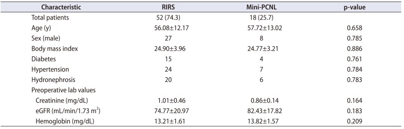
Table 1
Patient characteristics (n=70)
![]()
2. Complications
Transfusion was performed in a single case of mini-PCNL, and Foley catheter re-insertion as a result of blood clot obstruction and ureteral stent repositioning. No additional management was needed for these conditions.
3. Perioperative renal functional outcomes
About one-fifth of the total cases (13/70, 18.6%) were included in the group with normal renal function (Table 2). In the group with abnormal function, the mean Fn_Op was 55.5%. The preoperative GAP was 24.4%, 6.2%, and 28.6% in the total, normal, and abnormal groups, respectively. Among the patients in the abnormal group, more than two-thirds (40/57, 70.2%) of the patients were stable and seven patients (12.3%) were improved at postoperative 12 months (Table 2).
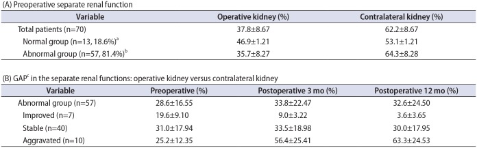
Table 2
Perioperative renal functional outcomes according to altered separate renal function
Values are presented as mean±standard deviation.
a:The difference between the preoperative separate renal function of the operative kidney and that of the contralateral kidney was <10%. b:The difference mentioned above was >10%. c:Defined as the separate renal function of the contralateral kidney minus that of the operative kidney.
![]()
4. Prediction of aggravated kidney function
Univariate analysis was conducted to estimate the significant predictors of postoperative renal aggravation. Age, BMI, comorbidities, previous stone procedure, stone volume, laterality, diameter, number, S-ReSC, Hounsfield unit, preoperative serum creatinine, and hydronephrosis were assessed. At 3 months postoperatively, none of the predictors showed p-values <0.05. At postoperative 12 months, the preoperative creatinine level (p=0.079) and a history of previous stone procedures (p=0.079) showed borderline significance (Supplementary Table 1). In a multivariate analysis, preoperative creatinine level (p=0.060) and a history of previous stone procedures (p=0.051) showed borderline significance at postoperative 12 months (Supplementary Table 1).
5. Prediction of renal function improvement at postoperative follow-up
Univariate analysis was performed to determine the significant predictors of postoperative renal function improvement. Operative time and presence of remnant stones were additional factors assessed to predict aggravated kidney dysfunction. None of the predictors showed p-values lower than 0.05 at 3 and 12 months postoperatively and were thus not included in the multivariate analysis (Supplementary Table 2).
6. Comparison of abnormal renal function on the basis of surgical method
Age, creatinine, eGFR, and hemoglobin level did not differ significantly between the RIRS and mini-PCNL groups (Table 3). Stone characteristics between the two procedures differed significantly. Although the number of stones was not significantly different between the two procedures, the total volume of the stone and S-ReSC scores were larger in the mini-PCNL group than in the RIRS group. The operative time was significantly longer in the mini-PCNL group than in the RIRS group. The stone-free rates were 75.6% and 75.0% in the RIRS and mini-PCNL groups (p=0.714), respectively, and the complication rate and perioperative renal functional outcomes did not differ significantly according to the surgical method.
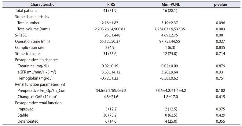
Table 3
Comparative analysis of patients with abnormal preoperative relative renal function according to the surgical method (n=57)
Values are presented as number (%) or mean±standard deviation.
RIRS, retrograde intrarenal surgery; mini-PCNL, miniaturized percutaneous nephrolithotomy; S-ReSC, Seoul National University Renal Stone Complexity; eGFR, estimated glomerular filtration rate.
a:Defined as separate renal function of the contralateral kidney minus that of the operative kidney.
![]()
DISCUSSION
Recent studies have recommended RIRS and mini-PCNL as minimally invasive surgical procedures for the removal of kidney stones [67]. Previous investigations have estimated the effect of minimally invasive surgery on perioperative renal functional outcomes. For example, Akman et al. [16] reported improved or stable eGFR in patients treated with conventional PCNL for staghorn calculi. Recently, kidney function was assessed using renal scintigraphy. Quantitative analysis using 99mTc-DMSA SPECT-CT scans [17] demonstrated that PCNL has a minimal effect on global renal function 3 months after surgery. Piao et al. [9] reported perioperative renal functional outcomes and postoperative outcomes at 3 months using 99mTc-DMSA or 99mTc-DTPA. Despite these studies, however, the effects of minimally invasive surgery on separate renal function during long-term follow-up have rarely been reported. Information about separate renal function and predictors of postoperative outcomes are important for clinical decision-making.
In the present study, we assessed short-term and longterm postoperative separate renal function in patients who underwent minimally invasive surgery for kidney stone disease. In 57 patients included in the abnormal group, the paired t-test revealed a mean change of 4.0% (±20.33) in GAP between preoperatively and 12 months postoperatively, which was not a significant (p=0.145; 95% confidence interval, −1.41 to −9.38). Ten patients showed aggravation with substantial mean changes in GAP between preoperatively and 12 months postoperatively. However, considering that the three biggest changes were 73.6%, 63.6%, and 58.4%, and the sample size was small, the mean change in GAP might not be substantially meaningful. These patients generally exhibited high levels of creatinine (mg/dL) and low eGFR (mL/min/1.73 m2) preoperatively (1.5 and 36.2, 3.0 and 16.0, and 1.28 and 42.0, respectively). Two of these three cases had previously undergone stone management procedures.
The positive relationship between surgery and postoperative separate renal function in the present study is consistent with previous studies. Kukreja et al. [18] and Canes et al. [11] reported improved or similar renal function after PNL during long-term follow-up. In another long-term study of staghorn stones managed by PCNL, radioisotope scanning revealed a lack of adverse renal effects in 91.5% of cases [16].
Several significant predictors of postoperative deterioration and recovery of renal function have been reported. A high preoperative serum creatinine level, renal cortical atrophy, larger stone burden, proteinuria greater than 300 mg/d during follow-up, history of nephrolithiasis, presence of hydronephrosis, and recurrent urinary infection have been reported as significant predictors of deteriorating renal function [218]. A stone number > 3, clear urine in the collecting system, and lack of renal sepsis are significant predictors of improvement of abnormal separate renal function [1519]. In a previous study [9], the authors reported preoperative hydronephrosis as a predictor of deterioration and more than three stones as a predictor of both deterioration and improvement during 3 months of follow-up. However, in the present investigation, none of the factors showed any statistical significance. The preoperative serum creatinine level (p=0.060) and history of previous stone procedures (p=0.051) showed borderline significance. However, analysis of the perioperative renal functional outcomes according to the Kidney Disease Outcomes Quality Initiative CKD classification system in this study revealed no stage-related differences.
This study did not show any significant differences in perioperative renal functional outcomes between RIRS and mini-PCNL. In a previous study of conventional PCNL, the different methods of nephrostomy tract dilatation [20] and multiple-tract access [21] did not exacerbate the loss of postoperative renal function. However, the functional volume at the entry site decreased significantly during regional assessment using SPECT measurement of DMSA uptake [22]. In mini-PCNL, a lower degree of kidney damage than with conventional PCNL might be attributed to the smaller nephrostomy tract. In this study, the PCNL cases were performed with a single tract of 16.5 to 18 Fr in 90% of cases or with two tracts, with the first tract being 16.5 to 18 Fr and the second being 12 Fr to minimize the effect of the tracts on renal function. Additionally, we excluded cases with three or more tracts. Therefore, the effect of these small tracts on renal function would be minimal. Note that perioperative renal functional outcomes were similar for RIRS and mini-PCNL in the present study. During RIRS, acute excessive intrarenal pressure threatens tubular function, and the authors used ureteral access sheaths in most cases. Additional studies are needed to evaluate the consequences of excessive renal pressure and the effects of using ureteral access sheaths on postoperative renal function.
This study had several limitations. First, this was an observational study conducted at a single institution, and the results were derived from a relatively small sample size. Second, although the baseline serum creatinine level was presented as a predictor of postoperative aggravation, a definitive cutoff level was not provided. Further study is needed to assess the cutoff level of preoperative serum creatinine in the evaluation of renal function using 99mTc-DMSA or 99mTc-DTPA. Third, it is necessary to classify the patients' comorbidities, because various comorbidities can affect renal function. Moreover, the compensated contralateral kidney might represent a confounding factor. Although overall postoperative renal functional outcomes were stable, six patients in the RIRS and four patients in the mini-PCNL group showed renal deterioration. Because PCNL and RIRS are known to ensure the safety of kidney function postoperatively [1823], further evaluation of the safety and efficacy of minimally invasive renal stone surgery during a long-term follow-up is needed, especially in patients with preoperative renal insufficiency. Finally, it is hard to clarify whether the deterioration in renal function was induced by surgery or was associated with the natural history of stone disease. Another study with a larger sample size is needed to address these limitations. Nonetheless, this study is valuable because it represents the first investigation of minimally invasive renal stone surgery to assess perioperative separate renal functional outcomes using 99mTc-DTPA during a 1-year follow-up.
CONCLUSIONS
As minimally invasive renal stone surgeries, RIRS and mini-PCNL were effective and resulted in favorable renal functional outcomes during 1 year of follow-up. The effect on renal function of the two procedures was similar during the postoperative period. Clinicians should pay careful attention to patients showing high baseline serum creatinine levels and reporting a history of stone procedures.

 XML Download