Journal List > J Korean Orthop Assoc > v.54(6) > 1140265

선, 최, 고, Seon, Choi, and Ko: 골관절염에서 줄기세포를 이용한 연골 재생의 최신 지견

Abstract

골관절염은 관절연골 침식의 진행을 특징적으로 보이는 질환으로 관절운동 중에 통증을 증가시키고 기계적 스트레스를 견디는 능력을 감소시켜 결과적으로 관절의 가동성과 기능을 저하시킨다. 외상 또는 퇴행성으로 인한 관절연골의 손상이 일반적 관절염의 주요 원인으로 생각되며 이러한 관절연골 손상의 재생에 관한 수많은 연구와 시도들이 현재까지 진행되어 오고 있다. 현재까지 연골 손상의 경우 미세골절술과 자가연골세포 이식술이 일반적인 수술적 치료방법으로 제시되어 왔으나 비교적 양호한 임상 결과에도 불구하고 정상 유리연골의 생성이 미흡하여 시간이 경과하면서 결과가 악화되는 등 단점이 있다. 이를 보완하기 위하여 줄기세포 기반 치료법이 개발되었다. 이 종설에서는 현재 사용되는 다양한 연골 재생 방법들의 장단점 및 결과에 대해 요약하고 특히 중간엽 줄기세포(mesenchymal stem cells) 기반 연골 재생 치료법을 논하고 나아가 이상적인 미래 연골 재생 치료법에 대해서도 고민해보고자 한다.

초록

Osteoarthritis is a disease characterized by the progression of articular cartilage erosion, that increases pain during joint motion and reduces the ability to withstand mechanical stress, which in turn limits joint mobility and function. Damage to articular cartilage due to trauma or degenerative injury is considered a major cause of arthritis. Numerous studies and attempts have been made to regenerate articular cartilage. In the case of partial degenerative cartilage changes, microfracture and autologous chondrocyte implantation have been proposed as surgical treatment methods, but they have disadvantages such as insufficient mutual binding to the host cells, inaccurate cell delivery, and deterioration of healthy cartilage. Stem cell-based therapies have been developed to compensate for this. This review summarizes the drawbacks and consequences of various cartilage regeneration methods and describes the various attempts to treat cartilage damage. In addition, this review will discuss cartilage regeneration, particularly mesenchymal stem cell engineering-based therapies, and explore how to treat future cartilage regeneration using mesenchymal stem cells.

서론

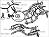
골관절염은 관절연골 침식(erosionl)의 진행을 특징적으로 보이는 질환으로 관절운동 중에 통증을 증가시키고 기계적 스트레스를 견디는 능력을 감소시켜 결과적으로 관절 기능을 저하시킨다. 관절염은 어느 관절에서나 발생이 가능하나 주로 슬관절, 고관절 그리고 수부 관절에서 발생한다.1) 미국에서의 2010년부터 2012년까지의 데이터에 따르면 21.4%, 약 5,200만 명의 성인에서 이 기간 동안 관절염 진단을 받았고 그 중 9.2%에서 관절염으로 인한 활동 제한이 있었으며2) 외상 또는 퇴행성으로 인한 관절연골의 손상이 일반적으로 관절염의 주요 원인으로 알려져 있다.관절 연골이란 양측 골단의 관절면을 덮고 있는 유리연골(hyaline cartilage)을 뜻한다. 관절연골은 외부충격을 흡수하고 두 골간에 마찰을 감소시켜 부드럽고 통증이 없는 관절운동을 가능하게 한다. 연골세포는 이러한 연골의 유일한 구성 세포이며 관절 연골의 1%–5%를 차지한다. 이 세포들은 콜라겐, 프로테오글리칸, 그리고 하이알루론산을 생산하여 세포외기질(extracellular matrix)을 구성하며 연골의 기계적 구성물의 기반을 이룬다. 이 중 콜라겐이 주요 성분으로 건조용량의 60% 정도를 차지한다. 제2형 콜라겐이 전체 콜라겐 중 90%–95% 정도를 차지하고 있으며 이러한 섬유들은 프로테오글리칸 복합체(proteoglycan complex)가 얽혀서 구성되어 있다(Fig.1).3)
그동안 진행된 관절연골의 재생에 관한 수많은 연구와 시도들이 현재까지도 진행되고 있다. 관절연골의 치료 방법은 환자의 상태 및 연골손상의 정도에 따라 달라진다. 광범위한 연골의 퇴행 변화는 인공관절 치환술의 적응증이 되지만 광범위하지 않은 국소 부위의 연골 손상의 경우 미세골절술(microfracture)과 자가연골세포 이식술(autologous chondrocyte implantation)이 수술적 치료 방법으로 사용되어 왔다. 중등도 범위의 퇴행성 변화에 의한 연골손상 환자들의 경우 미세골절술이나 자가연골세포 이식술보다 세포치료와 지지체(scaffold)를 이용한 조직공학적인 접근이 연골 재생에 있어서 효과적인 방안으로 떠오르고 있다. 조직공학적 치료를 위한 지지체의 사용은 숙주세포로의 불충분한 상호결합, 부정확한 세포 전달, 건강한 연골의 악화 등 세포기반 치료와 관련된 단점들을 보완하는 역할을 한다. 지지체기반 치료법은 연골 병변 부위에 3차원 환경을 제공함으로써 연골세포의 탈분화(dedifferentiation)가 적어지고 더 많은 유리연골(hyaline cartilage) 형태를 생성하여 보다 정상적인 연골 재생을 얻을 수 있다(Fig. 2).4)
자가연골세포를 포함하는 히알루론산(hyaluronic acid) 지지체를 병변 부위에 이식할 경우 유리연골 생성량은 증가하지만5) 생물학적 반응물, 외부적 기계 자극, 생화학적 자극을 이용한 여러 노력에도 불구하고 체내에서 동일한 구성을 가진 건강한 유리연골은 생성하지 못한다. 게다가 한정된 수의 세포들로는 이 치료 효과가 감소하므로 이러한 자가연골세포 치료의 단점을 보완하고자 줄기세포 기반 치료법이 개발되었다. 이 종설에서는 현재 사용되고 있는 다양한 연골 재생 방법들의 장단점 및 결과에 대해 기술하고자 한다. 특히 중간엽 줄기세포(mesenchymal stem cells, MSCs) 기반의 연골 재생 치료법을 알아보고 이상적인 연골 재생 치료법에 대해서도 고민해보고자 한다.

현존하는 연골 재생 치료의 종류와 한계점

1. 미세골절술

1994년부터 보고된 미세골절술은 연골 병변의 치료에 있어서 표준치료법으로 고려되는 치료법 중 하나이다.6) 미세골절술은 슬관절 외상 후 발생하는 완전 연골 결손 병변에 대한 치료법으로 고안되었으며 적절한 관상면상 하지 정렬을 가지고 있는 슬관절에서 연골하골을 불안정하게 덮고 있는 연골 병변 또한 미세골절술의 적응증이 될 수 있다. 이 기술은 연골 조직을 재표면화 함으로써 조직 재생을 위한 풍부한 환경을 제공하고 환자 본인의 치유능력을 이용한다는 장점이 있다. 증상을 보이는 연골 병변을 가진 슬관절 환자에서 미세골절술 후 2–5년 추시상 기능 및 임상적 결과에서 유의한 호전을 보였으며7) 그리고 퇴행성 슬관절 병변을 보인 환자에서도 2–6년 추시한 결과 유의한 호전을 보였다.8) 미세골절술 후 재생된 연골의 정확한 질은 자기공명영상(magnetic resonance imaging)으로는 충분히 평가할 수 없었다.9) 미세골절술 후 재생된 조직의 구성성분은 제2형 콜라겐의 비중이 적고 정상 관절연골인 유리연골과는 다른 섬유연골 형태로 재생된다고 알려져 있다. 그러나 이러한 섬유연골도 슬관절 통증 및 기능에 있어서는 도움이 되는 것으로 보고되었다.7) 하지만 미세골절술의 경우 정상 유리연골보다 생역학적으로 열등한 섬유연골로 재생되므로10) 기계적 강도가 낮아 18-24개월 후에 재생된 조직이 퇴화되고 25%–50%에서는 연골하골의 천공으로 인해 부골이 발생했다는 보고도 있다.11) 게다가 미세골절술은 병변의 크기에 있어서도 3 cm2 이상의 병변에서는 효과가 떨어진다는 한계점이 있으나 여전히 크기가 작은 연골 병변에서 미세골절술은 효과적인 치료방법 중 하나로 시행되고 있다.12)

2. 자가연골세포 이식술

자가연골세포 이식술은 현재 20년 이상 시행되어온 방법으로 병변의 크기, 나이, 일상으로의 복귀, 재수술에서의 사용, 실패율, 기능적 결과, 영상 및 조직학적 결과 등에 대한 다양한 결과가 보고되고 있으나13) 현재 자가연골세포 이식술은 장기 연골 재생 효과를 보이는 확실한 방법 중 하나이다.14) 이 방법은 체중부하를 덜 받는 부위에서 펀치 생검(punch-biopsy)을 통해 연골조직을 얻은 후 효소를 이용해 조직으로부터 연골세포 자체만 분리해 배양과정을 거쳐 증폭시킨 뒤 병변에 이식하는 방법이다. 미세골절술과는 달리 자가연골세포 이식술의 경우 3 cm2 이상의 큰 병변에서도 효과적인 것으로 알려져 있다. 자가연골세포 이식술은 10년 이상의 장기 추시 결과에서도 만족스러운 임상적 및 기능적 결과가 보고되고 있으며 게다가 환자 본인의 세포를 이용하는 이점으로 인해 잠재적인 면역 반응 합병증 등을 피할 수 있다는 장점이 있다.15)
하지만 자가연골세포 이식술의 경우 몇 가지 한계점들이 있다. 첫째, 체중부하 부위는 아니지만 정상조직을 손상시켜야 한다는 단점이 있다. 또한 골막을 이용했던 초기 자가연골세포 이식술의 경우엔 병변 부위에 연골의 과형성(hypertrophy) 등으로 인한 재수술 및 실패가 보고되었다. 자가연골세포 이식술 후에 시행한 조직학적 검사상 유리연골의 구성비율이 미세골절술에 비해 우월한 것으로 판단되지만 대부분의 보고에서 정상적인 유리연골로는 재생되지 않는다고 보고되고 있다.16) 자가연골세포 이식술의 주요 한계점은 장기간의 재활 기간으로 몇몇 연구에서는 재생된 조직이 적절하게 리모델링되고 성숙되는 데 길게는 18개월까지도 걸린다고 보고하고 있다.17) 그러나 최근 임상 연구에 따르면 점점 운동으로 복귀하는 재활기간이 빨라진다는 보고도 있었다.18)

3. 지지체 기반 치료

세포만을 단독적으로 이용한 세포치료는 여러가지 한계점을 보인다. 기술 자체의 높은 난이도와 실패율, 연골세포 배양 및 증폭 등의 어려움, 마지막으로 그렇게 배양된 세포를 이식할 때 병변부위에 고르지 않게 분포되는 등의 단점이 있다.19) 이러한 문제점들을 해결하고자 세포 이식술 시 함께 사용할 수 있는 지지체에 대한 연구가 지속되어 왔으며 조직공학의 발달로 10년 전부터 지지체를 이용한 매트릭스-기반 자가연골세포 이식술이 임상에 도입되어 왔고 최근에는 one-step 지지체 기반 기술 등이 개발되었다.20)
지지체를 이용한 치료는 살아있는 세포를 생분해성 3차원 지지체에 섞은 뒤 이를 병변 부위에 이식하는 방식이다. 3차원 구조의 지지체는 분화된 연골세포의 표현형을 유지해주는 이점을 가지고 있으며21) 균일한 세포의 분포를 통해 향상된 연골생성을 촉진하고 연골세포가 누출되는 위험도 낮출 수 있다는 장점이 있다. 수술적인 장점으로는 용액으로 되어있는 세포를 이식한 후에 어려운 봉합을 할 필요가 없어 과도한 관절노출이 필요하지 않으며 조직공학적으로 안정된 상태로 훨씬 편하게 수술이 이뤄지도록 도와준다는 장점이 있다.
연골 재생을 위한 다양한 지지체들의 개발이 시도되었으며 그 재료들로는 다양한 물질뿐만 아니라 여러 형태(섬유, 망, 겔)들이 사용되고 있다.19) 천연 물질로는 히알루론산(hyaluronic acid), 콜라겐(collagen) 유도체, 아가로즈(agarose), 아르기네이트(arginate), 피브린글루(fibringlue), 키토산(chitosan) 등이 있으며 이들은 생적합성이 뛰어날 뿐만 아니라 세포분화도 촉진시킨다. 폴리글리콜산(polyglycolic acid, PGA)이나 폴리락틱산(polylactic acid, PLA) 등의 합성 폴리머 지지체들은 초기에는 합성물 자체의 기능 저하나 이식된 세포에 대한 독성 등의 문제들이 제기되었지만 최근에는 활발한 개발 연구가 진행되면서 생물학적인 특성을 증진시키고 생적합성이 향상되었다.19)
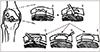
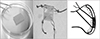
합성 폴리머 지지체 중에 PLA, PGA, 폴리디옥사논(polydioxanone)으로 이루어진 BioSeed C (BioTissue Technologies GmbH, Freiburg, Germany)는 현재 가장 많이 사용되는 합성 지지체 중 하나로 2001년부터 임상적으로 사용되어 왔다(Fig. 3).22) 그 외 상품화된 제품들로는 3차원적 제1형 콜라겐 겔에 자가연골세포가 이식되어 있는 NeoCart (Histogenics Corporation, Waltham, MA, USA), Novocart 3D (B. Braun-Tetec, Reutlingen, Germany), CaReS (Ars Arthro, Esslingen, Germany)등의 다양한 지지체들이 사용되고 있다(Fig. 4).23)
최근에는 지지체와 세포를 한 번의 수술적 치료로 제공하는 다양한 방법들이 고안되고 있다.24) 이환된 관절의 손상 받지 않은 부위의 건강한 연골조직에서 세포를 얻어 3차원 폴리머의 흡수성 지지체에 넣어 연골 손상 부위에 이식시키는 방법으로 2년간 미세골절술과 비교한 무작위 연구 결과에서 더 나은 결과를 보였다.25) 3차원적 제1형 콜라겐 지지체와 함께 골수농축액(bone marrow aspirate concentrate, BMAC)을 사용할 수 있고 피브린(fibrin)이나 봉합사를 이용하여 고정하며 일부 연구에서 좋은 결과를 보고하고 있다.
하지만 지지체를 이용한 방법 또한 단점은 있다. 지지체 기반 연골 재생법에서 연골세포 탈분화, 자멸세포 누출 등이 보고되었다. 또한 부적절한 세포 분포, 낮은 분화도, 조직 세포와 부적절한 세포 교합 등이 지지체를 이용한 세포 이식의 흔한 문제점으로 보고되었다.26)

보완책으로 떠오르는 중간엽 줄기세포

자가연골세포 이식술은 임상적으로 심각한 안정성 문제는 보이지 않지만 가용세포의 한정성, 여러 번의 수술적 단계가 필요한 점, 연골세포의 탈분화, 연골조직 채취로 인한 공여부의 손상 등의 단점이 있다.27) MSCs는 다양한 형질의 간질세포로서 골수 자체, 골격근, 지방세포 등에서 얻어질 수 있고 활액막 및 활액, 관절연골 등 다른 여러 결합조직에서도 얻어질 수 있다. 성인 MSCs는 1999년 Pittenger 등28)에 의해 처음으로 분리되었고 그들은 줄기세포의 다방향성 분화능력과 잠재능력을 보고하였다. MSCs는 부착력으로 인해 배양접시에서 잠재능력을 유지한 채 배양 및 증폭이 가능하며29) 중간엽 계열로 분화되어 나아가 다양한 결합조직(골조직, 지방 조직, 연골, 추간판, 인대, 근육 등)으로 분화된다.3031)

골관절염에서 줄기세포 기반 치료법

1. 골수유래 줄기세포(bone marrow-derived mesenchymal stem cell, BMSC)
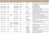
BMSC는 여러 in vivoin vitro 연구에서 지방조직 유래 줄기세포(adipose tissue-derived stem cell, ASC)에 비해 연골세포로의 분화능이 뛰어나고 자가 골수에서 줄기세포를 얻을 수 있다는 장점 때문에 MSCs를 이용한 골관절염 치료로 가장 활발히 사용 연구되고 있다(Table 1).323334353637383940414243444546474849505152) BMSC는 BMAC의 유핵세포를 배양하여 다량의 줄기세포를 얻어 관혈적 이식술이나 관절내 주사요법으로 골관절염의 치료에 사용되고 있으며 주로 자가 BMSC가 주로 이용되고 있다.
Yamasaki 등53)과 Wong 등49)은 골관절염 환자의 치료로 근위 경골 절골술 시행 시 자가 BMSC 이식술을 시행하여 줄기세포를 시행하지 않은 군에 비해 유의하게 좋은 연골 재생 효과를 보고하였다.1354) Ha 등54)은 BMSC를 관절 내 주사요법으로 관혈적 이식술과 비슷한 정도의 연골 재생 효과 및 임상적 결과를 보고하고 관절 내 주사도 관혈적 이식술만큼 안전하고 유용한 치료 방법이라 보고하였다. 그러나 BMAC의 유핵세포 중 단지 0.001%만 MSCs라는 단점이 있으며,55) BMSC는 증폭이 되면서 분화 능력이 떨어지고 골수 공여부의 통증 등의 단점으로 인해 여전히 사용에 제한이 많은 실정이다.
최근에는 배양된 BMSC 대신에 줄기세포의 양은 적지만 비교적 경비가 저렴하며 한 번의 마취로 수술이 가능하다는 장점 때문에 유핵세포 및 다양한 사이토카인을 함유하고 있는 BMAC가 골관절염의 치료에 널리 사용되고 있다.1356) 최근 다양한 연구에서 BMAC를 얻는 기술, 준비 방법, 그리고 다양한 정형외과 영역에서의 적용에 대해 보고하고 있으며 골손실이나 불유합, 골괴사나 연골병변의 치료, 스포츠 손상으로 인한 인대의 재건 및 치료 등 다양한 곳에 적용되고 있다.
Gobbi 등24)에 의해 BMAC를 이용한 연골 손상 병변 치료의 많은 연구가 이루어졌다. 15명의 환자에서 BMAC을 콜라겐 매트릭스에 도포한 후 Grade 4의 연골 손상에 대해 치료 후 24개월동안 추시 연구를 시행하였다. 환자들은 시각통증점수(visual analogue scale, VAS), International Knee Documentation Committee (IKDC), Knee Injury and Osteoarthritis Outcome Score (KOOS) 점수에 있어서 유의한 호전을 보였으며 작거나 고립된 병변을 가진 환자에서 더 월등한 결과를 보였다. 자기공명영상 및 조직학적인 검사를 통해서도 병변에 재생된 조직이 정상 유리연골과 비슷한 조직으로 구성되어 있는 것을 확인하였다. 최근에 BMAC를 이용한 연골 재생 치료 결과에 대해 훌륭한 임상적 효과를 보인다는 연구가 많이 보고되고 있고 수술 시 적용 뿐만 아니라 단지 관절 내 주입만으로도 좋은 결과를 보인다고도 보고되었다.57) Shapiro 등58)은 25명의 양측 슬관절 관절염 환자를 대상으로 한 전향적 연구에서 BMAC를 주입 받은 군과 위약군 간 모두에서 통증은 시술 후 1주, 3개월, 6개월까지 호전되었으나 두 군 간에 차이는 보이지 않았다고 보고하였다. 또 다른 슬관절염에서 BMAC를 사용한 두 연구에 따르면 대조군이 부족한 한계점이 있으나 통증 및 기능에서 BMAC이 유의한 효과를 보인다고 하였다.59)

2. 지방조직 유래 줄기세포(adipose tissue-derived mesenchymal stem cell, ASC)

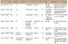
쉽게 조직으로부터 얻을 수 있는 또 다른 형태의 MSCs는 ASC이다(Table 2).136061626364656667) 2016년부터 ASC를 이용한 연골 재생 치료의 임상적 시도가 진행되어 왔다. Fodor와 Paulseth68)는 지방조직으로부터 자가 ASC를 추출한 후 슬관절염 환자에게 관절내 주입의 안정성과 양호한 임상 결과를 보고하였다.60) Jo 등61)은 전향적 코호트 연구를 통해 18명의 슬관절염 환자에서 ASC를 이용한 효과를 보고하였으며1369) 임상적 결과는 모든 환자에서 2년까지 호전되었으나 통계적으로 유의한 호전은 고용량군에서만 나타났다. 또한 저용량 및 중용량군에선 시술 후 1년째부터 임상적으로 악화되는 양상을 보였고 이는 결국 위약군과 2년째 동일한 수준이었으며 구조적인 결과도 비슷한 양상을 보였다. 이 연구 결과는 다량의 MSCs가 주입될 경우 손상된 병변 부위로 더 많은 수의 줄기세포가 부착하게 되고 더 많은 양의 영양 인자를 생산하여 더 많은 양의 연골 재생을 유발하게 됨을 증명하였다.
현재 여러 연구에서 슬관절염의 치료에 ASC 대신 간질혈관분획(stromal vascular fraction, SVF) 형태로 사용하기도 하고 있다. 슬관절염에 있어서 SVF 형태의 접목은 합병증을 최소화하면서 원하는 임상적 호전을 보이는 장점이 있으나 연구 데이터마다 다양한 결과를 보이고 같이 사용된 생물학적 보조약 때문에 정확한 효과는 아직 밝혀지지 않은 상태이다.7071) 또한 SVF를 이용한 치료는 엄격한 의미의 MSCs 치료라고 할 수 없다.

3. 인체제대혈 유래 줄기세포(human umbilical cord blood-derived mesenchymal stem cell, hUCB-MSC)

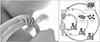
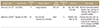
최근에 연골 재생에 hUCB-MSC의 임상적 적용이 보고되고 있는데(Fig. 5, Table 3)13607273) hUCB-MSC는 비침습적 방법으로 얻어지며 면역 반응이 적다는 장점이 있다.74) 또한 hUCB-MSC는 높은 증폭 능력으로 세포 치료의 적용에 있어서 충분한 세포량을 공급할 수 있는 장점이 있다. Park 등75)은 슬관절염 환자를 대상으로 hUCB-MSC를 이용한 임상적 치료 결과를 발표하였다. 관절경을 이용한 1년 추시 결과, 유리연골과 비슷한 연골로 병변 부위의 표면이 재형성됨을 확인할 수 있었다. 재생된 연골은 부드러운 곡면을 보이고 강도 또한 단단하였으며 주변 연골과의 상호 결합도 잘 이루고 부골형성이나 이상증식은 보이지 않았다. 생검(biopsy) 결과에 있어서도 정상 유리연골과 조직학적으로 매우 비슷함을 보여주었다. 환자의 임상적 결과에서도 VAS 및 IKDC 점수가 모두 유의하게 호전됨을 확인하였고 호전 정도는 7년까지도 유지된다고 보고되었다. 이 연구는 hUCB-MSC가 임상적 적용에 있어서 효과적이며 특별한 부작용 없이 사용될 수 있음을 확인하였다.
하지만 현재 이러한 줄기세포를 이용한 연골세포 재생에 있어서 아직 그 증명 자료는 부족한 것이 사실이다. 줄기세포를 이용한 결과의 혼선을 피하기 위해서는 정확한 세포 용량 및 용법에 대한 명확한 데이터를 기반으로 하는 연구들과 다기관 연구들이 지속적으로 필요할 것으로 생각된다.

결론

MSCs는 최근 줄기세포 연구에 있어서 가장 각광받는 주제이다. 관절 재생에 있어서 이러한 줄기세포의 적용은 많은 시도가 진행되고 있으나 현재까지는 연골 재생 효과가 연구마다 일정하지 않다. 또한 어떤 조직 유래 줄기세포, 어떤 용법 및 용량의 줄기세포가 골관절염의 치료에 이상적일 것인지에 대해서 더 많은 연구가 필요할 것으로 생각된다. 미래에는 이러한 줄기세포가 신뢰성 있는 치료 영역으로 인정받기 위해 효과적으로 분리시키고 배양하는 방법이 개선되어야 할 것으로 생각되며 이렇게 배양된 줄기세포들을 병변으로 잘 전달하는 시스템 구축과 안정성 및 효과에 대한 더 정확한 평가가 이루어져야 될 것으로 생각된다. 이번 종설에서 우리는 MSCs의 임상적 적용 및 연구를 포함한 가장 최신 정보에 대해서 간단히 리뷰해 보았다. 향후 MSCs를 이용한 치료가 좀 더 임상적으로 적용되고 근골격계 질환 전반적인 치료에서 사용되기를 기대한다.

Figures and Tables

Figure 1

Extracellular matrix of articular cartilage.

jkoa-54-478-g001
Figure 2

Cartilage regeneration method. (A) Focal cartilage damage, (B) chondroplasty, (C) microfracture, (D) autologous chondrocyte implantation, (E) matrix assisted autologous chondrocyte implantation.

jkoa-54-478-g002
Figure 3

BioSeed C (BioTissue Technologies GmbH).

jkoa-54-478-g003
Figure 4

NeoCart (Histogenics Corporation), Novocart (B. Braun-Tetec), CaReS (Ars Arthro, Esslingen).

jkoa-54-478-g004
Figure 5

Human umbilical cord blood-derived stem cell therapy.

jkoa-54-478-g005
Table 1

Clinical Studies of Bone Marrow-Derived Stem Cells

jkoa-54-478-i001

BMSC, bone marrow-derived mesenchymal stem cell; MSC, mesenchymal stem cell; OA, osteoarthritis; HSS, Hospital for Special Surgery Score; CD, chondral defect; MFC, medial femoral condyle; OD, osteochondral defect; P-F, patellofemoral; IKDC, International Knee Documentation Committee; MRI, magnetic resonance imaging; ON, osteonecrosis; VAS, visual analogue scale; ROM, range of motion; ACI, autologous chondrocyte implantation; ICRS-CIEP, International Cartilage Repair Society Cartilage Injury Evaluation Package; WOMAC, Western Ontario and McMaster Universities Osteoarthritis Index; KOOS, Knee injury and Osteoarthritis Outcome Score; EQ5D, EuroQoL 5-Dimension Health Questionnaire.

Table 2

Clinical Studies of Adipose Tissue-Derived Stem Cells

jkoa-54-478-i002

ASC, adipose tissue-derived mesenchymal stem cell; OA, osteoarthritis; VAS, visual analogue scale; KSS, Knee Society Score; WOMAC, Western Ontario and McMaster Universities Osteoarthritis Index; MRI, magnetic resonance imaging; ICRS, International Cartilage Repair Society; KOOS, Knee Injury and Osteoarthritis Outcome score; EKG, electrocardiogram; ADSVF, adipose tissue-derived stromal vascular fraction; ON, osteonecrosis; PRP, platelet-rich plasma; ADSC, adipose-derived stem cell, MFX, microfracture; CD, chondral defect; MS, marrow stimulation.

Table 3

Clinical Studies of Human Umbilical Cord Blood-Derived Stem Cells

jkoa-54-478-i003

UCB-MSCs, umbilical cord blood-derived mesenchymal stem cells; MFC, medial femoral condyle; LFC, lateral femoral condyle; VAS, visual analogue scale; IKDC, International Knee Documentation Committee; ICRS, International Cartilage Repair Society; MRI, magnetic resonance imaging; UC-MSC, umbilical cord mesenchymal stem cells; WOMAC, Western Ontario and McMaster Universities Osteoarthritis Index; QOL, quality of life; WORMS, Whole-Organ Magnetic Resonance Imaging Score.

Notes

CONFLICTS OF INTEREST The authors have nothing to disclose.

References

1. Gupta S, Hawker GA, Laporte A, Croxford R, Coyte PC. The economic burden of disabling hip and knee osteoarthritis (OA) from the perspective of individuals living with this condition. Rheumatology (Oxford). 2005; 44:1531–1537.
crossref
2. Barbour KE, Helmick CG, Boring M, Zhang X, Lu H, Holt JB. Prevalence of doctor-diagnosed arthritis at state and county levels - United States, 2014. MMWR Morb Mortal Wkly Rep. 2016; 65:489–494.
crossref
3. Sophia Fox AJ, Bedi A, Rodeo SA. The basic science of articular cartilage: structure, composition, and function. Sports Health. 2009; 1:461–468.
crossref
4. Makris EA, Gomoll AH, Malizos KN, Hu JC, Athanasiou KA. Repair and tissue engineering techniques for articular cartilage. Nat Rev Rheumatol. 2015; 11:21–34.
crossref
5. Kon E, Filardo G, Berruto M, et al. Articular cartilage treatment in high-level male soccer players: a prospective comparative study of arthroscopic second-generation autologous chondrocyte implantation versus microfracture. Am J Sports Med. 2011; 39:2549–2557.
6. Rodrigo JJ, Steadman JR, Silliman JF, Fulstone HA. Improvement in full-thickness chondral defect healing in the human knee after debridement and microfracture using continuous passive motion. Am J Knee Surg. 1994; 7:109–116.
7. Knutsen G, Drogset JO, Engebretsen L, et al. A randomized trial comparing autologous chondrocyte implantation with microfracture. Findings at five years. J Bone Joint Surg Am. 2007; 89:2105–2112.
8. Miller BS, Steadman JR, Briggs KK, Rodrigo JJ, Rodkey WG. Patient satisfaction and outcome after microfracture of the degenerative knee. J Knee Surg. 2004; 17:13–17.
crossref
9. Kreuz PC, Erggelet C, Steinwachs MR, et al. Is microfracture of chondral defects in the knee associated with different results in patients aged 40 years or younger? Arthroscopy. 2006; 22:1180–1186.
crossref
10. Kreuz PC, Steinwachs MR, Erggelet C, et al. Results after microfracture of full-thickness chondral defects in different compartments in the knee. Osteoarthritis Cartilage. 2006; 14:1119–1125.
crossref
11. Mithoefer K, McAdams T, Williams RJ, Kreuz PC, Mandelbaum BR. Clinical efficacy of the microfracture technique for articular cartilage repair in the knee: an evidence-based systematic analysis. Am J Sports Med. 2009; 37:2053–2063.
12. Gudas R, Gudaitė A, Mickevičius T, et al. Comparison of osteochondral autologous transplantation, microfracture, or debridement techniques in articular cartilage lesions associated with anterior cruciate ligament injury: a prospective study with a 3-year follow-up. Arthroscopy. 2013; 29:89–97.
crossref
13. Park YB, Ha CW, Rhim JH, Lee HJ. Stem cell therapy for articular cartilage repair: review of the entity of cell populations used and the result of the clinical application of each entity. Am J Sports Med. 2018; 46:2540–2552.
crossref
14. Knutsen G, Drogset JO, Engebretsen L, et al. A randomized multicenter trial comparing autologous chondrocyte implantation with microfracture: long-term follow-up at 14 to 15 years. J Bone Joint Surg Am. 2016; 98:1332–1339.
15. Ruta DJ, Villarreal AD, Richardson DR. Orthopedic Surgical Options for Joint Cartilage Repair and Restoration. Phys Med Rehabil Clin N Am. 2016; 27:1019–1042.
crossref
16. Riboh JC, Cvetanovich GL, Cole BJ, Yanke AB. Comparative efficacy of cartilage repair procedures in the knee: a network meta-analysis. Knee Surg Sports Traumatol Arthrosc. 2017; 25:3786–3799.
crossref
17. Krych AJ, Pareek A, King AH, Johnson NR, Stuart MJ, Williams RJ 3rd. Return to sport after the surgical management of articular cartilage lesions in the knee: a meta-analysis. Knee Surg Sports Traumatol Arthrosc. 2017; 25:3186–3196.
crossref
18. Ebert JR, Fallon M, Wood DJ, Janes GC. A prospective clinical and radiological evaluation at 5 years after arthroscopic matrix-induced autologous chondrocyte implantation. Am J Sports Med. 2017; 45:59–69.
crossref
19. Kon E, Verdonk P, Condello V, et al. Matrix-assisted autologous chondrocyte transplantation for the repair of cartilage defects of the knee: systematic clinical data review and study quality analysis. Am J Sports Med. 2009; 37 Suppl 1:156S–166S.
20. Kon E, Delcogliano M, Filardo G, Montaperto C, Marcacci M. Second generation issues in cartilage repair. Sports Med Arthrosc Rev. 2008; 16:221–229.
crossref
21. Grigolo B, Lisignoli G, Piacentini A, et al. Evidence for redifferentiation of human chondrocytes grown on a hyaluronan-based biomaterial (HYAff 11): molecular, immunohistochemical and ultrastructural analysis. Biomaterials. 2002; 23:1187–1195.
22. Irion VH, Flanigan DC. New and emerging techniques in cartilage repair: other scaffold-based cartilage treatment options. Oper Tech Sports Med. 2013; 21:125–137.
crossref
23. Crawford DC, Heveran CM, Cannon WD Jr, Foo LF, Potter HG. An autologous cartilage tissue implant NeoCart for treatment of grade III chondral injury to the distal femur: prospective clinical safety trial at 2 years. Am J Sports Med. 2009; 37:1334–1343.
24. Gobbi A, Karnatzikos G, Scotti C, Mahajan V, Mazzucco L, Grigolo B. One-step cartilage repair with bone marrow aspirate concentrated cells and collagen matrix in full-thickness knee cartilage lesions: results at 2-year follow-up. Cartilage. 2011; 2:286–299.
25. Cole BJ, Farr J, Winalski CS, et al. Outcomes after a single-stage procedure for cell-based cartilage repair: a prospective clinical safety trial with 2-year follow-up. Am J Sports Med. 2011; 39:1170–1179.
26. Rai V, Dilisio MF, Dietz NE, Agrawal DK. Recent strategies in cartilage repair: a systemic review of the scaffold development and tissue engineering. J Biomed Mater Res A. 2017; 105:2343–2354.
crossref
27. Matricali GA, Dereymaeker GP, Luyten FP. Donor site morbidity after articular cartilage repair procedures: a review. Acta Orthop Belg. 2010; 76:669–674.
28. Pittenger MF, Mackay AM, Beck SC, et al. Multilineage potential of adult human mesenchymal stem cells. Science. 1999; 284:143–147.
crossref
29. De Bari C, Dell'Accio F, Tylzanowski P, Luyten FP. Multipotent mesenchymal stem cells from adult human synovial membrane. Arthritis Rheum. 2001; 44:1928–1942.
crossref
30. Guilak F, Estes BT, Diekman BO, Moutos FT, Gimble JM. 2010 Nicolas Andry Award: multipotent adult stem cells from adipose tissue for musculoskeletal tissue engineering. Clin Orthop Relat Res. 2010; 468:2530–2540.
crossref
31. Trubiani O, Orsini G, Caputi S, Piatelli A. Adult mesenchymal stem cells in dental research: a new approach for tissue engineering. Int J Immunopathol Pharmacol. 2006; 19:451–460.
crossref
32. Danišovič L, Lesný P, Havlas V, et al. Chondrogenic differentiation of human bone marrow and adipose tissue-derived mesenchymal stem cells. J Appl Biomed. 2007; 5:139–150.
crossref
33. Koga H, Shimaya M, Muneta T, et al. Local adherent technique for transplanting mesenchymal stem cells as a potential treatment of cartilage defect. Arthritis Res Ther. 2008; 10:R84.
crossref
34. Wakitani S, Imoto K, Yamamoto T, Saito M, Murata N, Yoneda M. Human autologous culture expanded bone marrow mesenchymal cell transplantation for repair of cartilage defects in osteoarthritic knees. Osteoarthritis Cartilage. 2002; 10:199–206.
crossref
35. Wakitani S, Mitsuoka T, Nakamura N, Toritsuka Y, Nakamura Y, Horibe S. Autologous bone marrow stromal cell transplantation for repair of full-thickness articular cartilage defects in human patellae: two case reports. Cell Transplant. 2004; 13:595–600.
crossref
36. Adachi N, Ochi M, Deie M, Ito Y. Transplant of mesenchymal stem cells and hydroxyapatite ceramics to treat severe osteochondral damage after septic arthritis of the knee. J Rheumatol. 2005; 32:1615–1618.
37. Kuroda R, Ishida K, Matsumoto T, et al. Treatment of a full-thickness articular cartilage defect in the femoral condyle of an athlete with autologous bone-marrow stromal cells. Osteoarthritis Cartilage. 2007; 15:226–231.
crossref
38. Wakitani S, Nawata M, Tensho K, Okabe T, Machida H, Ohgushi H. Repair of articular cartilage defects in the patello-femoral joint with autologous bone marrow mesenchymal cell transplantation: three case reports involving nine defects in five knees. J Tissue Eng Regen Med. 2007; 1:74–79.
crossref
39. Adachi N, Ochi M, Deie M, Ishikawa M, Ito Y. Osteonecrosis of the knee treated with a tissue-engineered cartilage and bone implant. A case report. J Bone Joint Surg Am. 2007; 89:2752–2757.
40. Centeno CJ, Busse D, Kisiday J, Keohan C, Freeman M, Karli D. Increased knee cartilage volume in degenerative joint disease using percutaneously implanted, autologous mesenchymal stem cells. Pain Physician. 2008; 11:343–353.
41. Centeno CJ, Busse D, Kisiday J, Keohan C, Freeman M, Karli D. Regeneration of meniscus cartilage in a knee treated with percutaneously implanted autologous mesenchymal stem cells. Med Hypotheses. 2008; 71:900–908.
crossref
42. Nejadnik H, Hui JH, Feng Choong EP, Tai BC, Lee EH. Autologous bone marrow-derived mesenchymal stem cells versus autologous chondrocyte implantation: an observational cohort study. Am J Sports Med. 2010; 38:1110–1116.
43. Haleem AM, Singergy AA, Sabry D, et al. The clinical use of human culture-expanded autologous bone marrow mesenchymal stem cells transplanted on platelet-rich fibrin glue in the treatment of articular cartilage defects: a pilot study and preliminary results. Cartilage. 2010; 1:253–261.
44. Wakitani S, Okabe T, Horibe S, et al. Safety of autologous bone marrow-derived mesenchymal stem cell transplantation for cartilage repair in 41 patients with 45 joints followed for up to 11 years and 5 months. J Tissue Eng Regen Med. 2011; 5:146–150.
crossref
45. Davatchi F, Abdollahi BS, Mohyeddin M, Shahram F, Nikbin B. Mesenchymal stem cell therapy for knee osteoarthritis. Preliminary report of four patients. Int J Rheum Dis. 2011; 14:211–215.
crossref
46. Emadedin M, Aghdami N, Taghiyar L, et al. Intra-articular injection of autologous mesenchymal stem cells in six patients with knee osteoarthritis. Arch Iran Med. 2012; 15(7):422–428.
47. Lee KB, Wang VT, Chan YH, Hui JH. A novel, minimally-invasive technique of cartilage repair in the human knee using arthroscopic microfracture and injections of mesenchymal stem cells and hyaluronic acid: a prospective comparative study on safety and short-term efficacy. Ann Acad Med Singapore. 2012; 41:511–517.
48. Teo BJ, Buhary K, Tai BC, Hui JH. Cell-based therapy improves function in adolescents and young adults with patellar osteochondritis dissecans. Clin Orthop Relat Res. 2013; 471:1152–1158.
crossref
49. Wong KL, Lee KB, Tai BC, Law P, Lee EH, Hui JH. Injectable cultured bone marrow-derived mesenchymal stem cells in varus knees with cartilage defects undergoing high tibial osteotomy: a prospective, randomized controlled clinical trial with 2 years' follow-up. Arthroscopy. 2013; 29:2020–2028.
crossref
50. Orozco L, Munar A, Soler R, et al. Treatment of knee osteoarthritis with autologous mesenchymal stem cells: a pilot study. Transplantation. 2013; 95:1535–1541.
51. Davatchi F, Sadeghi Abdollahi B, Mohyeddin M, Nikbin B. Mesenchymal stem cell therapy for knee osteoarthritis: 5 years follow-up of three patients. Int J Rheum Dis. 2016; 19:219–225.
52. de Windt TS, Vonk LA, Slaper-Cortenbach IC, et al. Allogeneic mesenchymal stem cells stimulate cartilage regeneration and are safe for single-stage cartilage repair in humans upon mixture with recycled autologous chondrons. Stem Cells. 2017; 35:256–264.
crossref
53. Yamasaki S, Mera H, Itokazu M, Hashimoto Y, Wakitani S. Cartilage repair with autologous bone marrow mesenchymal stem cell transplantation: review of preclinical and clinical studies. Cartilage. 2014; 5:196–202.
54. Ha CW, Park YB, Kim SH, Lee HJ. Intra-articular mesenchymal stem cells in osteoarthritis of the knee: a systematic review of clinical outcomes and evidence of cartilage repair. Arthroscopy. 2019; 35:277–288.e2.
crossref
55. Medical Advisory Secretariat. Osteogenic protein-1 for long bone nonunion: an evidence-based analysis. Ont Health Technol Assess Ser. 2005; 5:1–57.
56. Jäger M, Jelinek EM, Wess KM, et al. Bone marrow concentrate: a novel strategy for bone defect treatment. Curr Stem Cell Res Ther. 2009; 4:34–43.
57. Sampson S, Smith J, Vincent H, Aufiero D, Zall M, Botto-van-Bemden A. Intra-articular bone marrow concentrate injection protocol: short-term efficacy in osteoarthritis. Regen Med. 2016; 11:511–520.
crossref
58. Shapiro SA, Kazmerchak SE, Heckman MG, Zubair AC, O'Connor MI. A prospective, single-blind, placebo-controlled trial of bone marrow aspirate concentrate for knee osteoarthritis. Am J Sports Med. 2017; 45:82–90.
crossref
59. Centeno C, Pitts J, Al-Sayegh H, Freeman M. Efficacy of autologous bone marrow concentrate for knee osteoarthritis with and without adipose graft. Biomed Res Int. 2014; 2014:370621.
crossref
60. Shariatzadeh M, Song J, Wilson SL. The efficacy of different sources of mesenchymal stem cells for the treatment of knee osteoarthritis. Cell Tissue Res. 2019; 378:399–410.
crossref
61. Jo CH, Lee YG, Shin WH, et al. Intra-articular injection of mesenchymal stem cells for the treatment of osteoarthritis of the knee: a proof-of-concept clinical trial. Stem Cells. 2014; 32:1254–1266.
crossref
62. Pers YM, Rackwitz L, Ferreira R, et al. ADIPOA Consortium. Adipose mesenchymal stromal cell-based therapy for severe osteoarthritis of the knee: a phase I dose-escalation trial. Stem Cells Transl Med. 2016; 5:847–856.
crossref
63. Song Y, Du H, Dai C, et al. Human adipose-derived mesenchymal stem cells for osteoarthritis: a pilot study with long-term follow-up and repeated injections. Regen Med. 2018; 13:295–307.
crossref
64. Pak J, Chang JJ, Lee JH, Lee SH. Safety reporting on implantation of autologous adipose tissue-derived stem cells with platelet-rich plasma into human articular joints. BMC Musculoskelet Disord. 2013; 14:337.
crossref
65. Bui KH-T, Duong TD, Nguyen NT, et al. Symptomatic knee osteoarthritis treatment using autologous adipose derived stem cells and platelet-rich plasma: a clinical study. Biomed Res Ther. 2014; 1:2–8.
crossref
66. Koh YG, Kwon OR, Kim YS, Choi YJ, Tak DH. Adipose-derived mesenchymal stem cells with microfracture versus microfracture alone: 2-year follow-up of a prospective randomized trial. Arthroscopy. 2016; 32:97–109.
crossref
67. Kim YS, Lee M, Koh YG. Additional mesenchymal stem cell injection improves the outcomes of marrow stimulation combined with supramalleolar osteotomy in varus ankle osteoarthritis: short-term clinical results with second-look arthroscopic evaluation. J Exp Orthop. 2016; 3:12.
crossref
68. Fodor PB, Paulseth SG. Adipose derived stromal cell (ADSC) injections for pain management of osteoarthritis in the human knee joint. Aesthet Surg J. 2016; 36:229–236.
crossref
69. Moher D, Liberati A, Tetzlaff J, Altman DG. PRISMA Group. Preferred reporting items for systematic reviews and meta-analyses: the PRISMA statement. Ann Intern Med. 2009; 151:264–269. W64.
crossref
70. Kim YS, Koh YG. Injection of mesenchymal stem cells as a supplementary strategy of marrow stimulation improves cartilage regeneration after lateral sliding calcaneal osteotomy for varus ankle osteoarthritis: clinical and second-look arthroscopic results. Arthroscopy. 2016; 32:878–889.
crossref
71. Kim YS, Kwon OR, Choi YJ, Suh DS, Heo DB, Koh YG. Comparative matched-pair analysis of the injection versus implantation of mesenchymal stem cells for knee osteoarthritis. Am J Sports Med. 2015; 43:2738–2746.
crossref
72. Park YB, Ha CW, Lee CH, Yoon YC, Park YG. Cartilage regeneration in osteoarthritic patients by a composite of allogeneic umbilical cord blood-derived mesenchymal stem cells and hyaluronate hydrogel: results from a clinical trial for safety and proof-of-concept with 7 years of extended follow-up. Stem Cells Transl Med. 2017; 6:613–621.
crossref
73. Matas J, Orrego M, Amenabar D, et al. Umbilical cord-derived mesenchymal stromal cells (MSCs) for knee osteoarthritis: repeated MSC dosing is superior to a single MSC dose and to hyaluronic acid in a controlled randomized phase I/II trial. Stem Cells Transl Med. 2019; 8:215–224.
crossref
74. Flynn A, Barry F, O'Brien T. UC blood-derived mesenchymal stromal cells: an overview. Cytotherapy. 2007; 9:717–726.
crossref
75. Park YB, Ha CW, Lee CH, Yoon YC, Park YG. Cartilage regeneration in osteoarthritic patients by a composite of allogeneic umbilical cord blood-derived mesenchymal stem cells and hyaluronate hydrogel: results from a clinical trial for safety and proof-of-concept with 7 years of extended follow-up. Stem Cells Transl Med. 2017; 6:613–621.
crossref
TOOLS
ORCID iDs

Jong-Keun Seon
https://orcid.org/0000-0002-6920-5659

Similar articles