

 PDF
PDF ePub
ePub Citation
 Print
Print


INTRODUCTION
Cerebral small vessel disease (CSVD) is a disorder of the brain's small perforating arterioles, capillaries and venules, which causes various lesions observed on pathological examination, or on brain magnetic resonance imaging (MRI) or computed tomography.1 Patients with CSVD exhibit many clinical manifestations, including lacunar ischemic stroke,2 cognitive impairment, especially affecting executive function,3 depression,4 urinary incontinence,5 gait disturbance,6 and extrapyramidal symptoms.7 CSVD is the attributable cause of 25% of strokes, and more than doubles the risk for recurrent stroke.8 CSVD is the most common cause of vascular dementia and a major contributor to mixed dementia.9 It is, therefore, important to treat CSVD to prevent and/or treat vascular and mixed dementias.
Features of CSVD evident on neuroimaging include recent small subcortical infarcts, lacunes, white matter changes (WMC), enlarged perivascular spaces, cerebral microbleeds (CMBs), and brain atrophy.10 Increased mean diffusivity (MD) and reduced fraction anisotropy (FA) on diffusion tensor imaging (DTI) are also observed in WMC and normal-appearing white matter (NAWM) in patients with severe WMC.1112 In a previous study, impaired microstructural integrity preceded conversion to WMC visible on conventional neuroimaging.11
The mechanisms that link CSVD to parenchymal damage are heterogeneous. Pathological changes in CSVD have both ischemic and hemorrhagic consequences. Lacunar infarcts can be caused by acute closure of cerebral small blood vessels, and WMC may be due to incomplete infarction or selective necrosis by chronic low perfusion caused by stenosis of small blood vessels.9 In addition, disrupted autoregulation and damage to the blood-brain barrier (BBB) could be involved in CSVD.13 The endothelium plays a crucial role in processes including the autoregulation of blood flow and function of the BBB.9 Derangement of the BBB, however, may begin some years before the first symptoms of ischemic forms of CSVD manifest, and lead to structural, vascular, and perivascular changes in the small vessels of the brain, resulting in edema, enlarged perivascular spaces, and tissue damage.91013
In the Cilostazol for Prevention of Secondary Stroke (CSPS 2) study, cilostazol appeared be superior to aspirin in the prevention of stroke after an ischemic stroke,14 in which two-thirds of the subjects experienced lacunar strokes due to CSVD.14 Cilostazol is a selective phosphodiesterase III inhibitor and inhibits platelet aggregation similar to aspirin.15 In addition, cilostazol exerts a vasodilating effect,16 and has the effect of inhibiting proliferation of smooth muscle cells,17 as well as a favorable effect on re-endothelialization mediated by hepatocyte growth factor18 and endothelial precursor cells.19 Therefore, cilostazol may be more effective for CSVD than drugs that exert only an antiplatelet effect.20 If primary outcome is all stroke events in a comparative study of cilostazol and aspirin in CSVD, thousands of subjects will be needed. However, when the primary outcome is a change in WMC on brain MRI, the number of subjects can be significantly reduced.21 To the best of our knowledge, there have been no studies comparing the effects of cilostazol and aspirin using changes in WMC volume as the primary outcome measure.
The comparison study of Cilostazol and aspirin on cHAnges in volume of cerebral smaLL vEssel disease white matter chaNGEs (CHALLENGE) aims to compare changes in WMC volume on MRI between aspirin and cilostazol in patients with CSVD. Our hypothesis is that there will be differences in changes in WMC volume from baseline to 24 months between the cilostazol and aspirin groups.
METHODS
Design
CHALLENGE is a multicenter, double blind, randomized trial with a two parallel-arm design. One arm is assigned to cilostazol as the experimental arm and the other arm is assigned to aspirin as the active comparator using a blinded randomization method. Subject enrollment are performed in 19 hospital neurology clinics across South Korea. This investigator-initiated clinical trial has received funding support from Korea Otsuka Pharmaceutical Co. Ltd. (Seoul, Korea). However, the sponsor are not involved in the study design or operations, such as the selection and management of sites, data management, or the drafting of any manuscripts submitted for publication. The study is conducted in accordance with the International Harmonization Conference guidelines on Good Clinical Practice, and the institutional review board of each center approves the study before initiation. Written informed consent is obtained from all potential subjects before participation in the study. The consent document includes the risks and potential benefits of treatment, the trial process, and alternatives to participation. The trial was registered with Clinical Trials.gov (NCT01932203).
Participants
The inclusion and exclusion criteria are summarized in Table 1. Briefly, the inclusion criteria included: 1) age between 50 and 85 years; 2) presence of CSVD based on brain MRI, including moderate or severe WMC (deep WMC ≥grade 2 and periventricular WMC ≥grade 2 according to the modified Fazekas criteria)22 and ≥1 lacunar infarction; 3) able to walk to hospital (use of a walker or cane is permissible); and 4) agreement to give the written informed consent. Key exclusion criteria include history of recent cerebral infarction within 3 months and conditions with contraindications to long-term antiplatelet therapy. In addition, patients with cortical infarction or subcortical infarction >1.5 cm in size, Parkinson's disease or Alzheimer's disease (AD), cardioembolic heart disease of high- and medium-risk sources by the Trial of Org 10172 in Acute Stroke Treatment classification,23 chronic liver disease, chronic renal failure, or specific white matter involving disease, such as multiple sclerosis or sarcoidosis, are excluded. Patients with severe or unstable medical disease that may prevent completion of study requirements, such as recent intracerebral hemorrhage, subdural hematoma, or intraventricular hemorrhage are also excluded.
Table 1
Inclusion and exclusion criteria
| Criteria | |
|---|---|
| Inclusion criteria | |
| 1. 50 to 85 years of age | |
| 2. Able to walk to the hospital (walker or cane is permissible) | |
| 3. Cerebral small vessel disease observed on brain MRI confirmed by the following: presence of ≥1 lacunar infarction (s); and moderate or severe confluent WMC (defined as grade 2 or 3 on the modified Fazekas scale22) of periventricular WMC with cap or rims >5 mm and deep subcortical WMC >10 mm in maximum diameter | |
| 4. Agreement to give the written informed consent | |
| Exclusion criteria | |
| 1. Contraindication (s) to antiplatelets | |
| 2. Cardioembolic source | |
| 3. Carotid bruit or large cerebral artery stenosis >50% | |
| 4. Cortical infarction or subcortical infarction >1.5 cm | |
| 5. Bleeding tendency | |
| 6. Chronic liver disease (AST or ALT >100 IL/L) | |
| 7. Chronic renal disease (creatinine >3.0 mg/dL) | |
| 8. Active gastrointestinal ulcer | |
| 9. Severe or unstable medical disease that may prevent completion of study requirements (i.e., unstable or severe asthma) | |
| 10. Anemia (hemoglobin <10 g/dL) or thrombocytopenia | |
| 11. Cardiac pacemaker or contraindication to MRI | |
| 12. Pregnancy or breast-feeding | |
| 13. Drug or alcohol addiction | |
| 14. Any other white matter disease (i.e., multiple sclerosis, sarcoidosis, or brain irradiation), or brain tumor | |
| 15. Parkinson's disease, Alzheimer's disease or any other neurodegenerative disease | |
| 16. Any hearing or visual impairment that can disturb efficient evaluation of the patient | |
| 17. Recent cerebral infarction within 3 months | |
MRI: magnetic resonance imaging, WMC: white matter changes, AST: aspartate aminotransferase, ALT: alanine aminotransferase.
![]()
Randomization
During the screening period, anonymized baseline clinical and neuroimaging data are collected to assess potential eligibility. After the confirmation of inclusion and exclusion criteria, participants are randomly assigned to the cilostazol or aspirin group in a 1:1 ratio. A study identification number generated electronically by a central randomization service is assigned to each patient, and the centralized block randomization, stratified by the center, is performed by the interactive web response system. After randomization, all participants are instructed to take cilostazol slow release (SR) 200 mg in the evening or aspirin 100 mg in the morning once daily, with a simultaneously provided placebo as a double dummy.
Treatment
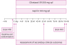
After randomization, the participants in the cilostazol group take cilostazol SR 200 mg (100 mg [2 capsules]) in the evening and a placebo aspirin tablet in the morning for 104 weeks, and those in the aspirin group take aspirin 100 mg in the morning and placebo cilostazol (2 capsules) in the evening (Fig. 1). If a participant experiences an adverse drug reaction (ADR), the drug can be temporarily suspended or reduced at any time during the study period. After the ADR is resolved, the dose can be increased. If a subject is unable to tolerate cilostazol SR 100 mg or aspirin 100 mg, the subject is dropped from the study. Temporary suspension of the trial treatment is permitted for invasive treatments, such as surgery, but cannot exceed two weeks. During the study period, additional anticoagulants or antiplatelet agents (e.g., ticlopidine, clopidogrel, dipyridamole, triflusal), except for antiplatelet agents, which are given as investigational products, are prohibited.
Follow-up
All participants visit four weeks after randomization to assess adverse events and compliance with the study drug. Thereafter, they visit every 12 weeks, up to 88 weeks and, finally, at 104 weeks to assess adverse events and compliance with the study drug. Brain MRI is performed using a 3.0 Tesla magnetic resonance (MR) scanner at baseline or within 4 weeks before baseline, and at week 104. Blood tests, including blood urea nitrogen, creatinine, hemoglobin (Hb), platelets, aspartate aminotransferase, alanine aminotransferase, albumin, glucose, glycated HbA1C (HbA1C), low density lipoprotein-cholesterol, high density lipoprotein-cholesterol, triglycerides, total cholesterol and uric acid, and urine microalbumin to creatinine ratio are evaluated at screening. Other clinical outcome assessments are also performed at baseline, week 52, and week 104 (Fig. 1).
Brain MRI examination
Brain MRI data are acquired from all participants at screening and at week 104 using a 3.0 Tesla MR scanner, which captures axial fluid-attenuated inversion recovery (FLAIR) imaging of 2 mm thickness without gap, three-dimensional (3D) T1-weighted imaging, axial T2-weighted imaging, DTI of 2 mm thickness without gap, and gradient echo imaging. The MR scanners used include the Discovery MR 750 and the Signa Hdxt (GE Healthcare, Milwaukee, WI, USA), the Achieva (Philips Healthcare, Andover, MA, USA), and the Skyra, Verio, and Trio Tim (Siemens, Washington, D.C., USA). The MRI protocols are based on the AD Neuroimaging Initiative phase 2 MRI protocols.24
MRI is performed using a standardized protocol provided to the centers. Prior to starting MRI acquisition in participants, centers are requested to test the protocol with a volunteer. Images are then sent to the laboratory at Hanyang University, a MRI coordinating center that provides the required certificates of quality. Once the quality of images is authorized, centers are requested to send the acquired images of a participant each time. Upon reception by the MRI coordinating center, quality controls of the acquired images are undertaken in order to detect whether a participant needed rescanning. The analysis of the images will be conducted at the laboratory of Hanyang University. To measure the volume of WMC, WMC on the FLAIR images will be segmented using the FMRIB Automatic Segmentation Tool (FAST) of the FMRIB Software Library (http://www.fmrib.ox.ac.uk/fsl/). The FAST segmentation tool is based on a hidden Markov random field model and an associated expectation-maximization algorithm.25 Hyperintensity signal artifacts included in the WMC mask will be edited manually using ITK-SNAP program (http://www.itksnap.org).
Outcome measures
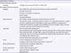
The primary outcome measure is change in WMC volume on brain FLAIR imaging from baseline to 104 weeks. Secondary imaging outcomes include changes in MD and FA for WMC and NAWM on DTI, number of lacunes, number of CMBs, brain volume, and cortical thickness. Secondary outcomes include all ischemic stroke events, including cerebral infarction and transient ischemic attack (TIA), and all vascular events including ischemic stroke, TIA, myocardial infarction, angina pectoris, cerebral venous thrombosis, pulmonary embolism, symptomatic deep vein thrombosis, symptomatic peripheral artery occlusion, other vascular occlusion, and any revascularization procedure. Changes in cognition, motor function, mood, urinary symptoms, and disability between the cilostazol and aspirin groups are also compared as secondary clinical outcomes. Detailed secondary outcome measures are summarized in Table 2.
Table 2
Primary and secondary outcome measures
| Instrument or method | ||
|---|---|---|
| Primary outcome measure | ||
| Imaging | Change in the volume of WMC on brain MRI | |
| Secondary outcome measure | ||
| Imaging | Mean diffusivity and fraction anisotropy on WMC and NAWM | |
| Number of lacunes | ||
| Number of cerebral microbleeds | ||
| Brain volume | ||
| Cortical thickness | ||
| Vascular event | All ischemic stroke including cerebral infarction and TIA | |
| All vascular events, including ischemic stroke, TIA, myocardial infarction, angina pectoris, cerebral venous thrombosis, pulmonary embolism, symptomatic deep vein thrombosis, symptomatic peripheral artery occlusion, other vascular occlusion, and any revascularization procedure | ||
| Cognition | Mini-Mental State Examination26 | |
| Neurocognitive tests including Seoul Verbal Learning Test, 15-item Boston Naming Test, Rey-Osterrieth Complex figure copy, animal fluency, controlled oral word association test, Stroop Color and Word test, Digit Symbol Substitution Test, and Trail Making Test Parts A and B | ||
| Clinical Dementia Rating scale-Sum of Boxes27 | ||
| Mood | Geriatric Depression Scale-15 items 28 | |
| Caregiver-Administered Neuropsychiatric Inventory29 | ||
| Motor performance | Pyramidal and Extrapyramidal Scale30 | |
| Timed “Up & Go” test31 | ||
| Urinary symptoms | King's Health Questionnaire32 | |
| Function | Bayer Activities of Daily Living33 | |
| Barthel Index34 | ||
WMC: white matter changes, MRI: magnetic resonance imaging, NAWM: normal-appearing white matter, TIA: transient ischemic attack.
![]()
Sample size estimates
The primary null hypothesis is that there will be no differences in change in WMC volume from baseline to study end between the cilostazol and aspirin groups. In a previous study, changes in WMC volume percentage from baseline to approximately 2 years were 0.11%±0.84% and 0.39%±0.48% for the aspirin and cilostazol groups, respectively.12 Based on a power of 0.8 for detecting a significant difference (p=0.05 [two-sided]) using a two-sample t-test using PASS 2008, 95 patients were required for each study group. Assuming a discontinuation rate of 25%,14 the calculated sample size is 254, with 127 participants in each group.
Statistical analysis
The end-points will be assessed using a modified intention-to-treat (ITT) population, defined as all randomized patients who receive at least one dose of study medication and undergo a baseline evaluation and at least one post-baseline assessment after beginning the study medication. Additional analyses on a per-protocol population will be also performed. The conclusion of the trial will use the modified ITT analysis. Analysis of covariance will be used to compare changes in imaging parameters from baseline to study end between the cilostazol and aspirin groups. Cox proportional hazard models will be used to calculate the hazard ratio of all ischemic stroke or all vascular events and corresponding 95% confidence intervals for the cilostazol group compared with the aspirin group. Linear mixed models will be used to compare changes in other secondary clinical outcomes between the cilostazol and aspirin groups. The safety analysis population will comprise participants who receive at least one dose of study medication and undergo at least one safety evaluation after baseline. The chi-squared test will be used to compare the prevalence of adverse events between the cilostazol and aspirin groups.
Preplanned substudies
Participants are tested to assess apolipoprotein E genotyping, renin-angiotensin system gene polymorphism (ACE I/D, AGT M235T), and C-peptide. Blood levels of markers of endothelial function, including von Willebrand factor, tissue factors, intercellular adhesion molecule-1 and vascular cell adhesion molecule-1 will be tested, as will anti-inflammatory molecules including interleukin (IL)-6, IL-18, and high sensitivity C-reactive protein. The risk and protective factors associated with progression of imaging parameters or clinical symptoms of CSVD will be evaluated.
DISCUSSION
CSVD is associated with stroke, vascular parkinsonism, and cognitive impairment.35 CSVD is also the most common accompanying pathology in AD.36 Mixed brain pathologies are common in most cases of dementia and in cognitively normal elderly individuals.3637 In a previous study, individuals with multiple pathologies were almost three times more likely to experience dementia compared with those with a single pathological diagnosis.36 It is important to control CSVD to prevent or delay the onset of dementia in patients with AD or other neurodegenerative disease (s) accompanied by CSVD. Therefore, this study aims to investigate antiplatelet agents that may be more effective at inhibiting the progression of CSVD. Aspirin is a popular drug used to treat patients with CSVD. Cilostazol exerts effects of vasodilation, inhibition of smooth muscle proliferation, endothelial protection, as well as an antiplatelet effect.151617181920 Therefore, cilostazol may be more effective in patients with CSVD than aspirin.
Most patients with CSVD have no clinical history of stroke. They usually experience slowly progressive cognitive impairment or neurological symptoms.38 Therefore, it may be inappropriate to consider symptomatic stroke incidence as a primary outcome for drugs that may inhibit the progression of CSVD. WMC is typically interpreted as a surrogate of CSVD.39
A recent study showed that advanced WMC in Asian patients with ischemic stroke and CSVD burden was associated with an increased risk of recurrent stroke.40 Overall, longitudinal studies have reported annual increases in WMC volume ranging from 4.4% to 37.2%.41 Although the subject pool in that study was not limited to those with CSVD, changes in WMC volume determined quantitatively using a semi-automatic method was used as an outcome in a post hoc analysis to investigate the effect of statins on WMC progression.42 Changes in WMC volume, graded according to a visual rating scale, were also used as an outcome in a study investigating the effects of blood pressure lowering on cerebral WMC.43 However, there have been no studies investigating the effect of any antiplatelet drugs on WMC progression in patients with CSVD. To our knowledge, therefore, the present study is the first to compare cilostazol and aspirin in terms of changes in WMC volume as the primary outcome measure in patients with CSVD.
In this study, we will investigate the effects of cilostazol and aspirin on all CSVD brain imaging markers by measuring changes in the number of lacunes, number of CMBs, MD and FA for WMC and NAWM on DTI, brain volume, and cortical thickness. More specifically, no study has evaluated the effect of any antiplatelet on brain atrophy and ultrastructural changes in white matter using DTI in patients with CSVD. Brain atrophy is an important measure to assess the burden of vascular damage in the brain, and atrophy is believed to mediate the effects of vascular lesions on cognition in CSVD.10 MD and FA for WMC or NAWM on DTI are correlated with cognitive impairment and disability, and cognitive and functional decline are associated with aggravated ultrastructural changes in white matter.1112 The number of incident new lacunes was related to cognitive decline in the Leukoaraiosis and Disability Study.44 Furthermore, the presence of multiple CMBs was associated with cognitive impairment in a previous cross-sectional study.45 Therefore, to fully evaluate CSVD progression, these imaging markers must be evaluated.
In addition to stroke recurrence, patients with CSVD experience a variety of symptoms, including cognitive impairment, depression, gait disturbance, extrapyramidal symptoms and signs, urinary incontinence, and disability. However, previous studies have only compared the efficacy of cilostazol and aspirin for the prevention of recurrent stroke or major vascular events.1446 In this trial, we plan to evaluate all CSVD symptoms as secondary outcomes. These clinical evaluations are aimed at investigating differences between the two drugs with regard to clinical symptoms that affect the daily lives and quality of life of the participants.
In conclusion, the CHALLENGE trial is the first to compare cilostazol and aspirin in terms of changes in WMC volume as the primary outcome, and changes in other CSVD imaging markers and various CSVD symptoms and vascular events as secondary outcomes. Our results will inform clinical decisions regarding the choice of long-term antiplatelet therapy in patients with CSVD, which is currently controversial in clinical practice.
 XML Download