

 PDF
PDF ePub
ePub Citation
 Print
Print


Introduction
Bisphosphonates are inhibitors of osteoclastic bone resorption that are useful for the treatment of osteoporosis and bone metastases.123456 However, they are also implicated in the onset of medication-related osteonecrosis of the jaw (MRONJ).78 The differential diagnosis for MRONJ includes osteoradionecrosis and osteomyelitis.9
The major symptom of osteoporosis is a loss of bone mass, and it poses a high risk of pathologic fractures.10 The medical community needs early diagnostic methods, because often, no signs of osteoporosis are evident until a pathologic fracture occurs.11 Osteoporosis involves a reduction of bone strength, which is correlated with bone mineral density (BMD).12 Dual X-ray absorptiometry is considered the gold standard for the quantification of BMD.13
Several studies have used panoramic radiography to investigate mandibular radiomorphometric indices as predictive factors for osteoporosis; these indices include the mental foramen index, the mandibular cortical index (MCI), and the panoramic mandibular index.14 These indices are potentially useful as predictors of decreased BMD,15 although substantial debate exists with regard to the diagnosis of osteoporosis using panoramic radiography.
In recent years, a computer programme (PanoSCOPE) that scans the mandibular inferior cortex and automatically assesses the MCI was developed.1617 This programme is a computer-aided detection (CAD) system for panoramic radiography, and it is useful for preliminary screening for osteoporosis in dental practice. However, few studies have evaluated the morphology of the mandibular cortex in the context of MRONJ using this programme. Furthermore, it is important for the medical management of MRONJ that clinicians differentiate cases involving osteoporosis from those involving bone metastases. We evaluated the morphology of the mandibular cortex using this computer programme in patients with osteoporosis or bone metastases.
Materials and Methods
This prospective study was approved by the ethics committee of The Nippon Dental University Niigata Hospital (ECNG-R-318), and all patients provided written informed consent. Fifty-four patients with MRONJ (4 men and 50 women; mean age, 73.9 years [range, 48–93 years]) were examined using panoramic radiography at our university hospital between July 2009 and January 2019. Of these patients, 35 had osteoporosis (0 men and 35 women; mean age, 80.1 years [range, 59–93 years]), and 19 had bone metastases (4 men and 15 women; mean age, 62.4 years [range, 48–73 years]). All patients were treated elsewhere for osteoporosis or metastasis of cancer to the bone. Patients were considered to have MRONJ if they met the criteria listed in the 2014 American Association of Oral and Maxillofacial Surgeons position paper.18
Panoramic radiographs were obtained with a panoramic machine (Veraviewepocs; J Morita Corp., Kyoto, Japan) using the maxillofacial protocol at our hospital, including a tube voltage of 70 kV and a tube current of 10 mA.
Computerized analysis of the morphology of the mandibular cortex19 was performed using a programme (PanoSCOPE, Media Co., Tokyo, Japan) that scanned the mandibular inferior cortex and assessed the MCI level automatically. The procedure used to determine the MCI classification was as follows. First, the mandibular contour was extracted from the images. Next, bilateral measurement points were set in the region of the mental foramen. Then, the thickness and roughness of the mandibular cortex were calculated to determine the MCI classification, as follows: normal (class 1), mildly to moderately eroded (class 2), or severely eroded (class 3). If the automatically-determined measurement point was incorrect, the observer manually chose an appropriate measurement point in the mandibular cortex.
The MCI classifications of MRONJ patients with osteoporosis or bone metastases were evaluated with the Pearson chi-square test. The analyses were performed using the statistical package SPSS Statistics version 24 (IBM Corp., Armonk, NY, USA) using a 5% significance level.
Results
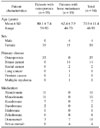
Table 1 shows the characteristics of the patients with MRONJ. For patients with bone metastases, breast cancer was the most common primary disease. Alendronate and minodronate were the most common medications in patients with osteoporosis, followed by risedronate, whereas zoledronate was the most common medication in patients with bone metastases, followed by denosumab.
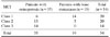
Table 2 presents a comparison of the MCI between MRONJ patients with osteoporosis and those with bone metastases. The MCI classifications of MRONJ patients with osteoporosis (class 1: 6, class 2: 15, class 3: 14) tended to be higher than those of patients with bone metastases (class 1: 14, class 2: 5, class 3: 0) (P=0.000).
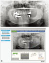
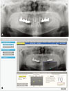
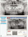
Figures 1, 2, 3 show the MCI classifications of 3 patients with MRONJ, as assessed with panoramic radiography and PanoSCOPE. Panoramic radiography (Fig. 1A) showed MRONJ in a 56-year-old woman with bone metastases of breast cancer. The PanoSCOPE software (Fig. 1B) indicated an MCI of class 1. Panoramic radiography (Fig. 2A) showed MRONJ in an 81-year-old woman with osteoporosis. The PanoSCOPE software (Fig. 2B) indicated an MCI of class 2. Panoramic radiography (Fig. 3A) showed MRONJ in a 77-year-old woman with osteoporosis. The PanoSCOPE software (Fig. 3B) indicated an MCI of class 3.
Discussion
Nakamoto et al.20 developed a CAD system based on a mathematical analysis of morphology for identifying post-menopausal women with low skeletal BMD or osteoporosis (based on World Health Organization criteria) by identifying whether the endosteal margin of the mandibular cortical bone was eroded. Their results suggested that a CAD system applied to dental panoramic radiographs may be useful for identifying post-menopausal women with low skeletal BMD or osteoporosis. Kavitha et al.21 developed a CAD system to make continuous measurements of mandibular inferior cortical width on dental panoramic radiographs, and they evaluated the system's efficacy in identifying postmenopausal women with low skeletal BMD. They concluded that their new CAD system was useful in screening for osteoporosis. Furthermore, Nakamoto et al.22 indicated that patients at risk for osteoporosis could be identified more rapidly using this new CAD system, which may contribute to earlier detection and intervention and improved medical care. We therefore considered that a CAD system using dental panoramic radiographs may be useful as a screening tool for osteoporosis.
Regarding PanoSCOPE, Muramatsu et al.16 have previously proposed an automated method for measuring the mandibular cortical width on dental panoramic radiographs for the early detection of osteoporosis. Furthermore, Muramatsu et al.17 have proposed a method for the computerized estimation of mandibular cortical degree and a new continuous measurement of MCI for osteoporosis risk assessment. We evaluated the morphology of the mandibular cortex in MRONJ patients with osteoporosis or bone metastases using the PanoSCOPE computer programme. In this study, the MCI of MRONJ patients with osteoporosis (class 1: 6, class 2: 15, class 3: 14) tended to be higher than that of patients with bone metastases (class 1: 14, class 2: 5, class 3: 0) (P=0.000). This significant difference in the MCI between patients with osteoporosis and those with bone metastases was identified using PanoSCOPE.
There are several limitations of this study. As a validation test, it is important to compare a new technique with the gold standard. Without this comparison, it is difficult to draw conclusions. Furthermore, considering the limited number of patients in this study, it would be difficult to generalize our findings to the broader population, with a more diverse range of factors such as age and gender. Therefore, further research on this topic is necessary to validate these results.
In conclusion, this study suggested that using computerized analysis to assess the morphology of the mandibular cortex may be an effective technique for the objective and quantitative evaluation of the MCI in MRONJ patients with osteoporosis or bone metastases.
 XML Download