

 PDF
PDF ePub
ePub Citation
 Print
Print


INTRODUCTION
Enteroviral infection is one of the most common viral infectious diseases in neonates.1) Enteroviral infection often leads to serious consequences because of the neonate's underdeveloped immune system and lack of serotype-specific maternal antibodies, especially in neonates less than 10 days old.2)
Enteroviral infections can occur through two major infection pathways in neonates. One pathway is infection through the blood, stool, and vaginal discharge of infected mothers at the time of delivery. The other is infection through contact with infected people (a parent, caregiver, or others) after delivery.3)
Enteroviral infections have various disease outcomes, ranging from asymptomatic infections to mild-to-serious illness or death.4) Common symptoms are fever, irritability, poor feeding, lethargy, and rashes. Some cases present with respiratory disorder. In severe cases, patients can have systemic enteroviral infections such as hepatitis, cardiovascular collapse, sepsis, and meningoencephalitis.5) Among the various disease outcomes, hepatitis, coagulopathy, and cardiomyopathy are highly associated with a poor prognosis or mortality.6) Therefore, early diagnosis and treatment of enteroviral infections are necessary to reduce mortality and morbidity in neonates.
Here, we report the early detection and successful treatment of a vertically transmitted severe enteroviral infection associated that hepatitis with coagulopathy and myocarditis along with various forms of arrhythmia.
CASE
The baby was born at a gestational age of 37 weeks and 6 days by vaginal delivery on June 25, 2017, with a body weight of 3,365 g and an Apgar score of 8 at 1 and 5 minutes. He was admitted to the neonatal intensive care unit (NICU) for ventilator care because of respiratory distress. His mother had a mild cough and fever of up to 38.7°C with a C-reactive protein (CRP) level of 4.7 mg/dL on the day of delivery, which lasted for 3 days,
The patient's 36-month-old sibling had an upper respiratory infection and fever for about 1 week before the delivery of the baby.
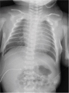
In the beginning, the baby's respiratory problem rapidly improved, so the mechanical ventilator was removed on the second day of life and feeding was started on the third day of life. The chest radiograph of the baby on the first day of life is shown in Fig. 1.
However, on the 4th day of life, the patient's body temperature abruptly increased to 38.5°C. His condition deteriorated and looked acutely ill but there was no cyanosis. The levels of CRP, aspartate aminotransferase (AST), alanine aminotransferase (ALT), total bilirubin, and direct bilirubin were 4.5 mg/dL, 220 IU/L, 33 IU/L, 8.7 mg/dL, and 0.3 mg/dL, respectively. Microbiological (bacterial) urine and blood tests were negative for both baby and mother. Cerebrospinal fluid (CSF) analysis revealed leukocytes of 2 /mm3, a protein level of 55 mg/dL and a glucose level of 68 mg/dL. Bacterial examination by culture of the CSF was negative in the baby. And enterovirus polymerase chain reaction (PCR) in CSF was negative.
Although his body temperature decreased on the 6th day of life, a follow-up blood test revealed severe thrombocytopenia. His laboratory data were as follows: platelet, 11,000 cells/mm3; prothrombin time, 20.1 seconds; activated partial thromboplastin time, 104.5 seconds; fibrinogen, 111 mg/dL; D-dimer, 6.97 µg/mL; and fibrin degradation product, 27.2 µg/mL. Meanwhile, in the liver function test, the levels of AST and ALT were 968 IU/L and 124 IU/L, respectively. Abdominal sonography revealed some ascites. Under the impression of disseminated intravascular coagulation (DIC) and hepatitis, transfusion of platelet concentrates, antithrombin III (240 IU/kg/day for 3 days), intravenous immunoglobulin (IVIG) (1 g/kg), and fresh frozen plasma treatment were administered to the patient.
On the 7th day of life, his heart rate increased abruptly up to 220 beats/min, and tachyarrhythmia, including a high degree of arrhythmia irregularities, was detected by electrocardiogram (ECG) (Fig. 2). No cardiomegaly was observed on chest radiography. Under the impression of paroxysmal supraventricular tachycardia (PSVT), up to 200 mcg/kg adenosine was administered 4 times, but there was no response. Then, direct current cardioversion (1 J/kg) was performed 2 times, and a β-blocker (1 mg/kg/day) was administered, which reduced his heart rate to 180 beats/min.
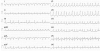 | Fig. 2The electrocardiogram on the 7th day of life showing a high-degree of heart rate irregularity of more than 200 beats/min.Abbreviations: aVR, augmented vector right; aVL, augmented vector left; aVF, augmented vector foot.
|
Laboratory tests revealed the following values: creatine kinase (CK), 199 U/L; creatine kinase muscle brain (CK-MB), 24.5 ng/mL; troponin I, 6.49 ng/mL; and brain natriuretic peptide (BNP), 2612 pg/mL, indicating myocarditis. Levels of AST, ALT, total bilirubin, and direct bilirubin were 594 IU/L, 109 IU/L, 14.5 mg/dL, and 2.3 mg/dL, respectively. Since myocarditis was suspected, hydrocortisone (10 mg/kg/day) was administered. Furthermore, since the patient was born during the peak season when enterovirus infection was prevalent, and the patient's mother and his 36-month-old sibling had a high fever around the time of delivery, we suspected enteroviral infection. The results of the respiratory virus panel and enteroviral PCR tests using nasal secretion, serum, and stool were positive on the 9th day. However, enteroviral PCR results of stool and throat samples in mother were negative after she was discharged from the hospital.
On the 11th day of life, his BNP level increased to 4,245 pg/mL and his ECG findings did not return to normal. IVIG (400 mg/kg) was administered again, and hydrocortisone was changed to dexamethasone (0.5 mg/kg/day). Thereafter, the tachyarrhythmia gradually improved, and the BNP level decreased. On the 13th day of life, his level of BNP, AST, ALT, total bilirubin, and direct bilirubin were 3,347 pg/mL, 109 IU/L, 27 IU/L, 11.2 mg/dL, and 1.7 mg/dL.
Although the arrhythmia and the patient's condition had improved, various intermittent tachyarrhythmias, including junctional ectopic tachycardia, ectopic atrial tachycardia, ventricular tachycardia, ST segment change, premature atrial contracture, and premature ventricular contracture continued until the 15th day of life (Fig. 3A).
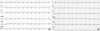 | Fig. 3(A) The ECG during the recovery phase from enterovirus myocarditis. Various ECG changes are shown, including supraventricular tachycardia, junctional ectopic tachycardia, and T-wave inversion. (B) The ECG shows complete recovery of normal heart rhythm.Abbreviations: ECG, electrocardiogram; aVR, augmented vector right; aVL, augmented vector left; aVF, augmented vector foot.
|
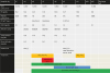
The ECG findings returned to normal after the 16th day of life (Fig. 3B). Thereafter, the steroid and β-blocker doses were gradually tapered, and steroid therapy was discontinued on the 24th day of life. On the 27th day of life, the patient was discharged home with β-blocker medication. The laboratory findings and course of treatment are shown in Fig. 4. Finally, at the 8-month follow-up, the baby showed normal development without any sequelae.
DISCUSSION
Enteroviral infection is one of the most common viral infections in neonates. The prevalence of enteroviral infection is seasonal, and it occurs more frequently during the summer and autumn months.3) The most important infection pathway is vertical transmission from an infected mother to her neonate. That is, neonates can be infected by enterovirus through the placenta in the uterus, or through the stool, blood, or vaginal discharge of infected mothers at the time of delivery.3) Perinatal vertical transmission can cause severe problems such as fetal or neonatal loss.7)
In many cases, enteroviral infections can cause severe diseases such as hepatitis, coagulopathy, pneumonia, myocarditis, meningitis, and meningoencephalitis,8) which may be because of the neonate's immature immune system.2) Therefore, a predictive factor is needed for severe illness due to enterovirus infection in neonates, which will make early detection and treatment possible. Based on our experience, we strongly recommend evaluating the possibility of enterovirus infection when the mother of a neonate shows symptoms of viral infection and the neonate shows signs of hepatitis, DIC, or myocarditis.
Our patient was admitted to the NICU because of respiratory distress immediately after birth. Although the breathing problem rapidly improved with the use of a mechanical ventilator, a sudden onset of high fever with a body temperature of 38.5°C occurred on the 4th day of life, and his condition deteriorated. He showed rapidly progressing severe symptoms of DIC, tachyarrhythmia, and myocarditis. Because it was the peak season for enteroviral infection, which is around June, and the patient's mother and his 36-month-old sibling had a high fever around the time of delivery, we suspected enteroviral infection and quickly began evaluating for it, along with a close observation and intensive care of the neonate.
Previous studies have reported that arrhythmias associated with enteroviral myocarditis often include supraventricular tachycardia, ventricular tachycardia, atrial flutter, atrioventricular (AV) block, and occasionally junctional ectopic tachycardia.69) Junctional ectopic tachycardia is a tachyarrhythmia that usually occurs after heart surgeries such as arterial switch operation, AV canal repair, or Norwood repair.10) Our case showed a sudden-onset tachyarrhythmia and myocarditis during treatment for DIC. Although only a few studies have reported predictive factors for the prognosis of pediatric myocarditis, tachyarrhythmia has been reported in a retrospective study to be associated with significant increases in mortality and resource utilization in children with myocarditis.11)
The patient's levels of AST and ALT were 968 IU/L and 124 IU/L, respectively, on 6th day of life. His serum aminotransferase levels indicated hepatocellular damage. One previous study reported that serum AST level may predict the severity of disease and the clinical outcome.4) As our case had myocarditis accompanied by various forms of arrhythmia and hepatitis with coagulopathy, we originally expected a poor prognosis. However, our patient recovered completely possibly because of the early detection and various supportive treatments.
No specific treatment has been established for enteroviral infection. The antiviral or immunomodulatory effect of IVIG remains unproven. Moreover, a drug called pleconaril acts on picornaviruses, including most non-polio enterovirus, but this drug still requires further research to determine its true effectiveness and safety.12)
In summary, we report a case of vertically transmitted enteroviral infection that showed serious symptoms such as hepatitis with coagulopathy and myocarditis with arrhythmia. However, with prompt and effective treatment using IVIG, β-blocker, and steroid, the neonate fully recovered without any sequelae.
We strongly recommend evaluation for the possibility of enteroviral infection in neonates when the mother of the neonate is suspected of having a viral infection (e.g., high fever) around the time of delivery and the neonate shows some early indicators of disease such as thrombocytopenia, DIC, hepatitis, or myocarditis. As delayed diagnosis of the enteroviral infection in neonates often leads to serious illness and high mortality, early detection of various symptoms associated with enteroviral infection and prompt implementation of proper treatment is key to reducing the risk of complications and mortality in neonates.
 XML Download