Journal List > Yonsei Med J > v.61(1) > 1139814

Kang, Lee, Chun, Choi, Moon, Lee, and Kang: Follistatin Mitigates Myofibroblast Differentiation and Collagen Synthesis of Fibroblasts from Scar Tissue around Injured Flexor Tendons

Abstract

Purpose

The aim of this study was to investigate the effect of FST gene on the inhibition of fibrosis in fibroblastic cells from scar tissue around repaired zone II flexor tendons.

Materials and Methods

Immunohistochemistry was conducted on fibroblast cells transfected with adenovirus-LacZ (Ad-LacZ) as a marker gene (control), or with adenovirus-FST (Ad-FST) as a therapeutic gene. Fibroblast cultures without adenoviral exposure served as controls.

Results

Fibroblastic cells transfected with Ad-FST demonstrated significant decrease in collagen type I, MMP-1, MMP2, and α-SMA mRNA expressions compared to those transfected with Ad-LacZ. In addition, fibroblastic cells transfected with Ad-FST exhibited significant decrease in MMP-1, TIMP-1, fibronectin, PAI-1, TRPV4, α-SMA, desmin, and PAX7 protein expressions.

Conclusion

Based on these findings, we conclude that FST may be a novel therapeutic strategy for preventing scar adhesions around repaired tendons by inhibiting fibroblasts from differentiating into myofibroblasts, in addition to producing type I collagen and regulating extracellular matrix turnover via the downregulation of MMP-1 and TIMP-1. FST may also decrease contracture of the scar by inhibiting Ca2+-dependent cell contraction.

INTRODUCTION

Although acute flexor tendon injuries are relatively uncommon, restoration of hand function following such trauma has been challenging.123 Adhesions may form between the repaired tendons and surrounding tendon sheath, especially in the tight fibro-osseous spaces of zone II injuries, despite meticulous surgical techniques and aggressive postoperative therapy.45 Several innovative approaches to prevent adhesion formation after tendon repair have been proposed, including anti-inflammatory agents, such as steroids and non-steroidal anti-inflammatory drugs;67 natural polymers, such as hyaluronic acid,8910 collagen solution,11 and amniotic fluid;11 anti-metabolites, such as 5-fluorouracil;1213 and barrier methods ,such as polytetrafluoroethylene membrane and porcine gelatin.1415 However, these anti-adhesion therapies remain unsuccessful and have not progressed beyond preclinical studies.
It has been shown that the transforming growth factor-beta (TGF-β) pathway is involved in the formation of adhesions.16 Studies on TGF-β pathway inhibition have reported an improvement in the range of digital motion as well as alteration of types I, III, and V collagen production.1718 In vitro studies using the natural TGF-β inhibitors decorin and mannose 6-phosphate (M6P) have reported a reduction in TGF-β induced collagen production, especially type I collagen.19 However, other studies have reported that TGF-β pathway inhibition is associated with a decrease in mechanical strength,2021 and a clinical trial on M6P did not demonstrate any beneficial effect on finger movements.22
It has been reported that activin A in fibroblasts, which is induced by TGF-β1, stimulates inflammation and fibrosis in many different models, including inflammatory bowel disease, rheumatoid arthritis, and wound healing, and following burn injuries.2324 A key regulator of activin is follistatin (FST), a glycoprotein that binds activin. This binding is virtually irreversible. The resulting complex targets a lysosomal degradation pathway.25 FST can block the fibrogenic actions of TGF-β despite its inability to bind TGF-β1, suggesting that TGF-β1 exerts fibrogenic actions through its stimulation of activin A.24 In addition, as FST binds to activin A, it attenuates the capacity of activin A to stimulate fibrosis, as demonstrated in hepatic fibrosis,26 pulmonary fibrosis,27 and radiation-induced fibrosis.28
In this study, we hypothesized that the activin/FST pathway offers a novel biological target for preventing the formation of tendon adhesions. We used an adenovirus system to transfer a follistatin gene construct into fibroblasts isolated from scar tissue around the repaired flexor tendons of patients who underwent tenolysis after repair of zone II tendon. The goal of this experiment was to investigate the effect of this gene on inhibition of fibrosis.

MATERIALS AND METHODS

The Human Subjects Institutional Review Board of Yonsei University Health System, Severance Hospital (4-2016-0174) approved all of the experimental protocols in this study.

Study design

To test the anti-fibrotic effect of adenovirus-FST construct (Ad-FST) on fibroblasts in vitro, we utilized cells from the scar tissue of patients who underwent tenolysis following repair of zone II flexor tendons. Fibroblasts were activated by TGF-β1 (2 ng/mL), exposed to Ad-FST as a therapeutic gene and adenovirus-LacZ construct as a marker gene, and cultured for 24 hours. The second set of fibroblast cultures that were not exposed to Ad-FST or Ad-LacZ served as the control. The mRNA expression levels of collagen types I, III, IV, V, and XI, as well as MMP-1, -3, -8, -9, and -13 were analyzed by reverse-transcription polymerase chain reaction (RT-PCR). In addition, protein expression levels of fibronectin, PAI-1, TRPV4, MMP-1, -2, -13, TIMP-1, -2, -4, and alpha-smooth muscle actin (α-SMA) were estimated using Western blot analysis.

Isolation and cell culture

Scar tissues around the repaired zone II flexors were harvested from seven patients, in whom adhesions had formed between the tendon and its sheath during tenolysis. Minced tissues were digested at 37℃ under gentle agitation with type IV collagenase (250 unit/mL; Sigma, St. Louis, MO, USA). Cells were placed in culture plates (TPP, Trasadingen, Switzerland) at 4×10⁵ cells/mL. Primary cultures were sustained for 2–3 weeks in Dulbecco's Modified Eagle Medium (DMEM; Life Technologies Corporation, Carlsbad, CA, USA) containing 10% fetal bovine serum, 1% v/v penicillin, streptomycin, and nystatin (Life Technologies Corporation) in a 37℃ incubator with 5% CO2 and humidity. The isolated primary cells at passage 1 were subcultured at a 1:3 ratio when they were 80–90% confluent, up to P3, which were stored at −80℃ until being used.

Follistatin constructs and transfections

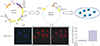
For this study, Ad-FST expressing the human follistatin gene was prepared, and Ad-LacZ expressing the LacZ gene was used as a viral control. Each recombinant adenoviral vector originated from replication-deficient adenovirus type 5 lacking E1 and E3 regions (Fig. 1). FST gene was cloned into the E1 region under the control of human cytomegalovirus early promoter. Recombinant virus was grown in transformed human embryonic kidney 293 (HEK293) cells and purified with Vivapure AdenoPACK 100 (Sartorius Stedim Biotech, Goettingen, Germany). Titers were determined by optical density at 260 nm. At confluence, fibroblastic cells were rinsed with Dulbecco's phosphate buffered saline (PBS; Life Technologies Corporation), and then exposed to DMEM containing one dose of Ad-LacZ or Ad-FST with a viral infection multiplicity of 80.

RT-PCR analysis for matrix metalloproteinases and collagens

Total RNA was isolated from the fibroblasts transfected with Ad-FST for 24 hours using QIAGEN RNeasy mini kit (QIAGEN, Germantown, MD, USA). In addition, cDNA was prepared using QuantiTect Reverse Transcription Kit (QIAGEN). Collagen and MMP genes were amplified, and relative expressions were normalized to the average beta-actin levels. Resulting data were analyzed using ImageJ software version 1.6 (National Institutes of Health, Bethesda, MD, USA).

Protein extraction and Western blot analysis

Fibroblasts at a density of 2×10⁵ cells per well were transfected with Ad-FST for 24 hours and lysed in RIPA lysis buffer (ATTO Corp., Tokyo, Japan) containing protease inhibitor (Pierce Mini Tablets, IL, USA). Meanwhile, the culture medium was collected to measure FST protein expression by Ad-FST. Lysates and culture medium were separated by sodium dodecyl sulfate-polyacrylamide gel electrophoresis, and proteins were then transferred to polyvinylidene difluoride membranes (Merck Millipore Ltd., Darmstadt, Germany) using a transfer system (Mini Trans-Blot Cell and systems, Bio-Rad, Hercules, CA, USA). The blots were incubated with specific antibodies against MMP-1, MMP-13, TIMP-1, TIMP-2, TIMP-4, fibronectin, PAI-1, TRPV4, and α-SMA (Abcam, Cambridge, UK). After reacting with secondary antibodies, immunoreactive bands were visualized by a Western blot detection system (EzWest-Lumi Plus, ATTO Corp., Japan). To verify the amounts of loaded proteins, blots were stripped of bound antibodies and reprobed using antibodies to actin (Abcam).

Immunocytochemistry

The cells were transfected with Ad-FST for 24 hours at a density of 2×10⁵ cells per well in culture medium, and fixed in 4% formaldehyde. The cells were then permeabilized in 0.1% Triton X-100 (SIGMA, St. Louis, MO, USA) for 20 minutes, and incubated in PBS with 1% bovine serum albumin for 1 hour. The cells were incubated with antibodies overnight at 4℃. The secondary antibodies (stained in red and green) were used at a 1:1000 dilution for 1 hour. DAPI was used to stain the cell nuclei (blue).

Statistical analyses

All data are expressed as the mean±standard deviation compiled from three independent experiments of separate cultures isolated from seven donors. A two-tailed Student's t-test was used to compare the means of the two groups. A p value<0.05 was considered statistically significant.

RESULTS

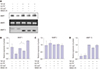
FST protein expression from Ad-FST transfected fibroblasts

Fibroblasts from the scar tissue of patients with adhesions around a repaired zone II flexor tendon that were transfected with Ad-FST showed increased staining of FST protein on confocal images compared to fibroblasts transfected with a control (Fig. 1), indicating that FST gene construct was efficiently transduced into the fibroblasts.

Expression of collagen type I, MMP-1, and MMP-2 mRNAs

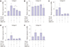
Fibroblasts transfected with Ad-FST exhibited a significant (25%) decrease in collagen type I mRNA expression at 24 hours compared to Ad-LacZ controls (TGF-β1+) (p=0.003) (Fig. 2). Fibroblasts transfected with Ad-FST also exhibited significant decrease in MMP-1 and MMP-2 mRNA expressions (31% and 59%, retrospectively) at 24 hours compared to Ad-LacZ controls (TGF-β1+) (Fig. 3). The expression of α-SMA mRNA also was significantly decreased (23%) in fibroblasts transfected with Ad-FST at 24 hours compared to Ad-LacZ controls (TGF-β1+) (p=0.001) (Fig. 4). The effect of TGF-β1 on these fibroblasts did not change when treated with a selective small molecule in hibitor, SB505124, of the canonical (i.e., Smad-dependent) TGF-β signaling pathway. These findings suggest that fibroblasts may be activated by TGF-β1 via the non-canonical TGF-β signaling pathway.

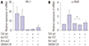
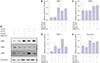
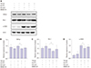
Expression of MMP-1, TIMP-1, fibronectin, PAI-1, TRPV4, and α-SMA

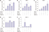
Fibroblasts transfected with Ad-FST exhibited significant decrease in MMP-1, TIMP-1, and fibronectin (24%, 23%, and 24%, retrospectively) at 24 hours compared to Ad-LacZ controls (TGF-β1+) (p<0.001, p=0.002, p<0.001, retrospectively) (Figs. 5 and 6). Fibroblasts transfected with Ad-FST also demonstrated significant inhibition of TRPV4, PAI-1, and α-SMA protein expressions (23%, 23%, and 28%, retrospectively) at 24 hours compared to Ad-LacZ controls (TGF-β1+) (p=0.002, p<0.001, p=0.002, retrospectively) (Fig. 7).

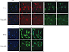
Immunocytochemistry analysis of α-SMA, desmin, and PAX7

Fibroblasts with Ad-FST exhibited decrease in α-SMA, desmin, and PAX7 expressions compared to Ad-LacZ controls (Fig. 8).

DISCUSSION

Following repair, tendons heal via both intrinsic and extrinsic pathways. Intrinsic healing involves the proliferation of tenocytes and the production of extracellular matrix (ECM).29 Extrinsic healing involves an invasion of cells from the surrounding sheath and synovium, which envelop the tendon structure and contribute to the formation of adhesions.29 Extrinsic healing dominates when the tendon or tendon sheath is injured, as the tendon has poor cell density and growth factor activity. Although several approaches have been proposed to prevent the formation of adhesions, there is limited evidence to prove that these approaches are successful, and none has been widely accepted for clinical use.30 Since tendon injury stimulates increased expression of TGF-β1 in the production of collagen types I and III, fibronectin, and glycosaminoglycans,31 blocking TGF-β1 has been reported to decrease tendon adhesion in animal tendon injury models. However, blocking TGF-β1 can also result in reduced tendon strength, as another role of TGF-β1 is to modulate immune responses and tissue repair.162032 Therefore, it is necessary to investigate methods that can selectively isolate and block the pro-fibrosis effects of TGF-β1.
FST, a systemically-expressed circulating glycoprotein, is the endogenous ligand-binding partner for activin A.33 It is well-recognized that FST can inhibit the activity of activin A, which is overexpressed in TGF-β1-induced fibrosis.2434 In the present study, the anti-fibrotic effect of FST was investigated via genetic modification of fibroblastic cells from patients with zone II flexor tendon adhesion after tendon repair. We found that fibroblastic cells from scar adhesion tissue transfected with Ad-FST showed decrease in expression levels of collagen type I mRNA, compared to fibroblasts transfected with Ad-LacZ controls. These findings indicate that FST gene inhibits the transcription of collagen type I. We also showed that the inserted FST gene decreased both transcription and translation of MMP-1 and TIMP-1. Tissue inhibitors of metalloproteinases (TIMPs) are pleiotropic extracellular proteins, and are recognized as endogenous regulators of matrix metalloproteinases (MMPs), which comprise a large family of extracellular enzymes with proteolytic activities that participate in cellular homeostasis, adaptation, and tissue remodeling.35 The findings of the present study suggest that FST restores the pathological imbalance of ECM turnover from zone II flexor tendon adhesions via the downregulation of MMP-1 and TIMP-1, which may contribute to the decrease in scar formation.
It is now accepted that myofibroblasts go through a precursor stage, in which large stress fibers are expressed. These large stress fibers are not observed in quiescent fibroblasts. These prominent bundles of microfilaments permit some contraction, pre-stressing, and remodeling of the surrounding ECM.36 At a later stage, fully differentiated myofibroblasts exhibit expression of α-SMA, which is usually an SM-specific cytoskeletal protein and is often used to define the myofibroblast phenotype.37 In the present study, the synthesis of fibronectin, a non-collagenous protein, as well as PAI-1 and α-SMA, were suppressed in fibroblasts transfected with Ad-FST compared to Ad-LacZ controls at protein level. The significant decrease in α-SMA expression suggests that FST may inhibit the transformation of fibroblasts to myofibroblasts. The decrease in transcription of collagen type I gene may also be associated with a decrease in the transformation of fibroblasts to myofibroblasts.
In the present study, the synthesis of TRPV4 was suppressed at protein level in fibroblasts transfected with Ad-FST compared to Ad-LacZ controls. It has been proposed that myofibroblasts contract via Ca2+-dependent mechanisms similar to those present in smooth muscle cells, with increased free Ca2+ regulating the phosphorylation of myosin light chain.38 The findings from our study suggest that FST may decrease the contraction of fibroblasts from tendon adhesion tissue by suppressing the translation of TRPV4.
In conclusion, FST may be a novel therapeutic strategy for preventing scar adhesions around repaired tendons by inhibiting fibroblasts from differentiating into myofibroblasts, as well as producing type I collagen and regulating ECM turnover via the downregulation of MMP-1 and TIMP-1. Due to the lack of knowledge regarding FST on tenocytes after tendon injury, further studies should be performed on how FST would affect tendon healing and strength when tenocytes are tested. FST may also decrease the contracture of the scar produced from myofibroblasts by inhibiting Ca2+-dependent contraction of the cells.

Figures and Tables

Fig. 1

Recombinant adenovirus was transfected in fibroblast from the scar tissue of patients with adhesions around zone II flexor tendon after tendon repair by immunocytochemistry analysis, indicating highly efficient transduction rate of adenoviral marker gene construct when activated by transforming growth factor-beta 1 (TGF-β1). ‘+’ means ‘treatment’ and ‘−’ means ‘no treatment’. Ad-FST, adenovirus follistatin gene construct; DAPI, 4′,6-diamidino-2-phenylindole.

ymj-61-85-g001
Fig. 2

Gene expressions of collagen I (A), collagen III (B), collagen IV (C), collagen V (D), and collagen XI (E) in fibroblast from adhesion tissue in patients with zone II flexor tendon repair and transfection with/without adenovirus follistatin gene construct (Ad-FST). Expression of collagen type I mRNA in fibroblast with Ad-FST showed a 25% decrease at 24 hours compared to control culture (TGF-β1+). ‘+’ means ‘treatment’ and ‘−’ means ‘no treatment’. *p<0.05. TGF-β1, transforming growth factor-beta 1; Ad-LacZ, adenovirus LacZ gene construct; SB505124, selective inhibitor of transforming growth factor-β type I receptor.

ymj-61-85-g002
Fig. 3

Gene expressions of MMP-1 (A), MMP-2 (B), MMP-3 (C), MMP-9 (D), and MMP-13 (E) in fibroblast from adhesion tissue in patients with zone II flexor tendon repair and transfection with/without adenovirus follistatin gene construct (Ad-FST). Expressions of MMP-1 and -2 mRNA in fibroblast with Ad-FST showed a 31% and 59% decrease, respectively, at 24 hours compared to control culture (TGF-β1+). ‘+’ means ‘treatment’ and ‘−’ means ‘no treatment’. *p<0.05. TGF-β1, transforming growth factor-beta 1; MMP, matrix metalloproteinase; Ad-LacZ, adenovirus LacZ gene construct; SB505124, selective inhibitor of transforming growth factor-β type I receptor.

ymj-61-85-g003
Fig. 4

Gene expressions of PAI-1 (A) and α-SMA (B) in fibroblast from adhesion tissue in patients with zone II flexor tendon repair and transfection with/without adenovirus follistatin gene construct (Ad-FST). Expression of α-SMA mRNA in the fibroblast with Ad-FST showed a 23% decrease at 24 hours compared to control culture (TGF-β1+). ‘+’ means ‘treatment’ and ‘−’ means ‘no treatment’. *p<0.05. TGF-β1, transforming growth factor-beta 1; Ad-LacZ, adenovirus LacZ gene construct; SB505124, selective inhibitor of transforming growth factor-β type I receptor.

ymj-61-85-g004
Fig. 5

Protein expressions of MMPs (A), MMP-1 (B), MMP-2 (C), and MMP-13 (D) in fibroblast from adhesion tissue in patients with zone II flexor tendon repair and transfection with/without adenovirus follistatin gene construct (Ad-FST). Expression of MMP-1 protein in fibroblast with Ad-FST showed a 24% decrease at 24 hours compared to control culture (TGF-β1+). ‘+’ means ‘treatment’ and ‘−’ means ‘no treatment’. *p<0.05. TGF-β1, transforming growth factor-beta 1; MMP, matrix metalloproteinase; Ad-LacZ, adenovirus LacZ gene construct; SB505124, selective inhibitor of transforming growth factor-β type I receptor.

ymj-61-85-g005
Fig. 6

Protein expressions of TIMP-1, -2, -4 (A–D) and fibronectin (E) in fibroblast from adhesion tissue in patients with zone II flexor tendon repair and transfection with/without adenovirus follistatin gene construct (Ad-FST). Expressions of TIMP-1 and fibronectin protein in fibroblast with Ad-FST showed a 23% and 24% decrease, respectively, at 24 hours compared to control culture (TGF-β1+). ‘+’ means ‘treatment’ and ‘−’ means ‘no treatment’. *p<0.05. TGF-β1, transforming growth factor-beta 1; Ad-LacZ, adenovirus LacZ gene construct; SB505124, selective inhibitor of transforming growth factor-β type I receptor.

ymj-61-85-g006
Fig. 7

Protein expressions of TRPV4, PAI-1, and α-SMA (A) in fibroblast from adhesion tissue in patients with zone II flexor tendon repair and transfection with/without adenovirus follistatin gene construct (Ad-FST). Expressions of TRPV4 (B), PAI-1 (C), and α-SMA (D) protein in fibroblast with Ad-FST showed a 23%, 23%, and 28% decrease, respectively, at 24 hours compared to control culture (TGF-β1+). ‘+’ means ‘treatment’ and ‘−’ means ‘no treatment’. *p<0.05. TGF-β1, transforming growth factor-beta 1; Ad-LacZ, adenovirus LacZ gene construct; SB505124, selective inhibitor of transforming growth factor-β type I receptor.

ymj-61-85-g007
Fig. 8

Fibroblasts with adenovirus follistatin gene construct (Ad-FST) showed decrease in α-SMA, desmin, and PAX7 expressions compared to control culture (TGF-β1+). TGF-β1, transforming growth factor-beta 1; DAPI, 4′,6-diamidino-2-phenylindole.

ymj-61-85-g008

ACKNOWLEDGEMENTS

This work was supported by a faculty research grant from Yonsei University College of Medicine (6-2017-0077) and Myung Sun Kim Memorial Foundation (2016).

Notes

The authors have no potential conflicts of interest to disclose.

AUTHOR CONTRIBUTIONS

  • Conceptualization: Young-Mi Kang, Su-Keon Lee, Yong-Min Chun, Seong-Hwan Moon, and Yun-Rak Choi.

  • Data curation: Young-Mi Kang, Su-Keon Lee, Yun-Rak Choi, and Seong-Hwan Moon.

  • Formal analysis: Young-Mi Kang, Su-Keon Lee, Yun-Rak Choi, and Seong-Hwan Moon.

  • Funding acquisition: Yun-Rak Choi.

  • Investigation: Young-Mi Kang, Su-Keon Lee, Yong-Min Chun, and Yun-Rak Choi.

  • Methodology: All authors.

  • Project administration: Yun-Rak Choi and Seong-Hwan Moon.

  • Resources: Yun-Rak Choi and Seong-Hwan Moon.

  • Software: Yun-Rak Choi, Seong-Hwan Moon, Hwan-Mo Lee, and Ho-Jung Kang.

  • Supervision: Seong-Hwan Moon, Hwan-Mo Lee, and Ho-Jung Kang.

  • Validation: Yun-Rak Choi, Seong-Hwan Moon, Hwan-Mo Lee, and Ho-Jung Kang.

  • Visualization: Young-Mi Kang, Su-Keon Lee, Yun-Rak Choi, and Seong-Hwan Moon.

  • Writing—original draft: Young-Mi Kang, Su-Keon Lee, and Yun-Rak Choi.

  • Writing—review & editing: Young-Mi Kang, Su-Keon Lee, Yun-Rak Choi, and Seong-Hwan Moon.

References

1. Clayton RA, Court-Brown CM. The epidemiology of musculoskeletal tendinous and ligamentous injuries. Injury. 2008; 39:1338–1344.
crossref pmid
2. Dy CJ, Daluiski A, Do HT, Hernandez-Soria A, Marx R, Lyman S. The epidemiology of reoperation after flexor tendon repair. J Hand Surg Am. 2012; 37:919–924.
crossref
3. Dy CJ, Hernandez-Soria A, Ma Y, Roberts TR, Daluiski A. Complications after flexor tendon repair: a systematic review and metaanalysis. J Hand Surg Am. 2012; 37:543–551.
crossref pmid
4. Hill C, Riaz M, Mozzam A, Brennen MD. A regional audit of hand and wrist injuries. A study of 4873 injuries. J Hand Surg Br. 1998; 23:196–200.
pmid
5. Tang JB. New developments are improving flexor tendon repair. Plast Reconstr Surg. 2018; 141:1427–1437.
crossref pmid
6. Ketchum LD. Effects of triamcinolone on tendon healing and function. A laboratory study. Plast Reconstr Surg. 1971; 47:471–482.
pmid
7. Kulick MI, Brazlow R, Smith S, Hentz VR. Injectable ibuprofen:preliminary evaluation of its ability to decrease peritendinous adhesions. Ann Plast Surg. 1984; 13:459–467.
crossref pmid
8. Amiel D, Ishizue K, Billings E Jr, Wiig M, Vande Berg J, Akeson WH, et al. Hyaluronan in flexor tendon repair. J Hand Surg Am. 1989; 14:837–843.
crossref pmid
9. Salti NI, Tuel RJ, Mass DP. Effect of hyaluronic acid on rabbit profundus flexor tendon healing in vitro. J Surg Res. 1993; 55:411–415.
crossref pmid
10. Wiig M, Abrahamsson SO, Lundborg G. Tendon repair--cellular activities in rabbit deep flexor tendons and surrounding synovial sheaths and the effects of hyaluronan: an experimental study in vivo and in vitro. J Hand Surg Am. 1997; 22:818–825.
crossref pmid
11. Nyska M, Porat S, Nyska A, Rousso M, Shoshan S. Decreased adhesion formation in flexor tendons by topical application of enriched collagen solution--a histological study. Arch Orthop Trauma Surg. 1987; 106:192–194.
crossref pmid
12. Akali A, Khan U, Khaw PT, McGrouther AD. Decrease in adhesion formation by a single application of 5-fluorouracil after flexor tendon injury. Plast Reconstr Surg. 1999; 103:151–158.
crossref pmid
13. Karaaltin MV, Ozalp B, Dadaci M, Kayikcioglu A, Kecik A, Oner F. The effects of 5-fluorouracil on flexor tendon healing by using a biodegradable gelatin, slow releasing system: experimental study in a hen model. J Hand Surg Eur Vol. 2013; 38:651–657.
crossref pmid
14. Bartle BK, Telepun GM, Goldberg NH. Development of a synthetic replacement for flexor tendon pulleys using expanded polytetrafluoroethylene membrane. Ann Plast Surg. 1992; 28:266–270.
crossref pmid
15. Golash A, Kay A, Warner JG, Peck F, Watson JS, Lees VC. Efficacy of ADCON-T/N after primary flexor tendon repair in Zone II: a controlled clinical trial. J Hand Surg Br. 2003; 28:113–115.
crossref pmid
16. Wynn TA, Ramalingam TR. Mechanisms of fibrosis: therapeutic translation for fibrotic disease. Nat Med. 2012; 18:1028–1040.
crossref pmid pmc
17. Chang J, Thunder R, Most D, Longaker MT, Lineaweaver WC. Studies in flexor tendon wound healing: neutralizing antibody to TGF-beta1 increases postoperative range of motion. Plast Reconstr Surg. 2000; 105:148–155.
crossref pmid
18. Chen CH, Zhou YL, Wu YF, Cao Y, Gao JS, Tang JB. Effectiveness of microRNA in Down-regulation of TGF-beta gene expression in digital flexor tendons of chickens: in vitro and in vivo study. J Hand Surg Am. 2009; 34:1777–1784.
pmid
19. Bates SJ, Morrow E, Zhang AY, Pham H, Longaker MT, Chang J. Mannose-6-phosphate, an inhibitor of transforming growth factor-beta, improves range of motion after flexor tendon repair. J Bone Joint Surg Am. 2006; 88:2465–2472.
crossref pmid
20. Katzel EB, Wolenski M, Loiselle AE, Basile P, Flick LM, Langstein HN, et al. Impact of Smad3 loss of function on scarring and adhesion formation during tendon healing. J Orthop Res. 2011; 29:684–693.
crossref pmid
21. Jørgensen HG, McLellan SD, Crossan JF, Curtis AS. Neutralisation of TGF beta or binding of VLA-4 to fibronectin prevents rat tendon adhesion following transection. Cytokine. 2005; 30:195–202.
crossref pmid
22. Lees VC, Warwick D, Gillespie P, Brown A, Akhavani M, Dewer D, et al. A multicentre, randomized, double-blind trial of the safety and efficacy of mannose-6-phosphate in patients having Zone II flexor tendon repairs. J Hand Surg Eur Vol. 2015; 40:682–694.
crossref pmid
23. Sugino K, Kurosawa N, Nakamura T, Takio K, Shimasaki S, Ling N, et al. Molecular heterogeneity of follistatin, an activin-binding protein. Higher affinity of the carboxyl-terminal truncated forms for heparan sulfate proteoglycans on the ovarian granulosa cell. J Biol Chem. 1993; 268:15579–15587.
crossref pmid
24. Hedger MP, Winnall WR, Phillips DJ, de Kretser DM. The regulation and functions of activin and follistatin in inflammation and immunity. Vitam Horm. 2011; 85:255–297.
crossref pmid
25. Hashimoto O, Nakamura T, Shoji H, Shimasaki S, Hayashi Y, Sugino H. A novel role of follistatin, an activin-binding protein, in the inhibition of activin action in rat pituitary cells. Endocytotic degradation of activin and its acceleration by follistatin associated with cell-surface heparan sulfate. J Biol Chem. 1997; 272:13835–13842.
crossref pmid
26. Patella S, Phillips DJ, Tchongue J, de Kretser DM, Sievert W. Follistatin attenuates early liver fibrosis: effects on hepatic stellate cell activation and hepatocyte apoptosis. Am J Physiol Gastrointest Liver Physiol. 2006; 290:G137–G144.
crossref
27. Aoki F, Kurabayashi M, Hasegawa Y, Kojima I. Attenuation of bleomycin-induced pulmonary fibrosis by follistatin. Am J Respir Crit Care Med. 2005; 172:713–720.
crossref pmid
28. Forrester HB, Li J, Leong T, McKay MJ, Sprung CN. Identification of a radiation sensitivity gene expression profile in primary fibroblasts derived from patients who developed radiotherapy-induced fibrosis. Radiother Oncol. 2014; 111:186–193.
crossref
29. Koob TJ, Summers AP. Tendon--bridging the gap. Comp Biochem Physiol A Mol Integr Physiol. 2002; 133:905–909.
crossref
30. Khanna A, Friel M, Gougoulias N, Longo UG, Maffulli N. Prevention of adhesions in surgery of the flexor tendons of the hand: what is the evidence? Br Med Bull. 2009; 90:85–109.
crossref pmid
31. Thomopoulos S, Parks WC, Rifkin DB, Derwin KA. Mechanisms of tendon injury and repair. J Orthop Res. 2015; 33:832–839.
crossref pmid pmc
32. Wu YF, Mao WF, Zhou YL, Wang XT, Liu PY, Tang JB. Adeno-associated virus-2-mediated TGF-β1 microRNA transfection inhibits adhesion formation after digital flexor tendon injury. Gene Ther. 2016; 23:167–175.
crossref pmid
33. DePaolo LV. Inhibins, activins, and follistatins: the saga continues. Proc Soc Exp Biol Med. 1997; 214:328–339.
crossref pmid
34. Harrington AE, Morris-Triggs SA, Ruotolo BT, Robinson CV, Ohnuma S, Hyvönen M. Structural basis for the inhibition of activin signalling by follistatin. EMBO J. 2006; 25:1035–1045.
crossref pmid pmc
35. Moore CS, Crocker SJ. An alternate perspective on the roles of TIMPs and MMPs in pathology. Am J Pathol. 2012; 180:12–16.
crossref pmid
36. Hinz B. Formation and function of the myofibroblast during tissue repair. J Invest Dermatol. 2007; 127:526–537.
crossref pmid
37. Sandbo N, Dulin N. Actin cytoskeleton in myofibroblast differentiation: ultrastructure defining form and driving function. Transl Res. 2011; 158:181–196.
crossref pmid pmc
38. Follonier Castella L, Gabbiani G, McCulloch CA, Hinz B. Regulation of myofibroblast activities: calcium pulls some strings behind the scene. Exp Cell Res. 2010; 316:2390–2401.
crossref pmid
TOOLS
ORCID iDs

Young-Mi Kang
https://orcid.org/0000-0002-7686-6316

Su-Keon Lee
https://orcid.org/0000-0003-3524-6345

Yong-Min Chun
https://orcid.org/0000-0002-8147-6136

Yun-Rak Choi
https://orcid.org/0000-0001-5197-3716

Seong-Hwan Moon
https://orcid.org/0000-0003-1121-2730

Hwan-Mo Lee
https://orcid.org/0000-0002-5405-3832

Ho-Jung Kang
https://orcid.org/0000-0003-0273-1264

Similar articles