

 PDF
PDF ePub
ePub Citation
 Print
Print


INTRODUCTION
Diffuse large B-cell lymphoma (DLBCL), the most prevalent type of non-Hodgkin lymphoma, constitutes 30–40% of all cases in different geographic regions.1 The two subtypes of DLBCL contained activated B cell-like and germinal center B cell-like, classified by distinct pathogenesis.2 Although the development of therapeutic methods has improved the survival of DLBCL patients, about 30% of patients still develop relapse/refractory disease.3 Therefore, it is important to search for new therapeutic markers for DLBCL treatment.
Curcumin, chemically called as diferuloylmethane, is a yellow-colored polyphenol that has been widely used as food additive and traditional medicine in Asian countries.4 The beneficial functions of curcumin are thought to attribute to its antimicrobial,5 anti-oxidant,6 anti-inflammatory,7 hepatoprotective,8 and hypoglycemia protective9 effects. Growing evidence have suggested that curcumin plays a protective role in preventing and treating numerous human cancers by inhibiting tumorigenesis, progression, metastasis, angiogenesis, chemoresistance, and radioresistance.101112
MicroRNAs (miRNAs), evolutionarily conserved singlestranded RNAs of about 22 nucleotides in length, play a critical role in a series of physiopathology processes.13 Mature miRNAs, loaded into RNA-induced silencing complex (RISC), negatively regulate target gene expression by binding to the 3′-untranslated region (3′-UTR) of target mRNAs, leading to translational repression and mRNA degradation.14 Recent studies have elucidated that miRNAs are involved in tumor growth, progression, metastasis, angiogenesis, and immune evasion.15 MiR-21, a commonly overexpressed miRNA, is a key oncomir in carcinogenesis of multiple human cancers, such as gastric cancer,16 breast cancer,17 and ovarian cancer.18 Moreover, circulating miR-21 was demonstrated as a new disease response biomarker with differential response stratified by interim-positron emission tomography/CT in DLBCL patients.19 High expression of miR-21 was associated with worse overall survival of DLBCL patients.20 Additionally, miR-21 contributed to DLBCL cells chemoresistance through targeting FOXO1 and regulating PI3K/AKT pathway.21 Downregulation of miR-21 promoted cell apoptosis and tumor-suppressor PTEN expression in DLBCL, highlighting its role as a potential useful approach for DLBCL treatment.22
In the present study, our data indicated that curcumin inhibited the proliferation, migration, and invasion, while promoting the apoptosis of DLBCL cell line. Moreover, curcumin suppressed miR-21 expression, and Von Hippel-Lindau (VHL) was a direct target of miR-21. In all, curcumin exerted its antiproliferation, anti-migration, anti-invasion, and pro-apoptosis functions, at least partly, by repressing miR-21 and regulating VHL expression in DLBCL cell line.
MATERIALS AND METHODS
Specimens and cells
Forty-five cases of lymphoma tissues were obtained from DLBCL patients who were diagnosed and had undergone surgical resection at Xianning Central Hospital (Hubei, China) between August 2010 to March 2016. Additionally, 23 cases of reactive lymphoid hyperplasia tissues were collected in the same period, and were selected as negative controls. No local or systemic therapy was conducted in these participators before operation. Primary CD19+ cells were obtained from the serum of healthy volunteers through positive selection with CD19+ MicroBeads antibody (Miltenyi Biotec, Auburn, CA, USA). Written informed consent was obtained from all participants, and the study was approved by Institutional Review Board of Xianning Central Hospital.
Three DLBCL cell lines (SU-DHL-8, OCI-LY1, and SU-DHL-10) were purchased from American Type Culture Collection (ATCC, Manassas, VA, USA), and were cultured in RPMI-1640 medium (Thermo Fisher Scientific, Tempe, AZ, USA) plus 10% fetal bovine serum (FBS, Thermo Fisher Scientific), 2 mmol/L L-glutamine (Thermo Fisher Scientific) and 100 units/mL penicillin/streptomycin (Thermo Fisher Scientific), at 37℃ in 5% CO2.
Cell transfection and treatment
The modified miR-21 mimics and the control (NC mimics); miR-21 inhibitor (anti-miR-21) and negative control (anti-NC); and siRNA targeting VHL (si-VHL) and corresponding control (si-NC) were all designed and synthesized by Sangon Biotech (Shanghai, China). 50 nM of the indicated oligonucleotide was transiently transfected into cells by Lipofectamine™ 2000 Transfection Reagent (Invitrogen, Breda, the Netherlands) according to the directions of manufacturers.
For curcumin treatment, SU-DHL-8 cells were treated with different concentrations (0, 5, 10, 20, 40, and 60 µmol/L) of curcumin (Sigma-Aldrich, St. Louis, MO, USA), or transfected cells were treated with 20 µmol/L of curcumin, followed by the detection of cell proliferation, migration, invasion, and apoptosis capacities.
MTT assay of cell proliferation ability
Cell proliferation ability was assessed by MTT assay. In brief, cells were treated with curcumin or transfected with miR-21 mimics in 96-well microplates. At 0, 24, 48, 72, and 96 h, a total of 50 µL of MTT solution (2 mg/mL, Sigma-Aldrich) was pipetted into each well, and incubated at 37℃ for 3 h to stain the living cells. After MTT solution was removed, 150 µL of dissolving solution (11 g SDS, 50 mL 0.02 M HCl, 50 mL isobutanol) was added to dissolve the formazan crystals. Absorbance at 490 nm was measeured with a microplate reader (Bio-Rad, Richmond, CA, USA).
Flow cytometric analysis of cell apoptosis
Cell apoptosis was determined by flow cytometry with Annexin V-FITC/PI Apoptosis Detection Kit (PharMingen, San Diego, CA, USA) according to the instruction of manufacturers. At 24 h of curcumin treatment or transfection, cells were harvested and washed twice with cold PBS. Following resuspension in 1× binding buffer at a concentration of 5.0×10⁵ cells/mL, 5 µL of Annexin V-FITC and 10 µL of PI were added to 100 µL of cells. Cell apoptosis rate was analyzed by FACScan (BD Biosciences, Franklin Lakes, NJ, USA) using CellQuest Pro (in vitro diagnostic) software (BD Biosciences).
Transwell assay of cell migration and invasion
Transwell assays were performed to detect cell migration and invasion capacities by using 8 µm polycarbonate nucleopore filter-containing Boyden chambers (Millipore, Billerica, MA, USA). For migration detection, a total of 2.0×10⁴ cells was plated in the upper chamber with non-coated membrance (Millipore) for 6–8 h to attach. For invasion detection, 2.0×10⁴ cells were seeded into the upper chamber with Matrigel-coated membrance (Millipore) to attach. In both assays, serum-free medium was changed in the upper chambers and complete medium containing 10% FBS was added to the lower chambers. Twenty-four hours later, migrated or invaded cells were fixed with 4% paraformaldehyde (Sigma-Aldrich), stained with 0.1% crystal violet (Sigma-Aldrich), and visualised under a microscope (Leica, Wetzlar, Germany) in random fields.
RNA isolation, reverse transcription PCR, and quantitative real-time PCR
Total RNAs were isolated from tissues and cells with Isogen II (Nippon Gene, Toyama, Japan). Concentration of RNA was measured by Nano-drop 2000 spectrophotometer (Thermo Fisher Scientific). To quantify VHL mRNA expression, 100 ng RNA was reversely transcribed into cDNA using iScript cDNA Synthesis Kit (Bio-Rad Laboratories, Hercules, CA, USA). Then, quantitative real-time PCR (qRT-PCR) was performed using SYBRTM Green qPCR Master Mix (Thermo Fisher Scientific) on a 7900HT system (Applied Biosystems, Foster City, CA, USA). Fold changes were calculated by 2−ΔΔCt method. For normalization, the transcript levels were compared to that of GAPDH. To quantify miR-21 expression, miScript II RT Kit (Qiagen, Hilden, Germany) was used for reverse transcription, and qRT-PCR was performed by miScript SYBR Green PCR Kit (Qiagen) and miScript Primer Assay specific for miR-21 (Qiagen), with U6 as endogenous control.
In situ hybridization of miR-21
The visualization of miR-21 expression in lymphoma tissues was detected using in situ hybridization (ISH) analysis with miRCURY LNATM microRNA ISH Detection Probes & Kit (Exiqon, Toronto, Canada). In brief, a 4-µm section of paraffin-embedded lymphoma tissues was dewaxed and hydrated, followed by the treatment with proteinase K (15 µg/mL, New England Biolabs, Ipswich, MA, USA) at 37℃ for 15 min. After being washed three times with PBS and dried with ethanol, the section was hybridized using 40 nM DIG-labeled LNA-miR-21-based probe at 55℃ for 1 h. After being washed with SCC buffer and PBST (PBS cotaining 0.1% Tween-20), the section was incubated with anti-DIG-AP (1:300) at 4℃ overnight. Then, the section was stained with NBT/BCIP (Thermo Fisher Scientific) substrates at 30℃ for 2 h, and the reaction was stopped with stop-buffer. When the section was dried with ethanol, the expression of miR-21 was determined based on the staining intensity using a microscope (Leica).
Western blot
Cells were lysed with lysis buffer comprised of pH 7.4 Tris-HCl (50 mM), NaCl (150 mM), EDTA (1 mM), Triton X-100 (1%), phenylmethylsulfonyl fluoride (PMSF; 1 mM), SDS (1%), Sodium deoxycholate (1%), and protease inhibitor cocktail (10 µM, Roche Applied Science, Mannheim, Germany). The concentration of protein extraction was determined by BCA Protein Assay Kit (Thermo Fisher Scientific). An equal amount of protein (50 µg) was separated on 10% SDS-PAGE and transferred onto ployvinylidene difluoride (PVDF, Millipore) membranes. Blocked by 5% non-fat milk, the membranes were incubated with anti-VHL (1:1000, Cell Signaling Technology, Danvers, MA, USA), anti-Ki-67 (1:5000, Abcam, Cambridge, UK), anti-caspase-3 (1:5000, Abcam), anti-cleaved caspase-3 (1:5000, Abcam), and anti-β-actin (1:1000, Cell Signaling Technology) overnight at 4℃, followed by incubation with HRP-labeled secondary antibody (1:1000, Cell Signaling Technology). Protein bands were visualized using Odyssey Imaging System (LI-COR, Lincoln, NE, USA).
Dual-luciferase reporter assay
The VHL wild-type reporter plasmid (VHL-WT) containing the predicted binding sites of miR-21, and its mutant-type (VHL-MUT) were designed and constructed by Sangon Biotech. Cells were cotransfected with 10 ng of VHL-WT or VHL-MUT constructs and 50 nM of miR-21 mimics or anti-miR-21 using Lipofectamine™ 2000 Transfection Reagent. The relative luciferase activities were evaluated using a dual-luciferase reporter assay system (Promega, Madison, WI, USA), and Renilla luciferase was used as an internal control.
RNA immunoprecipitation assay
RNA immunoprecipitation (RIP) assay was employed to determine the potentially endogenous interaction between miR-21 and VHL in RISC using Magna RIPTM RNA-Binding Protein Immunoprecipitation Kit (Millipore). In brief, cells were transfected with miR-21 mimics or NC mimics. Then, cells lysates were incubated with magnetic beads conjugated with anti-Ago2 (Abcam, Cambridge, UK) or anti-IgG (Abcam). Magnetic bead bound complexes were digested with Dnase (New England Biolabs) and proteinase K, and the relative enrichment of VHL mRNA was measured by qRT-PCR.
Statistical analysis
All data were analyzed by SPSS 19.0 software (IBM Corp, Armonk, NY, USA) and expressed as means±SD at least separate experiments performed in triplicate. Differences between two groups were compared using the two-tailed Student's t-test. ANOVA was used for comparison differences among groups. p-values <0.05 were considered statistically significant.
RESULTS
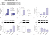
Curcumin inhibited proliferation and promoted apoptosis of SU-DHL-8 cells
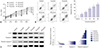
SU-DHL-8 cells were treated with different concentrations (0, 5, 10, 20, 40, and 60 µmol/L) of curcumin, followed by the detection of cell proliferation by MTT assay. These data revealed that curcumin suppressed the proliferation of SU-DHL-8 cells in a dose-dependent manner (Fig. 1A). Then, flow cytometric analysis was performed to detect the apoptosis rate of curcumin-treated (0, 5, 10, 20, 40, and 60 µmol/L) SU-DHL-8 cells. Results presented that curcumin treatment led to a significant promotion of cell apoptosis in a dose-dependent manner (Fig. 1B). Moreover, western blot results showed that curcumin treatment in SU-DHL-8 cells resulted in a decrease of proliferation marker Ki-67 expression and an increase of cleaved caspase-3 level (Fig. 1C).
Curcumin inhibited migration and invasion of SU-DHL-8 cells
Next, the migration and invasion abilities of curcumin-treated (0, 5, 10, 20, 40, and 60 µmol/L) SU-DHL-8 cells were evaluated by transwell assay. These results indicated that, compared to negative control, curcumin treatment resulted in decreased migration and invasion abilities of SU-DHL-8 cells in a dose-dependent manner (Fig. 2).
Curcumin inhibited miR-21 expression of SU-DHL-8 cells
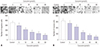
Previous studies have reported that curcumin downregulates miR-21 expression to repress tumor progression in human cancers.1023 Therefore, we further evaluated whether curcumin regulated miR-21 expression in SU-DHL-8 cells. Firstly, the expression levels of miR-21 in lymphoma tissues were evaluated by qRT-PCR and ISH assays. Results revealed that compared with lymphoid hyperplasia tissues, miR-21 was highly elevated in lymphoma tissues (Fig. 3A and B). Also, miR-21 expression was significantly upregulated in lymphoma cell lines compared to normal control (Fig. 3C). Then, miR-21 expression was examined in SU-DHL-8 cells after different concentrations (0, 5, 10, 20, 40, and 60 µmol/L) of curcumin treatment. As expected, curcumin dose-dependently inhibited miR-21 expression in SU-DHL-8 cells (Fig. 3D).
Curcumin exerted its anti-proliferation and pro-apoptosis effects by miR-21 in SU-DHL-8 cells
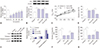
To further investigate the effect of miR-21 on the proliferation and apoptosis of SU-DHL-8 cells, gain-of-function experiments were performed by transfecting with miR-21 mimics into SU-DHL-8 cells. Subsequently, MTT assay showed that miR-21 mimics transfection resulted in a marked promotion of cell proliferation compared to negative control (Fig. 4A). Moreover, flow cytometric analysis revealed that the apoptosis of SU-DHL-8 cells was strikingly repressed in the presence of miR-21 mimics (Fig. 4B). Moreover, transfection of miR-21 mimics, but not NC control, significantly increased Ki-67 expression and decreased cleaved caspase-3 level in SU-DHL-8 cells (Fig. 4C).
Next, to provide further mechanistic insight into the link between curcumin and miR-21 on the proliferation and apoptosis of SU-DHL-8 cells, SU-DHL-8 cells were transfected with NC mimics or miR-21 mimics, and then treated with curcumin (20 µmol/L). Results demonstrated that, in comparison to NC mimics, the effects of curcumin-treated anti-proliferation and pro-apoptosis on SU-DHL-8 cells were antagonized by miR-21 mimics transfection (Fig. 4).
Curcumin-treated anti-migration and anti-invasion effects was mediated by miR-21 in SU-DHL-8 cells
Given the data on cell proliferation and apoptosis, we explored whether curcumin exerting its regulatory effect on cell migration and invasion was mediated by miR-21. Hence, SU-DHL-8 cells were transfected with NC mimics or miR-21 mimics, and then treated with or without curcumin (20 µmol/L). Transwell assays revealed that the numbers of migration and invasion cells were more in miR-21 mimics transfection group than those in control group (Fig. 5). Moreover, compared to NC mimics control, miR-21 mimics transfection in SU-DHL-8 cells remarkably abated the effects of curcumin-treated anti-migration and anti-invasion (Fig. 5).
VHL was a direct target of miR-21
Then, online software TargetScan (http://www.targetscan.org/) was used to predict the targets of miR-21. The data revealed that 3′-UTR of VHL mRNA harbored a putative binding site for miR-21 (Fig. 6A). To verify whether VHL was a target of miR-21, dual-luciferase reporter assay was performed by cotransfecting with VHL-WT or VHL-MUT constructs into SU-DHL-8 cells and miR-21 mimics or anti-miR-21. Results presented that the relative luciferase activity of VHL-WT was highly reduced by miR-21 mimics introduction, while it was strikingly increased when transfected with anti-miR-21 (Fig. 6B). However, the relative luciferase activity of VHL-MUT was almost at the same level, and it failed to respond to miR-21 expression alteration (Fig. 6B). In addition, RIP assay was performed to confirm the endogenous interaction between miR-21 and VHL in SU-DHL-8 cells. The data indicated that miR-21 mimics transfection resulted in a substantial enrichment of VHL mRNA with anti-Ago in SU-DHL-8 cells (Fig. 6C).
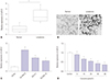
In addition, we observed whether miR-21 regulated VHL expression in SU-DHL-8 cells. qRT-PCR results revealed that VHL mRNA expression was significantly decreased by miR-21 mimics introduction, while it was strikingly increased following anti-miR-21 transfection (Fig. 6D). Consistently, western blot analysis demonstrated that VHL protein expression was inversely correlated with miR-21 level (Fig. 6E). Additionally, we assessed the expression of VHL protein in lymphoma tissues and cell lines. These results showed that, in comparison to its counterpart, VHL protein was highly downregulated in lymphoma tissues and cell lines (Fig. 6F and G).
Curcumin exerted its regulatory effects on the proliferation, migration, invasion, and apoptosis by VHL in SU-DHL-8 cells
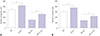
Lastly, we evaluated the expression levels of VHL mRNA in curcumin-treated SU-DHL-8 cells. As shown in Fig. 7A, curcumin treatment in SU-DHL-8 cells led to a significant promotion of VHL mRNA expression in a dose-dependent manner. To further investigate the molecular mechanism of curcumin on the progression of SU-DHL-8 cells, siRNA targeting VHL (si-VHL) were transfected into SU-DHL-8 cells prior to curcumin treatment (20 µmol/L). Western blot data presented that, compared to negative control, curcumin-treated promotion effect on VHL expression was strikingly reversed by si-VHL transfection (Fig. 7B). Subsequently, functional experiments results demonstrated that curcumin-treated anti-proliferation, antimigration, anti-invasion, and pro-apoptosis effects on SU-DHL-8 cells were highly antagonized by VHL expression restoration (Fig. 7C–G).
DISCUSSION
Accumulating evidence have suggested that curcumin possesses anti-cancer properties in a series of human cancers, including head and neck squamous cell carcinoma,24 non-small cell lung cancer,25 and advanced pancreatic cancer.26 Moreover, curcumin was found to function as a potential chemopreventive agents in tumorigenesis.27 Curcumin was also demonstrated to target cancer stem cells and specific miRNAs, and thus served as a promising adjunct to traditional cancer treatments.28 A previous document manifested that the combination of curcumin with epigallocatechin-3 gallate led to a synergistic anticancer activity by repressing angiogenesis, metastasis, and entering in complete remission in B cell lymphoma.29 In the present study, we firstly found that curcumin treatment resulted in a inhibition of the proliferation and a suppression of migration and invasion abilities, as well as a promotion of apoptosis in SU-DHL-8 cells. All these results suggested that curcumin might play anti-cancer roles in the development and progression of DLBCL.
Dysregulation of miRNAs has been widely acknowledged to implicate in cancer development and progression.14 Moreover, curcumin was verified to exert its anti-cancer effects partly by targeting special miRNAs in human cancers.28 For example, curcumin repressed bisphenol A-induced proliferation of MCF-7 cells through inhibition of miR-19 expression.30 Curcumin targets miR-21 expression to repress invasion and metastasis of colorectal cancer.10 Conversely, curcumin facilitated tumorsuppressor miR-203 expression, resulting in decreased proliferation and increased apoptosis of bladder cancer cells.31 In our study, we validated that curcumin treatment led to a reduction of miR-21 expression in SU-DHL-8 cells, consistent with former works.102332
MiR-21 has been demonstrated as a cancer-associated transcript of oncogenesis in DLBCL, providing a possibility of miR-21 as biomarkers of diagnose, therapy, and prognosis of DLBCL.203334 In the study, our data supported a significant upregulation of miR-21 in DLBCL tissues and cell lines, and miR-21 overexpression enhanced the proliferation, migration, and invasion, and inhibiting the apoptosis of SU-DHL-8 cells, in accordance with previous results.202234 In addition, we validated that curcumin exerted its anti-proliferation, anti-migration, anti-invasion, and pro-apoptosis effects by miR-21 in SU-DHL-8 cells. Similar with our findings, Mudduluru, et al.10 confirmed that curcumin repressed miR-21 expression, and thus inhibited invasion and metastasis of colorectal cancer cells.
Then, online software TargetScan was employed to predict the targets of miR-21. Among these candidates, VHL was selected for further study due to its important tumor suppressive role in a variety of cancers, including sporadic clear cell renal cancer35 and triple-negative breast cancer.36 Moreover, VHL hypermethylation was reported to be associated with aggressive phenotype and worse prognosis in DLBCL.37 In our study, we found a drastic downregulation of VHL protein in DLBCL tissues and cell lines. We also confirmed that VHL was a direct target of miR-21 in SU-DHL-8 cells. Similar with our finding, previous documents demonstrated that miR-21 targeted VHL in glioblastomas cells38 and head and neck squamous cell carcinomas cells.39 Lastly, our study was the first to verify that curcumin promoted VHL expression and curcumin exerted its anti-proliferation, anti-migration, anti-invasion, and proapoptosis effects by VHL in SU-DHL-8 cells.
Additionally, earlier document revealed that curcumin (50 µmol/L) inhibited the proliferation of rat thymocytes.40 In the present study, we demonstrated that curcumin repressed the proliferation of SU-DHL-8 cells at a concentration of 40–60 µmol/L. We speculated that higher concentrations of curcumin might have the capacity for inhibition of DLBCL and normal cells proliferation. Therefore, further studies about the effect of 20 µmol/L of curcumin on normal cells proliferation are needed. In conclusion, our study indicated that curcumin exerted its anti-proliferation, anti-migration, anti-invasion, and pro-apoptosis effects, at least partly, through miR-21/VHL axis. Our findings provided a possible molecular mechanism of curcumin-mediated anti-cancer effect.

 XML Download