

 PDF
PDF ePub
ePub Citation
 Print
Print


Hepatitis A virus (HAV) infection is one of the main causes of acute viral hepatitis. As reported by the World Health Organization in 2019, about 1.4 million new cases of acute hepatitis A (AHA) occur globally every year. According to the Korea Centers for Disease Control and Prevention, HAV has recently re-emerged, and although most of the patients are in their 20s and 40s, many are also reported to be in their 60s and above.1 In most patients, AHA is naturally recovered with lifelong protective immunity. However, about 10% of patients progress to atypical clinical courses such as prolonged hepatitis, relapsing hepatitis, cholestatic hepatitis, and autoimmune hepatitis (AIH).23 Several cases of AIH following AHA have been reported, but the mechanism and pathophysiology still remain unclear.45678
Recently, it has been shown that liver injury in patients with AHA is associated with quantitative and qualitative changes in the CD4+CD25+FoxP3+ regulatory T (Treg) cell population, including a decrease in their proportion9 and TNF production.10 Here, we presented a patient who underwent liver transplantation caused by autoimmune hepatic failure following AHA, and we performed an immunological analysis of Treg cells (IRB Number : 4-2015-0184, IRB Institution : Yonsei University Health System, Severance Hospital, Institutional Review Board).
A 68-year-old female with no relevant medical history visited our emergency room complaining of constant fatigue, poor oral intake, and jaundice lasting 2 weeks. The laboratory data were as follows: alanine aminotransferase 1095 IU/L, aspartate aminotransferase 740 IU/L, alkaline phosphatase 219 IU/L, gamma-glutamyl transpeptidase 330 IU/L, and total bilirubin 14.5 mg/dL (direct fraction 11.1 mg/dL). Abdominopelvic computed tomography (CT) revealed mild periportal edema and diffuse gallbladder wall edema, suggestive of AHA. Her immunoglobulin M (IgM) anti-hepatitis A virus (anti-HAV) antibody status was positive, but serological markers of viral hepatitis B and C infection were negative. In the diagnosis of AHA, with 3 days of conservative care, her symptoms and liver function test were improved and she was discharged from hospital.
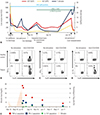
However, unlike with most AHA patients, during regular follow-up tests, her liver function had deteriorated repeatedly (Fig. 1A). Additional immunological tests revealed an antinuclear antibody titer of 1:640, and the presence of perinuclear anti-neutrophil cytoplasmic antibodies and anti-cardiolipin antibody. Although her IgM anti-HAV antibody status was positive, we suspected AIH according to the revised scoring system of the international AIH group, which was approved by the American Association for the Study of Liver Diseases (AALSD) in 2010 (score of 15).
Six months after the first hospitalization, the patient experienced recurrent abdominal distension, jaundice, and poor oral intake; as a result, she revisited our hospital. Laboratory tests revealed marked deterioration of liver function, and liver CT showed progression of liver cirrhosis affecting the collateral vessels, as well as extensive ascites. To determine the cause of hepatic function deterioration, we performed transjugular liver biopsy. In the results of liver biopsy, liver cirrhosis of stage 4c was confirmed, and typical histology of AIH was found. We diagnosed liver failure attributable to acute deterioration of her hepatitis, and started 60 mg of prednisolone daily. Ten days later, she underwent liver transplantation.
We performed intracellular cytokine staining and multi-color flow cytometry to measure tumor necrosis factor-α (TNF-α) production by peripheral blood Treg cells, since the relative proportion of TNF-α-producing Treg cells is reported to be increased (≤10% in average) and associated with liver injury in AHA patients.10 The proportion of TNF-α-producing Treg cells reached 18.6% on the day of liver transplantation (Fig. 1B). Treg cells also produced interferon-γ (IFN-γ) and interleukin-17A (IL-17A) (Fig. 1C and D). However, cytokine production by CD8+ T cells and non-Treg CD4+ T cells was not elevated (data not shown). After liver transplantation, the clinical symptoms improved, CT scan revealed no abnormal findings, and the liver function normalized rapidly. TNF-α, IFN-γ, and IL-17A production by Treg cells decreased to normal levels (Fig. 1E).
Since the 1990s, several cases of AIH developing after AHA have been reported. Individual genetic susceptibility to autoimmune disease may play a role, but the pathophysiology remains poorly understood. In our case, 68-year-old patient had suffered from AHA over the previous 6 months. Unlike typical AHA cases, she suffered from prolonged course of HAV infection with persisted IgM anti-HAV antibody. At the third worsening of liver function, additional laboratory tests were performed, and AIH was suspected (Fig. 1A). Although we did not examine autoimmune markers initially, considering that she had no past medical history of any autoimmune diseases, and initial radiologic tests and laboratory tests showed no suspected autoimmune diseases, AIH seemed to be triggered by prolonged HAV infection. During the course of the disease, the patient's IgM anti-HAV antibody remained constantly positive, and it only became negative after liver transplantation.
Recently, we showed that in AHA patients, Treg cells paradoxically produce inflammatory cytokines, and the TNF-α-producing Treg cells of AHA patients exhibit Th17-like properties and reduced suppressive function.11 Notably, the proportion of TNF-α-producing Treg cells in AHA patients was significantly correlated with the extent of liver injury, indicating that such cells play a role in the induction of immune-mediated liver injury. Such changes usually normalize within 2–4 weeks after recovery from AHA. However, in our case, the proportion of TNF-α-producing Treg cells remained high. The prolonged AHA course was accompanied by TNF-α production by Treg cells; conversion of these cells into mediators of inflammation may have played a role in the observed autoimmune pathogenesis. Further research is required to explore how changes in Treg cell populations trigger autoimmune complications after viral infection.
 XML Download