

 PDF
PDF ePub
ePub Citation
 Print
Print


INTRODUCTION
Papillary thyroid cancer (PTC) is the most common endocrine malignancy in the world, and its prevalence is continuously increasing (1). It has been claimed that compared with a high prevalence of thyroid cancer in South Korea, where an increased rate of screening is carried out, the incidence of thyroid cancer detected in other countries is just the tip of the iceberg (2). However, if other countries also had increased screening, PTC could be found to be even more prevalent worldwide to the extent of being considered an epidemic (3).
The size of thyroid cancer in patients with PTC does not always increase. According to observation trials, tumor enlargement by 3 mm or more occurred in 6.4%–6.7% and 8%–15.9% of patients with thyroid cancer at 5 and 10 years, respectively (456).
In observation trials tumor size, together with lymph node (LN) metastasis, objectively determines whether a patient should undergo surgery. Tumor size has been included in many risk stratifications for thyroid cancer, such as the Age, histologic Grade of the tumor, Extent of extrathyroidal invasion or distant metastases, and Size of the primary tumor (AGES), Age, presence of distant Metastases, Extent and Size of the primary tumor (AMES), Metastasis, patient Age, Completeness of resection, local Invasion, and tumor Size (MACIS), European Organization for Research on Treatment of Cancer (EORTC), and American Thyroid Association (ATA) classifications. Furthermore, it was reported to be the most important prognostic factor for recurrence and LN metastases. Finding a predictor of enlargement of tumor will therefore help in the treatment of PTC patients.
Among other variables, young age was reported to be an independent predictor of disease progression, which is defined as tumor enlargement or new LN metastases (7). However, other factors that may be related to tumor enlargement have not yet been investigated well in thyroid cancer. For example, among many other factors, diabetes mellitus (DM) has been reported to contribute to development of cancer through multiple mechanisms, including hyperglycemia, oxidative stress, and chronic inflammation. Also, use of metformin as a first-line treatment of type 2 DM is known to affect cancer biology through inhibiting the proliferation, metabolism, and angiogenesis of cancer cells (8910). Other clinical factors involving sex hormones have also been reported to influence tumor size (11).
We, therefore, investigated various clinicopathological factors correlated with tumor size to discern which group of patients may be observed, without the need for early surgery, so that the primary treatment will be optimally effective.
MATERIALS AND METHODS
The records of 1,401 patients treated for PTC at Ajou University Hospital between 2015 and 2017 were reviewed. Clinicopathological factors were divided in two groups, pre- and post-operation factors. Those are summarized in Table 1. The follicular variant of PTC was excluded, because discerning it from noninvasive follicular neoplasm with papillary-like nuclear features is challenging (12). Of the patients, 154 (11.0%) had one or more first-degree relatives with differentiated thyroid cancer. Among them, sixteen (1.1%) patients were diagnosed as having familial non-medullary thyroid cancer (FNMTC), this being more stringently defined as patients having three or more first-degree relatives with differentiated thyroid cancer. Patients with previous ovary-uterine surgery were defined as those having undergone ovarian or uterine surgery due to myoma, ovarian cystic torsion, endometriosis, and ovarian teratoma. The V600E B-type Raf kinase (BRAF) mutation was analyzed according to the procedure of Hayashida et al. (13), and Hashimoto's thyroiditis was diagnosed on pathological examination. DM patients were divided into 2 groups, according to whether they were taking metformin. Lymphovascular invasion (LVI) was defined as the presence of tumor cells within a vascular space, meeting the following criteria: 1) tumor cells surrounded by red cells or lymphocytes, 2) tumor cells surrounded by endothelial cell lining, and 3) attachment of tumor cells to vascular walls. It is difficult to prove LVI, because both the thinness of the lymphatic vascular wall and the invasiveness of tumor mask the evidence of lymphatic invasion (14).
Table 1
Clinical and pathological features of 1,401 patients
The Clinical and pathological features of 1,401 patients treated for PTC at Ajou University Hospital are summarized. Values are presented as the number of cases (%) or mean±standard error.
Op = operation; HTN = hypertension; DM = diabetes mellitus; Hx = history; FHx = familial history; PTC = papillary thyroid cancer; BRAF = B-type Raf kinase; LVI = lymphovascular invasion.
Comparisons of clinicopathological features were performed using Student's t-test and the χ2 test of proportions. The relationships between 2 variables were examined using correlation testing, and multivariate regression analysis was performed to control for baseline differences and confounding variables. All tests were 2-sided, with a Cronbach's α level of 0.05. All calculations were performed using SPSS 18.0 software (SPSS Inc., Chicago, IL, USA).
RESULTS
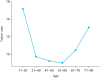
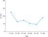
Tumor size was shown to be related to sex, age, hypertension, ovary-uterine surgery, psammoma bodies and LVI. However, after the effects of other factors were adjusted for, tumor size was found to be significantly related to both age and LVI (Table 2). Tumors increase in size at both younger age and older age, as opposed to in middle age (Table 3), as well as in cases with LVI. Age and LVI were also significantly related to each other (Tables 4 and 5). When both mean tumor size and proportion of LVI were plotted according to age, the two graphs showed a similar U shape (Figs. 1 and 2). In other words, at younger and older ages tumor sizes were larger and LVI was more frequent. Other factors, including DM and metformin usage, were not found to be related to size of tumor in the present study, although they have been reported to be related to the biological behavior of cancer cells in other studies (89).
Table 2
The relationship between tumor size and other clinical and pathological factors
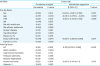
Tumor size was shown to be related to sex, age, HTN, ovary-uterine surgery, LVI, and psammoma bodies. However, after the effects of other factors were adjusted for, tumor size was found to be significantly related to both age and LVI.
CI = confidence interval; Op = operation; HTN = hypertension; DM = diabetes mellitus; Hx = history; FHx = familial history; LVI = lymphovascular invasion; BRAF = B-type Raf kinase.
Table 3
The comparison of tumor sizes according to age and LVI
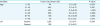
Table 4
The relationship between age and other clinical and pathological factors
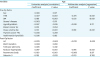
Table 5
The comparison of mean ages in significantly related factors
DISCUSSION
In PTC patients, tumor size was found to be a prognostic factor for survival, and a decisive factor for observation without early surgery. Although LN metastasis was also a decisive factor for observation, tumor size is known to be the strongest predictor of LN metastasis (15). Ultrasonography can be used to detect LN metastasis, but this modality comes with the problem of inter-institutional differences ranging from 55.8% to 89% (1617). Also, accuracy in detecting LN metastasis through ultrasonography at the central compartment of the neck is low, compared to accuracy at the lateral compartment, which follows the initial metastasis at the central compartment (18). Considering these limitations of the use of ultrasonography, tumor size can be considered the easiest and most useful indicator in observation trials.
When we investigated the relationship between suspicious clinicopathological features (Table 1) and tumor size, tumor size was not correlated with ovary-uterine surgery, DM, metformin use, familial thyroid cancer history, or BRAF mutations.
In this study, ovary-uterine surgery included hysterectomy, myomectomy, and ovarian cystectomy due to myoma, teratoma, or endometriosis. Uterine surgery for uterine myoma is most common, because this disease occurs in women of reproductive-age (19). Uterine myoma significantly increases concentrations of both estrogen and progesterone receptors, and both estrogen and progesterone promote myoma (2021). Estrogen receptors are also present in thyroid cancer, and sex hormones, especially estrogen, influence thyroid cancer cell growth (11). For this reason, this history was included in this study, together with breast cancer history. However, ovary-uterine surgery was not related to tumor size in PTC in the present study.
Epidemiological and biological data have indicated that many cancers are related to DM (22). Potential mechanisms include insulin resistance, hyperinsulinemia, insulin-like growth factor-1 (IGF-1), hyperglycemia, and dyslipidemia through mitogen-activated protein kinase, phostphoinositide-3-kinase/Akt, and Janus Kinase/signal transduction and/or activation of transcription 3 signaling pathways by IGF-1, adipokines, and cytokines (8). However, in the present study we could not confirm any association between tumor size and DM, nor did we see a decrease in tumor size in PTC with metformin usage. Even in the sub-analysis of patients older than 50 years, DM or use of metformin was not found to be related to tumor size. There may be other mechanisms that support and control tumor growth and progression in thyroid cancer as compared to other cancers.
FNMTC has been reported in about 5% of the cases, whereas its rate in the Korean population has been reported to be 9.6% in a previous study (23). However, its relatively low prevalence of 1.2% of the cases in our study can be explained by the more stringent definition of FNMTC as being present when a patient had three or more first-degree relatives with differentiated thyroid cancer. Generally, FNMTC is more aggressive than sporadic differentiated thyroid cancer, and also has more extrathyroidal extension, LN metastasis, multifocality, and recurrence than sporadic differentiated thyroid cancer (2425). In our study, tumor size in FNMTC was similar to that in sporadic differentiated thyroid cancer, as reported in previous studies (2425), despite FNMTC being more aggressive than the sporadic form.
BRAF mutation is the most frequent genetic mutation in PTC, present in more than 45% of cases (26). BRAF mutation is associated with aggressive behavior such as extrathyroidal extension, extent of tumor and extent of spread to lymph nodes, and increased number of metastatic LNs, but its poor prognostic effect is controversial (2627). Considering that BRAF mutation was present in a high prevalence of cases (87%), but not associated with tumor size in our study, it can be assumed that while BRAF mutation may be associated with the development of thyroid cancer, other events together with this oncogene may be necessary for cancer proliferation.
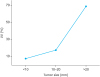
However, tumor size was found to be clearly associated with age at surgery and LVI in this study, and age was also correlated with LVI. The association between LVI and tumor size was similar to that reported in other forms of cancer (2829). We found LVI in 13.5% of patients with PTC, and as tumor size increased, the distribution of LVI also increased from 8.7% in patients with tumors ≤1 cm to 60.7% in those with tumors >3 cm (Fig. 3). Also tumor size was found to be related to extrathyroidal extension(ETE). As tumor size increased, the proportion of ETE also increased from 15.6% in patients with tumors ≤1 cm to 40% in those with tumors >3 cm.
LVI is well known to be associated with lymphangiogenesis and LN metastasis (30). Through the mechanisms of lymphangiogenesis and tumor cell entry into the lymphatic system, LVI is also related to tumor growth. Vascular endothelial growth factor C (VEGF-C)/VEGF-D is a significantly related mechanism in tumor growth. VEGF-C and VEGF-D are essential not only for blood vessel development, but also for lymphangiogenesis and LN metastasis (31). The blood vessels ensure effective oxygen diffusion to cancer cells, thereby promoting tumor growth and survival. Furthermore, VEGF attracts and stimulates growth of the endothelial cells that construct new blood vessels. Therefore, interruption of VEGF and its receptor's autocrine signal can induce the arrest of tumor growth and apoptosis (32). In addition, physiological chemokine receptor ligand interaction such as CXC chemokine receptor (CXCR) 4, C-C chemokine receptor (CCR) 7, C-X-C motif chemokine ligand (CXCL) 12 (also known as stromal-cell-derived factor-1), C-C motif chemokine ligand (CCL) 21 (also known as secondary lymphoid chemokine), and lymphoepithelial cyst (LEC) is involved in LVI (3334).
Our study has some of the limitations of any retrospective study as well as that of inter-institutional differences in definition of LVI. Diagnosing LVI is difficult, because mimickers of LVI are more common than LVI itself, which may account for its overdiagnosis (35). Other diagnostic difficulties in LVI are directly related to technical aspects of tissue preparation, such as staining techniques and number of blocks examined, and to pathologists' experience and specialization (2936). Therefore, a careful historical assessment is required to identify these features, and strict criteria should be followed to identify LVI (2836). Finally, LVI is not determined preoperatively in patients with thyroid cancer, unlike those with gastric cancer, and important decisive factor for selecting an additional surgical resection group. In future investigations, we hope to discover preoperatively available biomarkers for LVI. On the other hand, there is a statistical significance between young age and tumor size. It might be a selection bias because younger age group doesn’t undergo a health checkup routinely, which means at the time of diagnosis, younger group might be symptomatic like palpable mass.
In summary, of several suspicious preconditions which could affect tumor size, such as DM, breast cancer history, and familial cancer history, LVI was found to be significantly correlated with enlarge tumor size. Identification of a biomarker for LVI may be necessary to discern the low-risk group from high-risk group in PTC.
 XML Download