Journal List > J Endocr Surg > v.19(4) > 1139665

Pino, Parafioriti, Caruso, De Pasquale, Del Rio, Calò, Dionigi, and Stagno d'Alcontres: What You Need to Know about Mental Nerve Surgical Anatomy for Transoral Thyroidectomy
Transoral endoscopic thyroid surgery is known to be an innovative, effective and safe procedure, which is offered as a scarless technique to strongly motivated patients who want to obtain a good aesthetic result. However, evaluating the complications of this procedure, what we know is that only a few of these are related to thyroid surgery (bleeding, permanent or transient hypocalcemia, recurrent laryngeal nerve injury, seroma, surgical site infection) since more than 70% are specific of the transoral endoscopic technique such as: zygomatic bruising, chin flap perforation, oral commissure tearing, dimpling on the chin and mental nerve (MN) injury (1). The main cause of their occurrence is the placement of the trocars. Due to the fact that transoral endoscopic thyroidectomy with vestibular approach (TOETVA) is carried out through a 3-port technique placed at the oral vestibule, the incisions that are currently performed are one central of 10 millimeters (optical port for 30° endoscope) and 2 lateral of 5 millimeters (working ports for dissecting and coagulating instruments) (2). The central one is performed 1 cm above the inferior labial frenulum, meanwhile the laterals are performed in correspondence of the mental foramina (MFs), parallel to the first incision. After cutting mucosa and submucosa, we move on to the dissection of a flap, with a curved Kelly clamp, up to the lower edge of the mandible. The flap is set up along a plane that runs at the same level as the mental nerve, and its branches, and it will allow the insertion and passage of the trocars. It is clear that during this maneuver there are 2 main risks: 1) perforation of the chin flap; 2) MN injury. Therefore the precise location of the nerve is important in this type of surgery and its injury should be avoided.
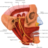
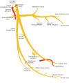
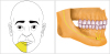
The MN is one of the branches of the inferior alveolar nerve, branch of the mandibular nerve. The mandibular nerve is the third branch of the trigeminal nerve (fifth cranial nerve), a mixed nerve consisting of somatic efferent (motor) and somatic afferent (sensory) fibers (Fig. 1). The afferent fibers of the mandibular nerve innervate several skin areas of the face, the oral mucosa, the lower teeth and gums. Instead, the motor branches supply muscles of the first branchial arch, including the masticatory muscles. The mandibular nerve emerges from the lateral part of the trigeminal ganglion and exits the middle cranial fossa via the foramen ovale, which is anterior to the foramen spinosum (3). After coming out of the foramen ovale, it splits into 2 large trunks, anterior and posterior: 1) the anterior trunk is a predominantly motor nerve and gives rise to the masseteric branch, pterygoid medial nerve, deep temporal nerve, and buccal nerve; 2) the posterior or posteromedial trunk is mainly sensitive nerve, it breaks down into the auriculo-temporal nerve and lingual nerve, and the inferior alveolar nerve. The inferior alveolar nerve has only-sensitive fibers that traverse the mandibular canal to exit via the MF (Fig. 2). Through this foramen, the MN exits, it supplies sensation to the skin and buccal mucosa of the lip and the skin of the chin (Fig. 3). In particular sensory innervation of the MN may have some overlap with the contralateral once. The sensory innervation of the MN divides among 3 smaller branches. One of the branches from the MN will innervate the skin on the chin. The other 2 nerves provide sensory innervation to the gingivae, the mucosa, and the lower lip (Fig. 3). The MN is also linked to the branches of the facial nerve (4). The MF is commonly located halfway between the upper alveolar crest and the lower edge of the mandibular bone, in direct line with the second premolar. As already mentioned, on a vertical plane, the foramen has been found above, at, or below the apex of the premolars. Most commonly, it lies below the midpoint of the distance between the lower border of the mandible and the alveolar margin. In the horizontal plane, it is one quarter of the distance from the mental symphysis to the posterior border of the ramus of the mandible (5) (Fig. 4). However exact MF location can be variable. According to the literature research in a very small percentage of people, the MN origin can be the first molar or the first premolar (Table 1) (6789). Moreover, in older or edentulous individuals, there is alveolar bone resorption which makes the location of the MF closer to the alveolar crest in most of the patients. The variations are obviously influenced by gender, age, race. In a study carried out on 525 dry mandibles, Gershenson et al. (10) reported that 4.3% of the mandibles had double MFs, 0.7% had triple MFs and that one mandible had 4 MFs on one side. Besides, the branching of the MN may vary slightly. In some individuals, there may be more branches that come from the MN. So the various foramina lead to variations in the branches of the exiting MN. Even with the differences in the branching of the MN, the sensory territory is consistent (11). Most common pattern is bifurcation (about 40%) followed by trifurcation (30%), single (20%), and quadrification (<10%) (Table 2) (12131415).
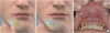
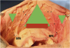
The above description of the anatomy of the MN is of remarkable importance for the surgeon performing a TOETVA. To improve the quality of TOETVA and prevent MN damage, accurate intraoperative anatomical orientation of MN is essential (16). Vestibular incisions must be made in safe areas to minimize the risk of MN involvement, which would lead to dysesthesias in the areas innervated by the MN. The current incisions are performed according to the projections of the branches of the MN and of the MF on the vestibular mucosa. But the dissection of the MN on cadaver model allowed to highlight the course of the MN and its ramifications (17). Zhang et al. (17) pointed out that the MN has a medial curvilinear course and MN soon branches out. So a triangular safety zone has been identified and the surgeon should consider that 1) a high median incision can damage the medial branches of the MN; 2) extending the median incision laterally, the MN injury can occur bilaterally; 3) a 1 cm high median incision, or a more inferiorly positioned median one, or a perpendicular opening may better preserve the MN. Instead, the 2 lateral 5 mm incisions are made at the most lateral-anterior aspect of the oral commissure in the mucosal border to avoid MN injury and instrument collision (Fig. 5) (18).
Transoral thyroid surgery represents the latest technological advancement in the approach to minimally invasive endocrine surgery. However, it is clear that conventional open thyroid surgery will still be performed. TOETVA advantages have been speculated and demonstrated in prospective studies, but so far, these procedures are still limited to a minority of patients. Several critical issues, including the MN injury and overall safety of the transoral approach and whether it will provide significant patient benefit in postoperative outcomes compared with open surgery and other endoscopic procedures, must be resolved before the new technique is successfully and responsibly introduced in clinical care. Additionally, it is recognised that the early use of this approach by surgeons, who might be relatively inexperienced in the particular skill sets required, might lead to new or serious complications, which should definitely be avoided during the start of this concept. Finally, TOETVA should provide all the advantages of other endoscopic surgeries and natural orifice transluminal endoscopic surgery (NOTES) concept. Certainly, there are significant differences between TOETVA and the other NOTES approaches (transgastric appendectomy, transvaginal cholecystectomy, cancer staging, percutaneous endoscopic gastrostomy salvage). Wound infection, incisional hernias and postoperative adhesions in abdominal surgery are common surgical complications and are eliminated with NOTES. However, these sequelae rarely belong to thyroid surgery. The advantages of NOTES for thyroid gland incision might still be less impressive than those in abdominal surgery.

Figures and Tables

Fig. 1

MN is a branch of the posterior trunk of the inferior alveolar nerve, which is itself a branch of the mandibular division of the trigeminal nerve. The MN emerges at the mental foramen in the mandible, and divides beneath the depressor anguli oris muscle into 3 branches: one descends to the skin of the chin, 2 ascend to the skin and mucous membrane of the lower lip. These branches communicate freely with the facial nerve.

MN = mental nerve.
jes-19-144-g001
Fig. 2

The mental foramen may be oval or round and is usually located apical to the second mandibular premolar or between apices of the premolars. However, its location can vary from the mandibular canine to the first molar. Very few variants of this nerve have been described in literature.

jes-19-144-g002
Fig. 3

mental nerve is a sensory nerve which provides sensation to the front of the chin and lower lip as well as the labial gingivae of the mandibular anterior teeth and the premolars.

jes-19-144-g003
Fig. 4

Landmarks for MN identification. (A) MN origin and foramen is 2 cm below the ipsilateral labial commissure, a finger nail above the jaw; (B) the nerve is 2.5 cm lateral to an imaginary median line of the face; (C) in the intra-oral perspective, the MN originates between the 3rd and 4th tooth.

MN = mental nerve.
jes-19-144-g004
Fig. 5

The dissection of the MN on cadaver allowed to highlight the course of the MN and its ramifications. The MN has a medial curvilinear course. MN soon branches out. The areas highlighted in green are zones with absence or less concentration of peripheral branches of the MN. A vestibular incision in these areas appears to be safe and prevents MN injury. Conventionally, the central incision is 1–2 cm in length and is placed beyond the cranial aspect of the buccal-mandibular frenulum, while 2 lateral 5 mm incisions are made at the most lateral-anterior aspect of the oral commissure in the mucosal border to avoid MN injury and instrument collision. It should be noted that a too inferior median incision, too close to the gingiva (red area) can interfere with the endoscopic instruments and furthermore create adhesions/sinechie with the gingival sulcus or lesion of the inferior labial frenulum.

MN = mental nerve.
*MN branches.
jes-19-144-g005
Table 1

MN origin/foramen location according to literature research

jes-19-144-i001
Authors Year Country Side MN origin/foramen location (%)
I II III IV V VI
Agarwal and Gupta (6) 2012 India Rt. 0 0 7.8 81.5 2.7 7.9
Lt. 0 0 7.6 81.5 3.1 7.8
Amorim et al. (7) 2008 Brazil Rt. 0 0 19.8 71.4 8.8 0
Lt. 0 0 23.1 68.1 8.8 0
Yeşilyurt et al. (8) 2008 Turkey Rt. 0 5.7 34.3 55.7 4.3 0
Lt. 0 7.1 25.7 61.4 5.7 0
Budhiraja et al. (9) 2013 India Rt. 0 3.8 20.0 61.0 14.3 0.9
Lt. 0 2.9 20.9 59.1 15.2 1.9
The most common position of MN origin/foramen according to literature research is the second premolar (position IV), followed by positions III (between first and second premolars), V (between second premolar and first molar), II (first premolar), and VI (first molar). The MN origin/foramen was not observed in position I (between canine and first premolar) in any dissected mandible. Variations of the mental foramen are often encountered ranging from difference in shape and positions, to presence of accessory foramen, or even complete absence in some cases.
MN = mental nerve; Rt. = right; Lt. = left.
Table 2

Prevalence of the different branching patterns of the MN in different populations

jes-19-144-i002
Authors Year Country MN dissected No. of MN branches (%)
1 2 3 4
Hu et al. (12) 2007 Korea 62 - 38.8 61.3 -
Alsaad et al. (13) 2003 Ireland 26 - 44.4 55.6 -
Alantar et al. (14) 2000 French 32 22.0 28.0 28.0 22.0
Loyal et al. (15) 2012 Kenya 64 19.0 39.0 34.0 8.0
Knowledge of the branching pattern of the MN is an important consideration during transoral thyroidectomy. With the recent increase in transoral interventions, a detailed description of mental nerve is warranted. Most common pattern is bifurcation (about 40%) followed by trifurcation (30%), single (20%), and quadrification (<10%).
MN = mental nerve.

Notes

Conflict of Interest The authors have no conflict of interest to disclose, and no other funding or financial relationship with the surgical industry.

Author Contributions

  • Conceptualization: Antonella Pino, Gianlorenzo Dionigi.

  • Data curation: Andrea Parafioriti, Ettore Caruso, Paolo Del Rio, Pietro Giorgio Calò, Francesco Stagno d'Alcontres.

  • Formal analysis: Antonella Pino, Andrea Parafioriti, Ettore Caruso, Maria De Pasquale, Paolo Del Rio, Pietro Giorgio Calò, Gianlorenzo Dionigi, Francesco Stagno d'Alcontres.

  • Investigation: Antonella Pino, Andrea Parafioriti, Ettore Caruso, Maria De Pasquale, Paolo Del Rio, Pietro Giorgio Calò, Gianlorenzo Dionigi, Francesco Stagno d'Alcontres.

  • Writing - original draft: Antonella Pino, Andrea Parafioriti, Ettore Caruso, Maria De Pasquale, Paolo Del Rio, Pietro Giorgio Calò, Gianlorenzo Dionigi, Francesco Stagno d'Alcontres.

  • Writing - review & editing: Antonella Pino, Andrea Parafioriti, Ettore Caruso, Maria De Pasquale, Paolo Del Rio, Pietro Giorgio Calò, Gianlorenzo Dionigi, Francesco Stagno d'Alcontres.

References

1. Anuwong A, Sasanakietkul T, Jitpratoom P, Ketwong K, Kim HY, Dionigi G, et al. Transoral endoscopic thyroidectomy vestibular approach (TOETVA): indications, techniques and results. Surg Endosc. 2018; 32:456–465.
crossref
2. Dionigi G, Lavazza M, Bacuzzi A, Inversini D, Pappalardo V, Tufano RP, et al. Transoral endoscopic thyroidectomy vestibular approach (TOETVA): from A to Z. Surg Technol Int. 2017; 30:103–112.
3. Charalampakis A, Kourkoumelis G, Psari C, Antoniou V, Piagkou M, Demesticha T, et al. The position of the mental foramen in dentate and edentulous mandibles: clinical and surgical relevance. Folia Morphol (Warsz). 2017.
crossref
4. Iwanaga J, Saga T, Tabira Y, Nakamura M, Kitashima S, Watanabe K, et al. The clinical anatomy of accessory mental nerves and foramina. Clin Anat. 2015; 28:848–856.
crossref
5. Mbajiorgu EF, Mawera G, Asala SA, Zivanovic S. Position of the mental foramen in adult black Zimbabwean mandibles: a clinical anatomical study. Cent Afr J Med. 1998; 44:24–30.
6. Agarwal DR, Gupta SB. Morphometric analysis of mental foramen in human mandibles of south Gujarat. People J Sci Res. 2011; 4:15–18.
7. Amorim MM, Prado FB, Borini CB, Bittar TO, Volpato MC, Groppo FC. The mental foramen position in dentate and edentulous Brazilian's mandible. Int J Morphol. 2008; 26:981–987.
8. Yeşilyurt H, Aydinlioǧlu A, Kavakli A, Ekinci N, Eroglu C, Hacialiogullari M, et al. Local differences in the position of the mental foramen. Folia Morphol (Warsz). 2008; 67:32–35.
9. Budhiraja V, Rastogi R, Lalwani R, Goel P, Bose SC. Study of position, shape, and size of mental foramen utilizing various parameters in dry adult human mandibles from north India. ISRN Anat. 2012; 2013:961429.
crossref
10. Gershenson A, Nathan H, Luchansky E. Mental foramen and mental nerve: changes with age. Acta Anat (Basel). 1986; 126:21–28.
crossref
11. Costa ED, Peyneau PD, Visconti MA, Devito KL, Ambrosano GM, Verner FS. Double mandibular canal and triple mental foramina: detection of multiple anatomical variations in a single patient. Gen Dent. 2019; 67:46–49.
12. Hu KS, Yun HS, Hur MS, Kwon HJ, Abe S, Kim HJ. Branching patterns and intraosseous course of the mental nerve. J Oral Maxillofac Surg. 2007; 65:2288–2294.
crossref
13. Alsaad K, Lee TC, McCartan B. An anatomical study of the cutaneous branches of the mental nerve. Int J Oral Maxillofac Surg. 2003; 32:325–333.
crossref
14. Alantar A, Roche Y, Maman L, Carpentier P. The lower labial branches of the mental nerve: anatomic variations and surgical relevance. J Oral Maxillofac Surg. 2000; 58:415–418.
crossref
15. Loyal PK, Butt F, Ogeng'o JA. Branching pattern of the extraosseous mental nerve in a Kenyan population. Craniomaxillofac Trauma Reconstr. 2013; 6:251–256.
crossref
16. Dionigi G, Chai YJ, Tufano RP, Anuwong A, Kim HY. Transoral endoscopic thyroidectomy via a vestibular approach: why and how? Endocrine. 2018; 59:275–279.
crossref
17. Zhang D, Fu Y, Dionigi G, Pontin A, Caruso E, Pino A, et al. Human cadaveric model for studying the preservation of mental nerve during transoral endoscopic thyroidectomy. Surg Radiol Anat.
crossref
18. Anuwong A, Ketwong K, Jitpratoom P, Sasanakietkul T, Duh QY. Safety and outcomes of the transoral endoscopic thyroidectomy vestibular approach. JAMA Surg. 2018; 153:21–27.
crossref
TOOLS
ORCID iDs

Antonella Pino
https://orcid.org/0000-0002-5159-1395

Ettore Caruso
https://orcid.org/0000-0003-4706-9315

Paolo Del Rio
https://orcid.org/0000-0002-6776-5441

Pietro Giorgio Calò
https://orcid.org/0000-0001-9637-1145

Similar articles