

 PDF
PDF ePub
ePub Citation
 Print
Print


INTRODUCTION
Thyroid nodules are the most common thyroid disorder. The reported prevalence of thyroid nodules is about 2% to 7% by palpation alone and up to 30%–50% by autopsy without clinical evidence of thyroid disease (12). Among them, malignant nodules are reported in up to 5%–10%, and papillary thyroid carcinoma (PTC) is the most common malignant neoplasm of the thyroid (34). Ultrasound imaging and cytology from fine-needle aspiration (FNA) are the main tools to decide whether thyroidectomy is indicated because PTC can usually be diagnosed by well-defined cytological features and ultrasound findings (56).
The results of the FNA are reported according to the Bethesda system, which recognizes 6 diagnostic categories and provides an estimation of cancer risk within each category (Table 1) (789). The American Thyroid Association guidelines recommend surgery if cytology is diagnostic or suspicious (Bethesda class VI or V) for PTC (7). However, misdiagnosis of PTC is one of the most problematic issues. The rates of false-positive and -negative FNA results for thyroid nodules have been reported as 2%–10% and 0%–14%, respectively (101112). Overdiagnosis resulting from cytology results may lead to excessive treatment, specifically, unnecessary thyroidectomy. Considering a high survival rate and a good prognosis of PTC, false positivity of FNA can be more problematic than false negativity (101314).
Table 1
The Bethesda system for reporting thyroid cytopathology: diagnostic categories and risk of malignancy*

Values are presented as median (interquartile range) or number (%).
*As reported in the Bethesda system by Cibas and Ali (9).
![]()
Several studies have been reported on false positivity of FNA (1314151617). Some reports show that benign thyroid nodules such as adenomatous goiters, follicular adenomas, and nodular chronic thyroiditis mimic the nuclear features of PTC and share specific histological features with PTC. Therefore, sometimes differentiating certain benign nodules from PTC is not easy by cytology alone (131516). Interestingly, there were some reports of true disappearance of thyroid tumors after FNA (1417).
The aim of this study was to evaluate the incidence and clinicopathologic characteristics of patients who underwent thyroidectomy due to consistent findings of PTC in FNA but turned out to have no evidence of malignancy in a surgically removed thyroid specimen.
MATERIALS AND METHODS
1. Patient selection
We retrospectively reviewed the electronic medical record of 13,760 patients who underwent thyroidectomy at the Thyroid Cancer Center of Samsung Medical Center, a tertiary referral center in Korea, from January 2009 to January 2019. If the patient had more than one operation, the first was selected and analyzed. We excluded patients who underwent only completion thyroidectomy after lobectomy at another hospital, who did not undergo preoperative FNA, those aged <20 years or >80 years, and those with malignancy other than PTC.
A total of 10,776 patients with Bethesda V and VI in preoperative FNA cytology were included in this study. Of these, 10,736 patients were identified as PTC (true positive group) by permanent pathology results, and 40 patients were identified as benign (false positive group).
The Institutional Review Board (IRB) at Samsung Medical Center approved this study, and informed consent was obtained from every patient (IRB No. SMC 2019-11-003).
2. Definitions
A positive cytology finding was defined as thyroid nodules with FNA findings of Bethesda category V or VI. Positive histopathologic findings are defined as PTC confirmed by permanent pathology findings. Patients who underwent thyroidectomy for positive cytologic finding but were confirmed to have negative histopathologic findings were assigned to the false-positive group.
3. Variables
The following variables were analyzed for 10,776 patients: sex, age, Bethesda category, and pathologic thyroiditis. Additional preoperative ultrasonographic findings were further analyzed for 40 patients in the false-positive group.
If FNA and ultrasonography was repeated several times, the results close to the day of surgery were analyzed. If the test was conducted at another hospital and re-examined at SMC, the higher category was selected and classified. All FNA slides from outside institutions were re-reviewed. If the final report of outside FNA were unclear, repeat FNAs were done at Samsung Medical Center.
Most of the FNA results were reported according to the Bethesda system. If the FNA results were not categorized, we reviewed results and classified them into the most appropriate category.
The Radiology Department of SMC has used the Korean thyroid imaging reporting and data system (K-TIRADS) since 2016 (Table 2) (18). Results prior to 2016 were reviewed and classified based on K-TIRADS.
Table 2
Malignancy risk stratification according to Korean thyroid imaging reporting and data system*

*As reported in revised Korean Society of Thyroid Radiology consensus statement and recommendations by Shin et al. (18); †Microcalcification, nonparallel orientation (taller-than-side), spiculated/microlobulated margin.
![]()
The case specified as background thyroiditis on final permanent biopsy was classified as pathologic thyroiditis.
4. Statistical analysis
All statistical analyses were performed using SPSS version 22.0 software (IBM Corp., Armonk, NY, USA). Continuous variables are presented as mean±standard deviation, and categorical variables are presented as number and percentage of cases. The χ2 test and linear-by-linear association were used to evaluate the differences between the clinicopathologic features of the groups. The associations of potential risk factors were tested by logistic regression analysis. Variables with P<0.05 on univariate analysis were included in multivariate analysis. P<0.05 represented statistical significance.
RESULTS
1. Clinicopathologic characteristics of 10,776 patients who underwent thyroidectomy
The clinicopathologic characteristics results are summarized in Table 3. Among 10,776 patients, 2,545 (23.6%) were male and 8,231 (76.4%) were female. The mean age was 46.1 years. Patients younger than 55 years were more numerous than older patients (8,286, 76.9% vs. 2,490, 23.1%). Preoperative FNA showed that 3,828 (35.5%) patients were class V and 6,948 (64.5%) patients were class VI.
Table 3
Analysis clinical characteristics between the true-positive group and the false-positive group
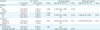
Data are shown as mean±standard deviation or number (%).
OR = odds ratio; CI = confidence interval; FNA = fine needle aspiration.
![]()
2. Comparison between the false-positive FNA and true-positive FNA groups
Of the total 10,776 patients, 10,736 (99.6%) were included in the true-positive group and 40 (0.4%) in the false-positive group. Females were predominant in both groups. Age at the time of surgery was statistically significantly higher in the false-positive group than in the true-positive group (51.4±9.2 vs. 46.1±11.4; P=0.003; Student's t-test). Age over 55 was a risk factor for false-positive FNA compared to age under 55 (adjusted OR, 1.988; P=0.037). There were more cases of FNA category VI in the true-positive group (35.5% vs. 64.5%) and more cases of FNA category V in the false-positive group (90.0% vs. 10.0%). This difference was statistically significant (P<0.001). FNA category V is a risk factor for false-positive FNA compared to FNA category VI (adjusted OR, 34.776; P<0.001). There was no difference between the 2 groups in presence of pathological thyroiditis (P=0.816).
3. Clinicopathologic findings of the false-positive FNA group
The clinicopathologic characteristics of false-positive group are summarized in Table 4. We compared the differences between 2 subgroups of FNA category V and VI. Among 40 patients, 9 (22.5%) were male and 31 (77.5%) were female.
Table 4
Characteristics of the 40 cases with positive cytology findings and a negative histological diagnosis of papillary thyroid carcinoma
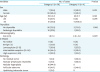
Data are shown as mean±standard deviation or number (%).
K-TIRADS = Korean thyroid imaging reporting and data system.
![]()
Preoperative thyroid ultrasonography showed that the average size of the nodules was 1.8±1.4 cm (0.3–6.4 cm). Preoperative thyroid ultrasonography, categorized using K-TIRADS, showed 9 cases (22.5%) with high suspicion, 18 cases (45.0%) with intermediate suspicion, 8 cases (20.0%) with low suspicion, and 5 (12.5%) cases that were benign.
The most common benign tumor was follicular adenoma (n=17, 42.5%), followed by nodular hyperplasia (n=16, 40.0%). Others were nodular lymphocytic thyroiditis (n=3, 7.5%) and hyalinizing trabecular tumor (n=2, 5.0%). Interestingly, 2 patients (5.0%) had no residual tumor.
If the FNA result was positive but the final biopsy was benign or no residual tumor, the pathologist reviewed the FNA slide again. All 40 slides were reviewed, of which 38 remained unchanged but 2 slides were confirmed as PTC.
One of these had a 0.5 cm low echoic, oval shaped thyroid nodule on US, and final biopsy confirmed nodular hyperplasia. The other had a 0.4 cm taller than wide hypoechoic nodule on US and was confirmed as no residual tumor on final biopsy. There was no underlying thyroiditis in either case.
DISCUSSION
In this study, we identified 10,776 patients who underwent thyroidectomy based on diagnosis of PTC by FNA. Forty patients were diagnosed with benign nodules, and 10,736 patients were diagnosed with PTC in the final biopsy. The false-positive rate was 0.4%. Patients in the false-positive group were significantly older. In addition, the ratio of age ≥55 in the false-positive group was high. Age ≥55 was a risk factor for false-positive FNA in multivariate analysis. Bethesda category V was also a risk factor for false-positive FNA compared to category VI.
Several studies have reported thyroiditis as a risk factor for false-positive FNA, but there was no significant difference between true-positive and false-positive groups in this study (1019).
FNA cytology has been established as a safe, reliable, and effective method for a diagnosis of thyroid cancer (51417). However, definite diagnosis of a thyroid nodule is provided by permanent pathology following surgical excision of the tumor. On occasion, the permanent pathology result can be different from initial diagnosis by FNA. The rate of false-positive FNA was reported as 2%–10% (111420).
There have been several reports about patients with positive FNA but negative tumor cells in permanent histology (10131417). One possibility is that cytological characteristics of some benign thyroid masses are very similar to those of PTC, making them difficult to be distinguished. Especially, in FNA cytology, diagnosis should be made based on cells, mainly the characteristics of the nucleus. Some benign masses show very similar nuclear features to those of PTC (81421). Some studies have reported that about 17% of cases with adenomatous goiter and some nodular hyperplasias had nuclear features similar to those of PTC, such as nuclear atypia including intranuclear grooves (21222324). It is also known that chronic lymphocytic thyroiditis is difficult to be distinguished from PTC in cytology (1019) because chronic lymphocytic thyroiditis can show atypical nuclear changes including nuclear enlargement and nuclear grooves (142526). In this study, most of the final biopsy results in the false-positive group were nodular hyperplasia or follicular adenoma. And 5 patients had nodular lymphocytic thyroiditis. Nuclear atypia was observed in these patients' cytology.
Another possibility is that PTC is so small that it is removed during FNA and not detected in the permanent biopsy. Several studies have reported not only cases of FNA showing microcarcinoma but negative histological diagnosis of PTC but also cases of histological alterations after FNA (1417). According to these studies, thyroid tumor can be completely replaced by reactive changes including fibrosis, infarction, and cystic degeneration. In the present study, 2 cases were identified as papillary microcarcinoma. It is correct to interpret these cases as the disappearance of PTC rather than the false positive of FNA.
Most of the 40 patients in this study with benign thyroid nodule showed nuclear features of PTC. The overlap of these pathological features remains a diagnostic dilemma. Therefore, surgeons should be aware of these circumstances, and it is important to inform patients about the possibility of a false positive result, especially in those patients over 55 years of age without FNA Bethesda category VI. In addition, if the K-TIRADS category of preoperative ultrasonography is low, surgeons can reduce unnecessary thyroidectomy by deciding to perform core needle biopsy or repeat FNA (27).
Recent studies have shown that immunochemical stains are being applied to overcome the diagnostic dilemma for PTC such as galectin-3, hector battifora mesothlia-1, cytokeratin 19, and CD 56 (2829). To increase the accuracy of FNA, a combination of morphological, immunohistochemical, and molecular biological approaches should be further studied.
CONCLUSION
Although the false positive rate is low (0.4%), surgeons should be aware of the possibility and inform patients, especially those aged >55 years, suspicious for malignancy (Bethesda category V) on FNA, and low suspicion/benign K-TIRADS. To reduce unnecessary thyroidectomy, core needle biopsy or repeat FNA should be considered in patients with these findings. Further large-scale studies are necessary to establish a firm conclusion.
 XML Download