

 PDF
PDF ePub
ePub Citation
 Print
Print


INTRODUCTION
Breast cancer is the most important cancer that affects procreation in women of reproductive age. Its incidence has markedly increased from 2002 to 2014 (crude incidence rate, from 29.5 to 83.4 per 100,000 individuals), making it the second most common cancer in Korea [12]. Along with the increase in the incidence, compared to the United States (5.5%), a greater proportion of women were diagnosed with breast cancer before the age of 40 years (11.5%) [123]. Moreover, younger women show poorer prognosis than older women do, and they are more likely to receive cytotoxic chemotherapy [4]. Therefore, a high number of Korean premenopausal women receive cytotoxic chemotherapy following the diagnosis of breast cancer.
The ovarian reserve, which is determined by the number of primordial follicles present in the ovaries, declines progressively with aging. Although chemotherapy was highly successful in reducing the risk of cancer recurrence and mortality, chemotherapeutic agents were also gonadotoxic and accelerated follicular depletion in the ovaries [2]. Consequently, most women experienced loss of menstruation during and after chemotherapy (treatment-related amenorrhea [TRA]), often resulting in permanent loss of ovarian function (chemotherapy-induced menopause [CIM]) [5]. The extent of ovarian damage depends on the total dosage and type of gonadotoxic agents, patient age, and baseline ovarian reserve before chemotherapy [6].
TRA has been widely used to assess ovarian reserve after chemotherapy, as the presence or absence of regular menstruation is readily recognizable without any tests. Meanwhile, serum follicle-stimulating hormone (FSH) is an objective surrogate marker for ovarian function, which gradually increases with age, and increases abruptly after menopause. Persistently high FSH levels combined with low estradiol levels are sufficient for the diagnosis of CIM. The National Comprehensive Cancer Network (NCCN) suggested assessing these hormones in women who are amenorrheic for 12 or more months, to identify CIM [7].
Diagnosing women with menopause or CIM is important not only for gynecologists to assess their fertility but also for oncologists to prescribe an appropriate agent for endocrine therapy (i.e. tamoxifen or aromatase inhibitors [AIs]), as AIs are recommended for postmenopausal women to reduce the risk of recurrence of breast cancer but are contraindicated in premenopausal women and in those with residual ovarian function [78]. Following these recommendations, medical oncologists periodically check FSH and estradiol levels in women who develop amenorrhea after chemotherapy.
In previous studies that investigated the frequency of CIM in patients with breast cancer, the frequency of CIM showed a wide variation (22.0%–91.2%) [910111213]. While the inconsistency may be attributed to several factors, differences in ethnicity may also have a role, and acquiring data from the Korean population is highly justified.
Fertility preservation has been gaining ground, and patients are increasingly concerned about their fertility after cancer treatment. When counseling these patients, the risk of developing CIM and TRA should be individualized according to age; however, there is paucity of data to predict the probabilities of developing CIM and TRA in women at a specific age. Although it is well known that CIM is more frequent in older women, the only study that reported age-related frequency of CIM was based on a small number of subjects in Malaysia (n = 54) [13]. Therefore, there is a need to establish a reference for the frequencies of CIM and TRA, especially in the Korean population.
This study aimed to investigate the age-specific frequencies of CIM and TRA at 12 months after chemotherapy in patients with breast cancer. The relationship between potential risk factors and the frequencies of CIM and TRA was also investigated.
METHODS
Study population and participants
This was a retrospective cohort study of patients with breast cancer who received chemotherapy at our institution (Seoul National University Hospital, Seoul, Korea) between May 1, 2004 and May 31, 2013. Only patients with operable breast cancer were selected, and both neoadjuvant- and adjuvant chemotherapies were included. The upper age limit of the patients was restricted to 44 years to reduce the odds of falsely diagnosing CIM in women approaching natural menopause. An initial cohort of 726 women aged 25–44 years was established from the hospital database (Figure 1).
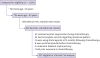 | Figure 1CONSORT diagram.FSH = follicle-stimulating hormone; GnRH = gonadotropin-releasing hormone; CIM = chemotherapy-induced menopause; CIA = chemotherapy-induced amenorrhea.
|
It was the institutional policy to recommend patients with breast cancer to undergo a blood test measuring FSH and estradiol prior to chemotherapy and periodically after the chemotherapy. These test results were used in our study. Both CIM and TRA were assessed at about 12 months after the last dosage of chemotherapy. Menstrual status was retrieved from medical records prepared either by medical oncologists or by gynecologists when patients were referred for routine gynecologic examination.
We used the values of FSH and estradiol tests in the laboratory reports ordered by medical oncologists. CIM was confirmed when women with TRA showed FSH levels above 40 IU/L and estradiol levels below 20 pg/mL. Patients who refused to undergo blood tests were excluded from our analysis (n = 393).
After a thorough review of medical records, patients who were undergoing concurrent gonadotropin-releasing hormone (GnRH) agonist therapy for fertility preservation (n = 45), those with incomplete records regarding their menstrual status (n = 24), those who were prescribed GnRH agonists at 12 months after chemotherapy (n = 15) by an oncologist, those who had attained menopause before chemotherapy (n = 8), those who had undergone bilateral oophorectomy with or without hysterectomy (n = 3), and those who had previously undergone chemotherapy for contralateral breast cancer (n = 1) were excluded (Figure 1).
Demographic data, information regarding the type of surgery, pathological reports including hormone receptor status, and information regarding the type and duration of chemotherapy and use of other adjuvant treatments were extracted from the medical records. Tumors were considered to be positive for estrogen receptor and progesterone receptor when the tumor nuclear staining was ≥ 10% on immunohistochemistry. Serum anti-Müllerian hormone (AMH) values were extracted from the medical records for analysis, when available.
The 4 major chemotherapy regimens were as follows: 4 cycles of doxorubicin (60 mg/m2) plus cyclophosphamide (600 mg/m2), followed by 4 cycles of docetaxel (75 mg/m2) every 3 weeks (AC-D); 4 cycles of doxorubicin (60 mg/m2) plus cyclophosphamide (600 mg/m2), followed by 4 cycles of paclitaxel (175 mg/m2) every 3 weeks (AC-T); 6 cycles of 5-fluorouracil (500 mg/m2) plus doxorubicin (50 mg/m2) plus cyclophosphamide (500 mg/m2) every 3 weeks (FAC); and 6 cycles of cyclophosphamide (50 mg per oral 3 times a day for 14 days) plus methotrexate (40 mg/m2 on Days 1 and 8) plus 5-fluorouracil (600 mg/m2 on Days 1 and 8) every 4 weeks (CMF).
Statistical analysis
Numerical variables are presented as the mean ± standard deviation, and categorical variables are presented as count (percent). Student's t-test was used to compare the continuous parametric variables. Pearson's χ2 test and Fisher's exact test were used to compare the proportions among different groups. The χ2 test for trend was used to test for linear trends in the CIM and TRA among women of different age groups. Multivariate logistic regression analysis was performed to identify the predictors of CIM and TRA. A p-value < 0.05 was regarded as statistically significant. Statistical analysis was performed using SPSS software version 22.0 (IBM corp., Armonk, USA).
Ethics statement
This study was reviewed and approved by the Institutional Review Board at Seoul National University Hospital (study registered as H-1512-091-728). The requirement for obtaining signed informed consent was waived because this was a retrospective analysis related to the follow-up of patients with breast cancer.
RESULTS
The patient characteristics are shown in Table 1. The mean age at diagnosis was 39.6 years, and the youngest patient in our cohort was 27 years old. The FSH and estradiol levels before chemotherapy were all within the premenopausal range. Most patients had early-stage breast cancer, and there was no patient with stage IV disease. The CMF was the least used regimen at our hospital and was administered to only 6 patients (2.5%). Cancer stage, tumor cell type, and hormone receptor status of the tumor had no influence on the choice of chemotherapeutic regimen (p = 0.491, 0.699, and 0.522, respectively)
Table 1
Patient characteristics
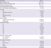
Data are shown as mean ± standard deviation or number (%).
IDC = invasive ductal carcinoma; ILC = invasive lobular carcinoma; ER = estrogen receptor; PR = progesterone receptor; FAC = 5-FU plus doxorubicin plus cyclophosphamide; AC-D = doxorubicin plus cyclophosphamide followed by docetaxel; AC-T = doxorubicin plus cyclophosphamide, followed by paclitaxel; CMF = cyclophosphamide plus methotrexate plus 5-FU.
![]()
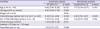
The overall frequency of TRA was 61.6% (Table 2). The frequency showed a gradual increase with age (p < 0.001, χ2 test for trend), and showed a significant difference between the different age groups (p < 0.001). Patients who received 6 or more cycles of chemotherapy were more likely to experience TRA (p = 0.001).
Table 2
Frequencies of CIM and TRA
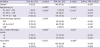
CIM = chemotherapy-induced menopause; TRA = treatment-related amenorrhea; FAC = 5-FU plus doxorubicin plus cyclophosphamide; AC-D = doxorubicin plus cyclophosphamide followed by docetaxel; AC-T = doxorubicin plus cyclophosphamide, followed by paclitaxel; CMF = cyclophosphamide plus methotrexate plus 5-FU.
*χ2 test; †χ2 test, test for trend.
![]()
CIM was found in 13.1% of the patients (Table 2). Although the age-specific frequency of CIM increased gradually with age, the trend for the increase was not significant (p = 0.096, χ2 test for trend). CIM was significantly less frequent than TRA in women of all age groups.
The frequency of CIM was higher in women who received the CMF regimen (33.3%) than in those who received the FAC (11.3%) or AC-D or AC-T (13.0%) regimens; however, the difference was not significant (p = 0.284). Unlike TRA, the frequency of CIM showed no association with the number of chemotherapy cycles (p = 0.690). CIM was less frequent in patients who were receiving tamoxifen than in those who were not receiving tamoxifen (9.3% and 37.5%, respectively, p < 0.001).
The mean age of women with CIM was 40.7 ± 2.8 years and that of women without CIM was 39.4 ± 3.7 years (p = 0.017). The mean age of women with TRA was 40.6 ± 2.9 years and that of women without TRA was 37.9 ± 3.9 years (p < 0.001). Women with CIM received 6.9 ± 1.3 chemotherapy cycles whereas those without CIM received 6.8 ± 1.1 chemotherapy cycles (p = 0.536). Women with TRA received 7.0 ± 1.2 chemotherapy cycles and those without TRA received 6.5 ± 0.9 cycles (p = 0.001).
The serum AMH values at 12 months were available for 45 patients. The AMH values were not significantly different between women with CIM (0.24 ± 0.38 ng/mL, n = 9) and those without CIM (0.39 ± 0.54 ng/mL, n = 36, p = 0.163 using Mann–Whitney U test). Similarly, there was no difference in AMH values between women with TRA (0.23 ± 0.34 ng/mL, n = 20) and those with regular menstruation (0.52 ± 0.64 ng/mL, n = 25, p = 0.114 using Mann–Whitney U test).
In multivariate analysis (Table 3), only tamoxifen administration was significantly associated with a reduced risk of CIM (odds ratio [OR], 0.10; 95% confidence interval [CI], 0.04–0.26). CIM did not have a significant association with age, chemotherapeutic regimen, or the number or duration of chemotherapy cycles.
Table 3
Multivariate model for 12-month CIM
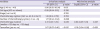
CIM = chemotherapy-induced menopause; OR = odds ratio; CI = confidence interval; IDC = invasive ductal carcinoma; FAC = 5-FU plus doxorubicin plus cyclophosphamide; AC-D = doxorubicin plus cyclophosphamide followed by docetaxel; AC-T = doxorubicin plus cyclophosphamide, followed by paclitaxel.
![]()
Table 4 shows the results of the multivariate analysis. Older age at diagnosis was significantly associated with a higher risk of TRA (OR, 7.77; 95% CI, 2.34–25.77). Compared with FAC regimen, AC-D or AC-T regimen was associated with a higher risk of TRA (OR, 4.42; 95% CI, 1.70–11.47). However, no significant association was found between TRA and tamoxifen administration.
Table 4
Multivariate model for 12-month TRA
TRA = treatment-related amenorrhea; OR = odds ratio; CI = confidence interval; IDC = invasive ductal carcinoma; AC-D = doxorubicin plus cyclophosphamide followed by docetaxel; AC-T = doxorubicin plus cyclophosphamide, followed by paclitaxel; FAC = 5-FU plus doxorubicin plus cyclophosphamide.
![]()
DISCUSSION
To the best of our knowledge, this is the largest study to assess CIM based on FSH measurements in patients with breast cancer. In addition, this is the first study to present the age-specific frequency of both CIM and TRA at 12 months after chemotherapy in Korean patients with breast cancer.
Identifying menopause in patients with breast cancer who develop amenorrhea after chemotherapy is challenging, and the tools available to identify menopause are not completely reliable; moreover, the definition of menopause is not consistent across studies [1415]. The ovarian reserve can be evaluated using several procedures including assessment of antral follicle count on ultrasound examination and blood tests to measure FSH, estradiol, inhibin B, and AMH. AMH and AFC are the most reliable in assessing the ovarian reserve; however, low-to-undetectable levels of AMH and low AFC do not predict imminent menopause but indicate a diminished ovarian reserve and earlier menopause in later life [16].
In the present study, we specifically investigated the frequencies of CIM and TRA at the one-year follow-up. In a previous study, FSH level increased at 6 months after chemotherapy and decreased later [17]. Thus, very early measurement of FSH may lead to an over-diagnosis of CIM. Hence, assessment of ovarian function at 1 year—as an arbitrary point—may strike balance between reducing the risk of false diagnosis of CIM and complying with the desire of the patients to determine their potential ability for conception in the near future.
The frequency of TRA after chemotherapy has been reported in several studies. However, the different definitions used in these studies render a comparison between them difficult. In a pooled analysis of these heterogeneous findings, the overall frequency of TRA was found to be 55%, whereas the age-specific frequencies were estimated as 26%, 39%, and 77% among patients aged < 35, 35−40, and > 40 years, respectively [18]. Our study showed a similarly high frequency of TRA (28.0%, 48.1%, and 75.9% among patients aged < 35, 35−39, and 40−44 years, respectively). The studies that described the frequency of TRA in the Korean population concentrated more on investigating the factors affecting this condition rather than on the rate of TRA in women of different age groups [1920]. The frequency of TRA and CIM varies greatly among different age groups, and we strongly suggest that the age distribution must be clearly stated in studies investigating these conditions.
The previous studies that assessed CIM based on FSH values are summarized in Table 5. The overall frequency of CIM was 14.7% in our study population, and the age range of the patients was 27−44 years. The frequency observed in the present study was lower than those described in previous studies (22.0%–91.2%) [910111213]. This difference may be explained by the following reasons: all the studies included a small number of subjects [910111213]; compared to the present study, the mean patient age was slightly higher in some other studies [1013]; amenorrheic women with missing FSH and estradiol values were included in the diagnosis of ovarian failure, and the FSH level was not measured at 12 months in one study [9]; the postmenopausal ranges of FSH and estradiol were not specified [912]; and the CIM was assessed at a time point other than 1 year after chemotherapy [1112]. The differences could also be attributed to the differences in ethnicity, age distribution, and treatment strategy (i.e. chemotherapeutic regimen and duration) used in the different studies.
Table 5
Previous studies that assessed CIM in breast cancer patients
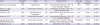
| Study | Publication year | Design | No. | Definition of CIM | Mean age (yr) | Time of assessment | CIM |
|---|---|---|---|---|---|---|---|
| Shin et al. (current study) | 2019 | Retrospective | 237 | Amenorrhea for 12 months + FSH > 40 IU/L + estradiol < 20pg/mL | 39.6 | 12 mon | 31 (13.1) |
| Moore et al. [12]* (POEMS study) | 2015 | Randomized controlled | 69 | Amenorrhea > 6 months + FSH in the postmenopausal range | 37.5 | 2 yr | 15 (22.0) |
| Tiong et al. [13] | 2014 | Retrospective and prospective | 102 | Amenorrhea for 12 months + FSH > 25.8 IU/L | 43.3 | 12 mon | 93 (91.2) |
| Song et al. [10]* | 2013 | Randomized controlled | 94 | Amenorrhea for 12 months + FSH > 40 IU/L + estradiol < 20pg/mL | 42.1 | 12 mon | 27 (28.7) |
| Del Mastro et al. [9]* (PROMISE-GIM6 study) | 2011 | Randomized controlled | 133 | Amenorrhea for 12 months + FSH and estradiol in the postmenopausal range | 39 (median) | 12 mon | 31 (25.6) |
| Badawy et al. [11] | 2009 | Randomized controlled | 39 | Amenorrhea + FSH > 40 IU/L | 29.2 | 8 mon | 21 (66.6) |
Values are presented as number (%).
CIM = chemotherapy-induced menopause; POEMS = Prevention Of Early Menopause Study; FSH = follicle stimulating hormone; PROMISE-GIM6 = PRevention Of Menopause Induced by chemotherapy: a Study in Early breast cancer patients-Gruppo Italiano Mammella 6.
*Data from the control arm of the clinical trial (women who did not use gonadotropin-releasing hormone agonist for fertility preservation).
![]()
CIM is expected to increase with age; however, there is paucity of evidence regarding its actual frequency. Only one randomized controlled trial showed CIM frequencies of 57%, 95%, and 97.9% in women aged < 35, 35−45, and > 45 years, respectively [13]. The higher frequencies observed in that study, compared to the present study, could be attributed to the lower cutoff values used for FSH (25.8 IU/L instead of 40 IU/L). Moreover, only 54 women were evaluated for CIM. Our study has a major advantage over this study because we included 237 patients, and FSH and estradiol levels were measured for all these patients at 12 months after the completion of chemotherapy.
As shown in our study, the frequency of CIM was significantly lower than that of TRA in all age groups (Table 2). Although TRA has been used to evaluate ovarian function in the majority of studies in the past, return of menses is a poor surrogate for ovarian reserve or ovarian failure [21]. Ovulation and spontaneous pregnancy have been reported in women with long-term amenorrhea and premature ovarian failure [22]. Meanwhile, patients with regular, albeit short, menstrual periods may have markedly diminished ovarian reserve, even years before menopause [2324]. The discrepancy between CIM and TRA should be well acknowledged, and future studies should use biochemical markers to evaluate ovarian reserve.
In multivariate analysis, age was a significant predictor of TRA, which was consistent with previous studies [1319]. In the present study, a specific chemotherapy regimen (AC-D or AC-T regimen) significantly affected the frequency of TRA but not the frequency of CIM. It should be noted that the duration of chemotherapy is not independent of the chemotherapy regimen, and hence, it may be inappropriate to compare just the regimen or the number of chemotherapy cycles separately. Both the AC-D and AC-T regimens require 8 cycles of administration, whereas both FAC and CMF require only 6 cycles, in addition to the different doses of cyclophosphamide used. In multivariate analysis, we found that CIM and TRA were not influenced by the chemotherapy regimen or the number of chemotherapy cycles.
The majority of patients were receiving tamoxifen hormone therapy at the time of assessment, and it was associated with a decreased frequency of CIM. Although tamoxifen itself was unlikely to affect the ovarian reserve [25], changes in the FSH and estradiol levels after adjuvant hormone therapy have been previously reported, and in one study, tamoxifen exerted different effects according to the menopausal status, with elevated FSH in premenopausal women and decreased FSH in postmenopausal women [1526]. The mechanism underlying the influence of tamoxifen on TRA/CIM remains unclear. In premenopausal women, elevated plasma estradiol levels that interfere with the hypothalamic–ovarian feedback loop have been reported [23]. Direct interaction of tamoxifen with granulosa cells, increased estrogen production due to increased FSH concentrations, or modified LH receptor expression may also occur [2627]. The use of tamoxifen after chemotherapy was associated with changes in the menstrual pattern, including amenorrhea, in certain studies [2128], but not in others [2930]. In the present study, the TRA was not affected by tamoxifen use, which is consistent with the latter studies.
The parameter that should be used to identify menopausal women who receive chemotherapy for breast cancer and who can continue receiving tamoxifen is debatable. In the most recent version of NCCN Breast Cancer Guidelines, namely version 3.2019, the NCCN panel defined menopause as FSH and plasma estradiol level in the postmenopausal range if the woman is aged below 60 years and is taking tamoxifen [7]. Within the same guideline, they stated that menopausal status cannot be assigned to women who are receiving GnRH analogs. It is highly unlikely that the guideline has been developed without considering the notion of the possible hormonal fluctuations caused by tamoxifen. Until a new definition of menopause is incorporated in the guidelines, it would be reasonable to use this definition not only for research purposes but also in clinical practice. Among the several parameters, the authors in the current study selected FSH and estradiol, as recommended by the NCCN Guideline.
Our study has some limitations. Only patients who received chemotherapy were included, and there were no control subjects for comparison. The hormones FSH and estradiol were not assessed in the early follicular phase. Hence, comparing the absolute values of these hormones could be misleading, and these hormones were only used to distinguish premenopause and menopause. In order to evaluate the role of FSH and estradiol in patients receiving tamoxifen, another marker of ovarian reserve should have been used, which was not the case in this study. The level of AMH, which is a widely used marker to assess ovarian reserve, was available in only 45 patients, and the statistical power was too small to draw a definite conclusion regarding the difference in AMH levels between women with and without CIM. Reproductive outcome is the most reliable marker of ovarian function; however, this information could not be ascertained from the medical records. Hence, further research is required to determine the reproductive outcome.
In this study, we present the frequency of CIM and TRA after chemotherapy in Korean women with breast cancer. Our findings would provide physicians with a practical estimate of the risk of CIM and TRA in patients who do not receive GnRH agonists for gonadal protection.
 XML Download