

 PDF
PDF ePub
ePub Citation
 Print
Print


Abstract
Purpose
Case summary
Figures and Tables
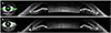
 | Figure 1Anterior segment photographs of the first visit. Right eye (A) and left eye (B) showed conjunctival injection and dilated pupils. Right eye (C) and left eye (D) showed shallow anterior chamber and mild nuclear sclerosis with anterior bowing of the iris.
|
 | Figure 2Anterior segment optical coherence tomography (AS-OCT) on the 5 days after the first visit. AS-OCT of the right eye (A) and the left eye (B). The scleral spurs are noted (arrows).
|
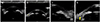 | Figure 3Images of ultrasound biomicroscopy of the right (A) and the left (B) eye on the 5 days after the first visit. The magnified view of the nasal side of the right eye (C) and the nasal side of the left eye (D). Crystalline lens moved anteriorly and anterior chamber depth and iridocorneal angle were decreased in both eyes. Ciliary effusion is observed in the left eye (yellow asterisk and arrow), while it is not obvious in the right eye.
|
Table 1
Summary of case reports of AACC induced by SNRIs
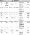
AACC = acute angle closure crisis; SNRIs = serotonin-norepinephrine reuptake inhibitors; IOP = intraocular pressure; F = female; OD = right eye; LI = laser iridotomy; OS = left eye; IV = intravenous; OU = both eyes; M = male; ACG = angle closure glaucoma; N-C = not checkable.
*Time intervals between the first drug intake and symptom onset; †other than age and sex; ‡angle closure crisis associated with venlafaxine treatment; only presented as a poster at 10th European Glaucoma Society Congress; §acute angle closure crisis induced by duloxetine, only presented as a poster at 2012 European Association for Vision and Eye Research Conference.
![]()
Notes
This study was presented as a poster at the 120th Annual Meeting of the Korean Ophthalmological Society 2018.
 XML Download