

 PDF
PDF ePub
ePub Citation
 Print
Print


Abstract
Case summary
A 32-year-old male presented with visual disturbance in both eyes. He had been diagnosed with testicular cancer and had undergone right orchiectomy 4 months prior. He completed adjuvant chemotherapy 2 weeks before presentation. His best-corrected visual acuities were 20/35 in both eyes. Fundus examination revealed multiple flame-shaped hemorrhages around the posterior pole, and boat-shaped hemorrhages on the macula in both eyes. Laboratory results showed that he had reduced hemoglobin (5.5 g/dL) and platelet counts (77,000/µL). After transfusion, visual acuities were improved and retinal hemorrhages were resolved along with normalization of the hematological conditions.
Conclusions
Many ocular and medical conditions can cause bilateral retinal hemorrhages. This case emphasizes that comprehensive history evaluation, systemic evaluation, and laboratory findings, as well as a detailed fundus examination, are important in the diagnosis of patients with bilateral retinal hemorrhages.
Figures and Tables
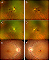 | Figure 1Fundus photographs at the initial presentation (A, B), at 1 month follow-up visit (C, D) and at 3 months follow-up visit (E, F). Fundus examination revealed multiple flame-shaped hemorrhages at the posterior pole in both eyes and premacular boat-shaped hemorrhage in the left eye (A, B). Retinal hemorrhages almost resolved in the right eye and small residual premacular hemorrhage remained in the left eye (E, F).
|
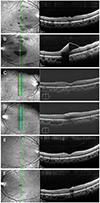 | Figure 2Spectral domain-optical coherence tomography (SD-OCT) images at the initial presentation (A, B), at 1 month follow-up visit (C, D) and at 3 months follow-up visit (E, F). Vertical macular scans of SD-OCT demonstrated premacular sub-internal limiting membrane (ILM) hemorrhage and multiple intra-retinal nerve fiber layer hemorrhage in both eyes. There was no definite outer retinal abnormality except optical shadowing effect due to hemorrhage (A, B). Vertical scans of SD-OCT demonstrated a small amount of sub-ILM hemorrhage on the macula in both eyes (E, F).
|
References
1. Carraro MC, Rossetti L, Gerli GC. Prevalence of retinopathy in patients with anemia or thrombocytopenia. Eur J Haematol. 2001; 67:238–244.
2. Duke-Elder S, Dobree J. Duke-Elder Sm, editor. System of ophthalmology (diseases of the retina). St. Louis: CV Mosby Co.;1967. p. 373–407.
3. Rubenstein RA, Yanoff M, Albert DM. Thrombocytopenia, anemia, and retinal hemorrhage. Am J Ophthalmol. 1968; 65:435–439.
4. Foulds W. The ocular manifestations of blood diseases. Trans Ophthalmol Soc U K. 1963; 83:345–367.
5. Kawai K, Ando S, Hinotsu S, et al. Completion and toxicity of induction chemotherapy for metastatic testicular cancer: an updated evaluation of Japanese patients. Jpn J Clin Oncol. 2006; 36:425–431.
6. Kacer B, Hattenbach LO, Hörle S, et al. Central retinal vein occlusion and nonarteritic ischemic optic neuropathy in 2 patients with mild iron deficiency anemia. Ophthalmologica. 2001; 215:128–131.
7. Celebi S, Kükner A. Photodisruptive Nd: YAG laser in the management of premacular subhyaloid hemorrhage. Eur J Ophthalmol. 2001; 11:281–286.
 XML Download