

 PDF
PDF ePub
ePub Citation
 Print
Print


Abstract
Purpose
We present four cases of welding arc maculopathy as observed using spectral-domain optical coherence tomography (SD-OCT).
Case summary
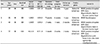
Four patients, who performed welding without wearing protective eye gear, presented to the hospital due to poor visual acuity. The mean visual acuity of the patients was 0.6. Fundus photographs of the four patients revealed a yellowish retinal scar at the fovea. SD-OCT images of the four patients showed photoreceptor inner segment/outer segment junction (IS/OS junction) disruption and retinal pigment epithelium injury. We diagnosed the patients with welding arc maculopathy, and three of them were treated with oral steroids or antioxidants. The IS/OS junctions were restored in two patients, who had short welding arc exposures. The disrupted IS/OS junction recovered partially in one of the other two patients, who had a longer duration of exposure, and the IS/OS junction disruption remained in another patient.
Figures and Tables
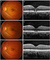 | Figure 1(Patient 1) Fundus photograph (A) and SD-OCT image obtained (B) at the first ophthalmic examination showing a yellowish retinal scar in the fovea and IS/OS junction disruption, hyperreflective band in the outer nuclear layer and RPE injury in the left eye. Fundus photograph (C) and SD-OCT image (D) after 1 month of treatment showing IS/OS junction restoration, but the retinal scar at the fovea and RPE injury persisted in the left eye. Fundus photograph (E) and SD-OCT image (F) after 1 year of treatment showing complete restoration of the IS/OS junction. SD-OCT = spectral domain optical coherence tomography; IS/OS junction = photoreceptor inner segment/outer segment junction; RPE = retinal pigmented epithelium.
|
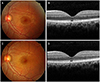 | Figure 2(Patient 2) Fundus photograph (A) and SD-OCT image (B) at the first ophthalmic examination showing yellowish retinal scar in the fovea and IS/OS junction disruption and RPE injury in the left eye. Fundus photograph (C) and SD-OCT image (D) after 1 year of treatment showing persistent IS/OS junction disruption, retinal scar in the fovea, and RPE injury. SD-OCT = spectral domain optical coherence tomography; IS/OS junction = photoreceptor inner segment/outer segment junction; RPE = retinal pigmented epithelium.
|
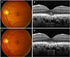 | Figure 3(Patient 3) Fundus photograph (A) and SD-OCT image (B) at the first ophthalmic examination showing yellowish retinal scar in the fovea and IS/OS junction disruption, hyperreflective band in the outer nuclear layer and RPE injury in the left eye. Fundus photograph (C) and SD-OCT image (D) after 1 years of treatment showing grater improvement in the IS/OS junction, RPE injury and retinal scar in the fovea. SD-OCT = spectral domain optical coherence tomography; IS/OS junction = photoreceptor inner segment/outer segment junction; RPE = retinal pigmented epithelium.
|
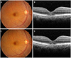 | Figure 4(Patient 4) Fundus photograph (A) and SD-OCT image (B) at the first ophthalmic examination showing yellowish retinal scar in the fovea and IS/OS junction disruption, RPE injury and hyporeflextive cystic lesion in the cone outer segment tips (interdigitation zone) in the right eye. Fundus photograph (C) and SD-OCT image (D) taken after 3 weeks, showing partially restored IS/OS junction and hyporeflextive cystic lesion with a persistent retinal scar in the fovea and RPE injury. SD-OCT = spectral domain optical coherence tomography; IS/OS junction = photoreceptor inner segment/outer segment junction; RPE = retinal pigmented epithelium.
|
References
1. Kim YJ, Chung IY, Kim SJ, et al. A case of maculopathy from handheld green laser pointer. J Korean Ophthalmol Soc. 2015; 56:447–451.
2. Choi SW, Chun KI, Lee SJ, Rah SH. A case of photic retinal injury associated with exposure to plasma arc welding. Korean J Ophthalmol. 2006; 20:250–253.
3. Karp KO, Flood TP, Wilder AL, Epstein RJ. Photic maculopathy after pterygium excision. Am J Ophthalmol. 1999; 128:248–250.
4. Ruiz-del-Río N, Moriche-Carretero M, Ortega-Canales I, et al. Photic maculopathy and iris damage in a psychotic patient. Arch Soc Esp Oftalmol. 2006; 81:165–168.
5. Maier R, Heilig P, Winker R, et al. Welder's maculopathy? Int Arch Occup Environ Health. 2005; 78:681–685.
6. Kim YK, Lee HK, Lee JH. An experimental study of the corneal epithelial damage by electric welding light on the rabbit cornea. J Korean Ophthalmol Soc. 1988; 29:61–67.
7. Stefaniotou M, Katsanos A, Kaloudis A, et al. Spectral-domain optical coherence tomography in lightening-induced maculopathy. Ophthalmic Surg Lasers Imaging. 2012; 43:E35–E37.
8. Stokkermans TJ, Dunbar MT. Solar retinopathy in a hospital based primary care clinic. J Am Optom Assoc. 1998; 69:625–636.
9. Würdemann HV. The formation of a hole in the macular: light burn from exposure to electric welding. Am J Ophthalmol. 1936; 19:457–460.
10. Yang X, Shao D, Ding X, et al. Chronic phototoxic maculopathy caused by welding arc in occupational welders. Can J Ophthalmol. 2012; 47:45–50.
11. Park DW, Alonzo B, Faridi A, Bhavsar KV. Multimodal imaging of phototic maculopathy from arc welding. Retin Cases Brief Rep. 2018; 09. 26. DOI: 10.1097/ICB.0000000000000823. [Epub ahead of print].
12. Zhang C, Dang G, Zhao T, et al. Predictive value of spectral-domain optical coherence tomography features in assessment of visual prognosis in eyes with acute welding arc maculopathy. Int Ophthalmol. 2019; 39:1081–1088.
13. Magnavita N. Photoretinitis: an underestimated occupational injury? Occup Med (Lond). 2002; 52:223–225.
14. Hirsch DR, Booth DG, Schocket S, Sliney DH. Recovery from pulsed-dye laser retinal injury. Arch Ophthalmol. 1992; 110:1688–1689.
15. Zwick H, Stuck BE, Dunlap W, et al. Accidental bilateral Q-switched neodymium laser exposure: treatment and recovery of visual function. In BiOS'98 International Biomedical Optics Symposium. Int Soc Opt Photonics. 1998; 3254:80–89.
 XML Download