

 PDF
PDF ePub
ePub Citation
 Print
Print


Abstract
Purpose
To present the first report describing lesions of osteoma cutis on the upper eyelid and medial canthus.
Case summary
A 4-year-old female complained of a right upper eyelid mass. The examination showed a well-delineated, mild bluish-colored, hard mass over the upper eyelid and the medial canthus measuring 10 × 10 mm and 2 × 2 mm. During the cutaneous examination, her forearm, left shin, right dorsum of the foot, neck, and abdominal wall also showed well-delineated, mild bluish-colored, immobile hard masses, similar to the upper eyelid mass. A right upper eyelid and medial canthus mass excision was performed and a biopsy specimen was collected. Hematoxyline and eosin staining showed a mature bone in the dermis with spicules of bone and osteoblasts. She was finally diagnosed with osteoma cutis on the upper eyelid and the medial canthus.
Figures and Tables
Figure 1
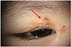
Anterior segment photographs of the mass and clinical photograph of patient at inital presentation. (A) There was well marginated, mild bluish-colored, hard mass was seen on right eyelid (arrow) and medial canthus (arrowhead). (B) The child showed rounded chubby face and flat nose. Informed consent was obtained from the study participant.
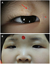
Figure 2
Orbit computed tomography scan with enhancement image. That showed bone density well-defined lesions in right eyelid (arrow) and medial cahthus (arrowhead). Axial view.
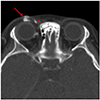
Figure 3
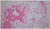
Overview dissection of biopsy of eyelid mass. Histopathology of the tissue biopsy demonstrated spicules of bone (red arrow) in the dermis and subcutaneous tissue (haematoxylin and eosin stain, ×12.5). Hair follicle (red arrowhead) and eccrine sweat gland (black arrowhead) are shown in dermis.
Notes
References
1. Roth S, Stowell RE, Helwige B. Cutaneous ossification. Report of 120 cases and review of the literature. Arch Pathol. 1963; 76:44–54.
2. Bang HD, Kim KH, Cho KH. A case of primary osteoma cutis. Annals of Dermatology. 1996; 8:121–124.
3. Mast AM, Hansen R. Multiple papules on the elbows. Arch Dermatol. 1997; 133:780.
4. Boyd AS, King LE Jr. Basal cell carcinoma with ossification. J Am Acad Dermatol. 1998; 38:906–910.
5. Gupta SR, Cogen MS, Metz TH Jr. Congenital osteoma cutis of the lateral canthus. J AAPOS. 2009; 13:410–412.
6. Fazeli P, Harvell J, Jacobs MB. Osteoma cutis (cutaneous ossification). West J Med. 1999; 171:243–245.
7. Monroe AB, Burgdorf WH, Sheward S. Platelike cutaneous osteoma. J Am Acad Dermatol. 1987; 16(2 Pt 2):481–484.
8. Jung JN, Cho YH, Seo JH, et al. Osteoma cutis in Albright's hereditary osteodystrophy. Korean J Dermatol. 2004; 42:493.
9. Kapoor S, Gogia S, Paul R, Banerjee S. Albright’s hereditary osteodystrophy. Indian J Pediatr. 2006; 73:153–156.
10. Prendiville JS, Lucky AW, Mallory SB, et al. Osteoma cutis as a presenting sign of pseudohypoparathyroidism. Pediatr Dermatol. 1992; 9:11–18.
11. Ryu DJ, Oh SH, Han EC, et al. A case of osteoma cutis, a diagnostic clue for albright's hereditary osteodystrophy. Korean J Dermatol. 2009; 47:435–438.
12. Aguinaga F, Trope B, Piñeiro-Maceira J, Ramos-e-Silva M. Miliary osteoma cutis: a case report. Case Reports in Dermatological Medicine. 2014; 2014:347829.
13. Hernandez-Martin A, Perez-Mies B, Torrelo A. Congenital Platelike Osteoma Cutis in an Infant. Pediatr Dermatol. 2009; 26:479–481.
14. Moreira Amorim G, Mastrangelo Marinho Falcão EMMF, Carvalho Quintella D, et al. Primary isolated osteoma cutis of the face. Dermatol Online J. 2017; 23:13030/qt4zz8d3tm.
15. Sanmartin O, Alegre V, Martinez-Aparicio A, et al. Congenital platelike osteoma cutis: case report and review of the literature. Pediatr Dermatol. 1993; 10:182–186.
 XML Download