

 PDF
PDF ePub
ePub Citation
 Print
Print


Abstract
Purpose
We investigated changes in postoperative refractive error after surgery to treat congenital ptosis and the clinical outcomes by surgical method.
Methods
The study was retrospective and interventional. We enrolled 73 patients in whom 86 eyes exhibited visual axis-obscuring congenital ptosis. All patients were under 8 years of age, with refractive errors or amblyopia, and underwent maximal levator resection or frontalis sling surgery with fascia lata preservation from January 2008 to January 2018; the minimum follow-up time was 6 months. Visual and surgical outcomes were assessed by reviewing clinical photographs taken before and 1 year after surgery. Refractive error changes were measured at these times.
Results
Maximal levator resection was performed on 42 of 86 eyes (48.8%) and frontalis sling surgery with preservation of the fascia lata on 44 eyes, 95.2% and 75.0% of patients, respectively, exhibited good or fair surgical outcomes. The preoperative mean astigmatisms of the ptotic and control eyes of those with unilateral disease did not differ significantly: −0.71 ± 0.85 D for ptotic eyes and −0.66 ± 0.97 D for control eyes. The mean astigmatism increased from −0.71 ± 0.85 D preoperatively to −1.27 ± 1.2 D postoperatively (p < 0.001). The postoperative MRD1 value correlated with the increase in postoperative astigmatism (p = 0.022, r = −0.261).
Conclusions
Maximal levator resection tended to afford better surgical outcomes than frontalis sling surgery with preservation of the fascia lata in patients with congenital ptosis. Patients in whom the postoperative eyelid position was good tended to exhibit higher refractive errors. Careful examination and treatment are recommended to ensure good visual outcomes.
Figures and Tables
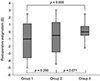 | Figure 1Postoperative astigmatism of patients divided into three groups: the highest (group 1), middle (group 2), and lowest (group 3) tertile of postoperative 1 year margin reflex distance 1 (MRD1). The patient group with the highest postoperative MRD1 tends to have higher astigmatism than the lowest one does (p = 0.005). D = diopters.
|
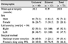

Table 3
Surgical results of frontalis sling surgery versus maximal levator resection in congenital ptosis patients
![]()
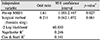
Table 4
Multivariate logistic regression analysis of facters affecting surgical outcomes in congenital ptosis patients
![]()


References
1. Anderson RL, Baumgartner SA. Amblyopia in ptosis. Arch Ophthalmol. 1980; 98:1068–1069.
2. Beneish R, Williams F, Polomeno RC, et al. Unilateral congenital ptosis and amblyopia. Can J Ophthalmol. 1983; 18:127–130.
3. Mokhtarzadeh A, Harrison AR. Controversies and advances in the management of congenital ptosis. Expert Rev Ophthalmol. 2015; 10:59–63.
4. Wagner RS, Mauriello JA Jr, Nelson LB, et al. Treatment of congenital ptosis with frontalis suspension: a comparison of suspensory materials. Ophthalmology. 1984; 91:245–248.
5. McNeil NL. Patterns on visual defects in children. Br J Ophthalmol. 1955; 39:688–701.
6. Harrad RA, Graham CM, Collin JR. Amblyopia and strabismus in congenital ptosis. Eye (Lond). 1988; 2 ( Pt 6):625–627.
7. Hornblass A, Kass LG, Ziffer AJ. Amblyopia in congenital ptosis. Ophthalmic Surg. 1995; 26:334–337.
8. Merriam WW, Ellis FD, Helveston EM. Congenital blepharoptosis, anisometropia, and amblyopia. Am J Ophthalmol. 1980; 89:401–407.
9. Anderson RL, Baumgartner SA. Strabismus in ptosis. Arch Ophthalmol. 1980; 98:1062–1067.
10. Cadera W, Orton RB, Hakim O. Changes in astigmatism after surgery for congenital ptosis. J Pediatr Ophthalmol Strabismus. 1992; 29:85–88.
11. Kao SC, Tsai CC, Lee SM, Liu JH. Astigmatic change following congenital ptosis surgery. Zhonghua Yi Xue Za Zhi (Taipei). 1998; 61:689–693.
12. Kim SK, Yoon JR, Chang HK. The clinical study of 33 cases of congenital blepharoptosis. J Korean Ophthalmol Soc. 1995; 36:1636–1642.
13. So JY, Woo KI, Chang HR. The amblyopia in congenital ptosis. J Korean Ophthalmol Soc. 2001; 42:1747–1752.
14. Oral Y, Ozgur OR, Akcay L, et al. Congenital ptosis and amblyopia. J Pediatr Ophthalmol Strabismus. 2010; 47:101–104.
15. Wang Y, Xu Y, Liu X, et al. Amblyopia, strabismus and refractive errors in congenital ptosis: a systematic review and meta-analysis. Sci Rep. 2018; 8:8320.
16. Crawford JS. Repair of ptosis using frontalis muscle and fascia lata: a 20-year review. Ophthalmic Surg. 1977; 8:31–40.
17. Lee JH, Aryasit O, Kim YD, et al. Maximal levator resection in unilateral congenital ptosis with poor levator function. Br J Ophthalmol. 2017; 101:740–746.
18. Mauriello JA, Wagner RS, Caputo AR, et al. Treatment of congenital ptosis by maximal levator resection. Ophthalmology. 1986; 93:466–469.
19. Press UP, Hübner H. Maximal levator resection in the treatment of unilateral congenital ptosis with poor levator function. Orbit. 2001; 20:125–129.
20. Gazzola R, Piozzi E, Vaienti L, Wilhelm Baruffaldi Preis F. Therapeutic algorithm for congenital ptosis repair with levator resection and frontalis suspension: results and literature review. Semin Ophthalmol. 2018; 33:454–460.
21. Hersh D, Martin FJ, Rowe N. Comparison of silastic and banked fascia lata in pediatric frontalis suspension. J Pediatr Ophthalmol Strabismus. 2006; 43:212–218.
22. Philandrianos C, Galinier P, Salazard B, et al. Congenital ptosis: Long-term outcome of frontalis suspension using autogenous temporal fascia or fascia lata in children. J Plast Reconstr Aesthet Surg. 2010; 63:782–786.
23. Ho YF, Wu SY, Tsai YJ. Factors associated with surgical outcomes in congenital ptosis: a 10-year study of 319 cases. Am J Ophthalmol. 2017; 175:173–182.
24. Ormond AW. Notes on three cases of acquired astigmatism associated with meibomian cysts. Br J Ophthalmol. 1921; 5:117–118.
25. Robb RM. Refractive errors associated with hemangiomas of the eyelids and orbit in infancy. Am J Ophthalmol. 1977; 83:52–58.
26. Uğurbaş SH, Zilelioğlu G. Corneal topography in patients with congenital ptosis. Eye (Lond). 1999; 13 ( Pt 4):550–554.
27. Savino G, Battendieri R, Riso M, et al. Corneal topographic changes after eyelid ptosis surgery. Cornea. 2016; 35:501–505.
28. Klimek DL, Summers CG, Letson RD, Davitt BV. Change in refractive error after unilateral levator resection for congenital ptosis. J AAPOS. 2001; 5:297–300.
29. Kumar S, Chaudhuri Z, Chauhan D. Clinical evaluation of refractive changes following brow suspension surgery in pediatric patients with congenital blepharoptosis. Ophthalmic Surg Lasers Imaging. 2005; 36:217–227.
30. Byard SD, Sood V, Jones CA. Long-term refractive changes in children following ptosis surgery: a case series and a review of the literature. Int Ophthalmol. 2014; 34:1303–1307.
31. Lee DS, Kim JM, Woo KI, Chang HR. Changes in astigmatism after surgery for congenital ptosis. J Korean Ophthalmol Soc. 2006; 47:1459–1464.
 XML Download