

 PDF
PDF ePub
ePub Citation
 Print
Print


Abstract
Methods
The medical records of patients who were presumed to have ocular ischemic syndrome by ophthalmic examination, and confirmed by carotid artery imaging, were retrospectively reviewed from 2010 to 2017.
Results
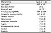
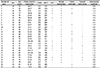
A total of 22 patients and 27 eyes were included in the study. Twenty patients were male. The average age was 64.2 years. Fifteen patients had hypertension and fifteen patients had diabetes mellitus. Twenty-one patients presented with acute visual impairment at the initial visit. The average best-corrected visual acuity (BCVA) was a LogMAR of 0.89 ± 0.65, with an average IOP of 16.1 ± 6.9 mmHg. Elevated IOP > 21 mmHg was noted in five eyes (18.5%). Iris neovascularization was the most common (13 eyes, 48.1%) feature in the anterior segment. Retinal hemorrhage was the most common feature in the fundus examination (23 eyes, 85.2%). The average central submacular thickness was 255.0 µm, and there was no macular edema except for one case with vitreomacular traction syndrome on optical coherent tomography.
Figures and Tables
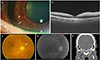 | Figure 1Representative case of ocular ischemic syndrome patient (case 10). (A) Anterior photo showed neovascularization of the iris (white arrows). (B) Optical coherent tomography showed hyperreflectivity suggesting inner retina ischemia, but there was no macular edema. (C) Fundus photo showed dot-shaped retina hemorrhage (white arrow) and neovascularization (red arrow). (D) Fluorescein angiography showed retinal artery filling delay (20 seconds). (E) Computed tomography angiography showed total occlusion of the right internal carotid artery (blue arrow).
|
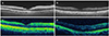 | Figure 2Optical coherence tomography of 4 representative cases with ocular ischemic syndrome patient at the initial visit. (A–C) There was no macular edema, and the choroid thickness was thinner than the normal eye. (D) Only 1 of 27 eyes showed macular edema. The macular thickening might be originated from the vitreomacular traction.
|
References
1. Kearns TP, Hollenhorst RW. Venous-stasis retinopathy of occlusive disease of the carotid artery. Proc Staff Meet Mayo Clin. 1963; 38:304–312.
2. Mendrinos E, Machinis TG, Pournaras CJ. Ocular ischemic syndrome. Surv Ophthalmol. 2010; 55:2–34.
3. Sivalingam A, Brown GC, Magargal LE, Menduke H. The ocular ischemic syndrome. II. Mortality and systemic morbidity. Int Ophthalmol. 1989; 13:187–191.
4. Han YS, Yoo WS, Chung IY, Park JM. Ocular ischemic syndrome successfully treated with carotid angioplasty and stenting. J Korean Ophthalmol Soc. 2010; 51:447–452.
5. Kim JW, Yoon IN. Acute visual loss after intravitreal bevacizumab injection in a patient with ocular ischemic syndrome. J Korean Ophthalmol Soc. 2012; 53:1893–1897.
6. Lee JH, Moon HS, Nam DH, Lee DY. Treatment of acute central retinal artery occlusion with ocular ischemic syndrome. J Korean Ophthalmol Soc. 2014; 55:1242–1247.
7. Lee SM, Lee JW. A case of neovascular glaucoma secondary to ocular ischemic syndrome in a patient with moyamoya disease. J Korean Ophthalmol Soc. 2012; 53:1712–1717.
8. Yamamoto T, Mori K, Yasuhara T, et al. Ophthalmic artery blood flow in patients with internal carotid artery occlusion. Br J Ophthalmol. 2004; 88:505–508.
9. Brown GC, Brown MM, Sharma S. The Ocular Ischemic Syndrome. In : Arévalo JF, editor. Retinal and Choroidal Manifestations of Selected Systemic Diseases. 1st ed. London: Springer;2014. chap. 24.
10. Sarkies NJ, Shilling JS, Russell RW. Fluorescein angiography in carotid disease. Trans Ophthalmol Soc U K. 1986; 105 ( Pt 4):489–493.
11. Taylor DC, Strandness DE Jr. Carotid artery duplex scanning. J Clin Ultrasound. 1987; 15:635–644.
12. Nederkoorn PJ, van der Graaf Y, Hunink MG. Duplex ultrasound and magnetic resonance angiography compared with digital subtraction angiography in carotid artery stenosis: a systematic review. Stroke. 2003; 34:1324–1332.
13. Koelemay MJ, Nederkoorn PJ, Reitsma JB, Majoie CB. Systematic review of computed tomographic angiography for assessment of carotid artery disease. Stroke. 2004; 35:2306–2312.
14. Brown GC, Magargal LE. The ocular ischemic syndrome. Clinical, fluorescein angiographic and carotid angiographic features. Int ophthalmol. 1988; 11:239–251.
15. Ino-ue M, Azumi A, Kajiura-Tsukahara Y, Yamamoto M. Ocular ischemic syndrome in diabetic patients. Jpn J Ophthalmol. 1999; 43:31–35.
16. Terelak-Borys B, Skonieczna K, Grabska-Liberek I. Ocular ischemic syndrome - a systematic review. Med Sci Monit. 2012; 18:Ra138–Ra144.
17. Mizener JB, Podhajsky P, Hayreh SS. Ocular ischemic syndrome. Ophthalmology. 1997; 104:859–864.
18. Sharma S. Ocular ischemic syndrome. Can Fam Physician. 1999; 45:901–909.
19. Chen CS, Miller NR. Ocular ischemic syndrome: review of clinical presentations, etiology, investigation, and management. Compr Ophthalmol Update. 2007; 8:17–28.
20. Choromokos EA, Raymond LA, Sacks JG. Recognition of carotid stenosis with bilateral simultaneous retinal fluorescein angiography. Ophthalmology. 1982; 89:1146–1148.
21. Dhooge M, de Laey JJ. The ocular ischemic syndrome. Bull Soc Belge Ophtalmol. 1989; 231:1–13.
22. Kerty E, Eide N, Horven I. Ocular hemodynamic changes in patients with high-grade carotid occlusive disease and development of chronic ocular ischaemia. II. Clinical findings. Acta Ophthalmol Scand. 1995; 73:72–76.
23. Kang HM, Lee CS, Lee SC. Thinner subfoveal choroidal thickness in eyes with ocular ischemic syndrome than in unaffected contralateral eyes. Graefes Arch Clin Exp Ophthalmol. 2014; 252:851–852.
24. Kim DY, Joe SG, Lee JY, et al. Choroidal thickness in eyes with unilateral ocular ischemic syndrome. J Ophthalmol. 2015; DOI: 10.1155/2015/620372.
 XML Download