

 PDF
PDF ePub
ePub Citation
 Print
Print


Abstract
Purpose
Methods
Results
Conclusions
Figures and Tables
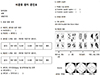 | Figure 1The characters of floaters. Questionnaire detailing their symptoms include onset, duration of symptoms, floaters with flash and associated another visual symptoms (e.g., headache, whiteout) and past medical histories had been performed. The characters of floaters were categorized by the pictures classified into four groups from the previous independent study of van Overdam et al6 (with permission of JAMA Ophthalmology).
|
Table 1
Comparison of demographics, previous eye status and past medical history between patients with retinal breaks and control
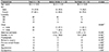
Values are presented as mean ± standard deviation or number (%) unless otherwise indicated.
OD = oculus dexter; OS = oculus sinister; OU = oculus unitas; D = diopters; DM = diabetes mellitus.
*Statistical significance was determined using the Fisher exact test, chi-square test from cross-table analysis; †odd ratio, 4.48; ‡statistical significance was determined using the t-test.
![]()
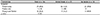
Table 2
Comparison of characteristics of vitreous floater symptoms
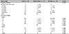
Values are presented as number (%) unless otherwise indicated.
*Statistically significance is p < 0.05 chi-square test from cross-table analysis; †group A indicates 1 to 3 floaters; group B, 4 to 10 floaters; group C, more than 10 floaters; and group D, a curtain or cloud; ‡odd ratio, 26.6; §statistically significance is p < 0.05 chi-square test and Fisher's exact test from cross-table analysis.
![]()

 XML Download