

 PDF
PDF ePub
ePub Citation
 Print
Print


Abstract
Purpose
To compare the in vitro toxicity of commercial olopatadine anti-allergic ophthalmic agents on cultured rabbit's conjunctival cells according to concentrations and exposure times.
Methods
Rabbit conjunctival cells were exposed to anti-allergic olopatadine ophthalmic agents, (Patanol® [0.1% olopatadine hydrochloride; Alcon, Fort Worth, TX, USA], Pataday® [0.2% olopatadine hydrochloride; Alcon], and Pazeo® [0.7% olopatadine hydrochloride; Alcon]) at concentrations of 5%, 10%, and 15% for periods of 30 minutes, 1, 2, 3, and 6 hours, respectively. Cell proliferation and injury assays were performed using the methylthiazoltetrazolium and lactate dehydrogenase (LDH) leakage assays. We checked the composition of the three anti-allergic agents, and performed light and transmission electron microscopy to compare the morphological changes in cells.
Results
The conjunctival cell proliferation was inhibited after 1 hour exposure to each olopatadine ophthalmic agent, with significant cell proliferation inhibited using 15% of each drug. The proliferation of conjunctival cells was inhibited during 6 hours of drug exposure at all concentrations of Pataday® and Pazeo®. The titer of LDH increased from 3 hours after drug exposure, but 15% Pazeo® significantly increased the LDH titer at 2 hours after drug exposure. As the concentration of the drug increased, the LDH titer also significantly increased. The cellular morphological changes of conjunctival cells were in the increasing order of Pazeo®, Pataday®, and Patanol® with a high concentration of olopatadine hydrochloride.
References
1. Bielory L. Allergic and immunologic disorders of the eye. Part I: immunology of the eye. J Allergy Clin Immunol. 2000; 106:805–16.

2. Bielory L. Therapeutic targets in allergic eye disease. Allergy Asthma Proc. 2001; 22:25–8.
3. Kidd M, McKenzie S, Steven I, et al. Efficacy and safety of abdominal eye drops in the treatment of seasonal allergic conjunctivitis. Br J Ophthalmol. 2003; 87:1206–11.
4. Abelson MB, Ferzola NJ, McWhirter CL, Crampton HJ. Efficacy and safety of single‐ and multiple‐ dose ketotifen fumarate 0.025% ophthalmic solution in a pediatric population. Pediatr Allergy Immunol. 2004; 15:551–7.
5. Kempuraj D, Asadi S, Zhang B, et al. Mercury induces inflammatory mediator release from human mast cells. J Neuroinflammation. 2010; 7:20.
6. Yamaguchi M, Azuma H, Fujihara M, et al. Generation of a considerable number of functional mast cells with a high basal level of Fcɛ RI expression from cord blood CD34+ cells by co‐ culturing them with bone marrow stromal cell line under serum‐ free conditions. Scand J Immunol. 2007; 65:581–8.
7. Yanni JM, Miller ST, Gamache DA, et al. Comparative effects of topical ocular anti-allergy drugs on human conjunctival mast cells. Ann Allergy Asthma Immunol. 1997; 79:541–5.
8. Byon IS, Park JS, Lee JE, Lee JS. The effects of abdominal ophthalmic agents on the cultured rabbit conjunctival cells. J Korean Ophthalmol Soc. 2006; 47:1340–8.
9. Mosmann T. Rapid colorimetric assay for cellular growth and abdominal: application to proliferation and cytotoxicity assays. J Immunol Methods. 1983; 65:55–63.
10. Kim SJ, Yoon TJ, Lee JE, Lee JS. Comparison of the efficacy of topical antihistamine/mast cell stabilizers in vitro. J Korean Ophthalmol Soc. 2010; 51:406–17.
11. Wee WR, Wang XW, McDonnell PJ. Effect of artificial tears on cultured keratocytes in vitro. Cornea. 1995; 14:273–9.
12. Park Y. Physiology of body fluid. Kang DH, editor. Physiology. 5th ed.Seoul: Seoul;2000. chap. 5.
13. Kim K. Acid-base balance. Sung HK, KIM KH, editors. Physiology. 5th ed.Seoul: Seoul;1996. p. 326–7.
14. Freshney R. The culture environment: substrate, gas phase, medium and temperature. Freshney R, editor. Culture of animal cells: a manual of basic technique. 3rd ed.New York: New York;1994. p. 71.
15. Burstein NL, Klyce SD. Electrophysiologic and morphologic abdominals of ophthalmic preparations on rabbit cornea epithelium. Invest Ophthalmol Vis Sci. 1977; 16:899–911.
16. Pfister RR, Burstein N. The effects of ophthalmic drugs, vehicles, and preservatives on corneal epithelium: a scanning electron microscope study. Invest Ophthalmol. 1976; 15:246–59.
17. Lemp MA, Zimmerman LE. Toxic endothelial degeneration in abdominal surface disease treated with topical medications containing benzalkonium chloride. Am J Ophthalmol. 1988; 105:670–3.
18. Green K, Tonjum A. Influence of various agents on corneal permeability. Am J Ophthalmol. 1971; 72:897–905.
19. Lee JS, Oum BS, Kim CD. Cytotoxicity of benzalkonium chloride on the corneal epithelial cell of rabbit. J Korean Ophthalmol Soc. 1998; 39:1326–33.
Figure 1.
Metabolic activities of rabbit's epithelial cells as determined by Methylthiazoltetrazolium assay after 30 minutes or 1, 2, 3 or 6 hours of treatment with Patanol® (0.1% olopatadine; Alcon, Fort Worth, TX, USA), Pataday® (0.2% olopatadine; Alcon) or Pazeo® (0.7% olopatadine; Alcon) in concentrations of 5, 10 and 15%. * p < 0.05 by student's t-test.
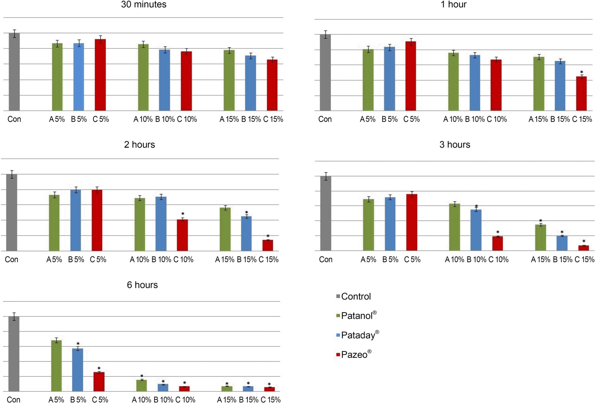
Figure 2.
LDH levels started to increase rapidly after 3 hours in groups exposed to 5%, 10% and 15% Patanol® (0.1% olopatadine; Alcon, Fort Worth, TX, USA), Pataday® (0.2% olopatadine; Alcon) or Pazeo® (0.7% olopatadine; Alcon). Only 15% concentration of Pazeo® increased LDH titer after 2 hours. * p < 0.05 by analysis of LDH = lactate dehydrogenase. variance.
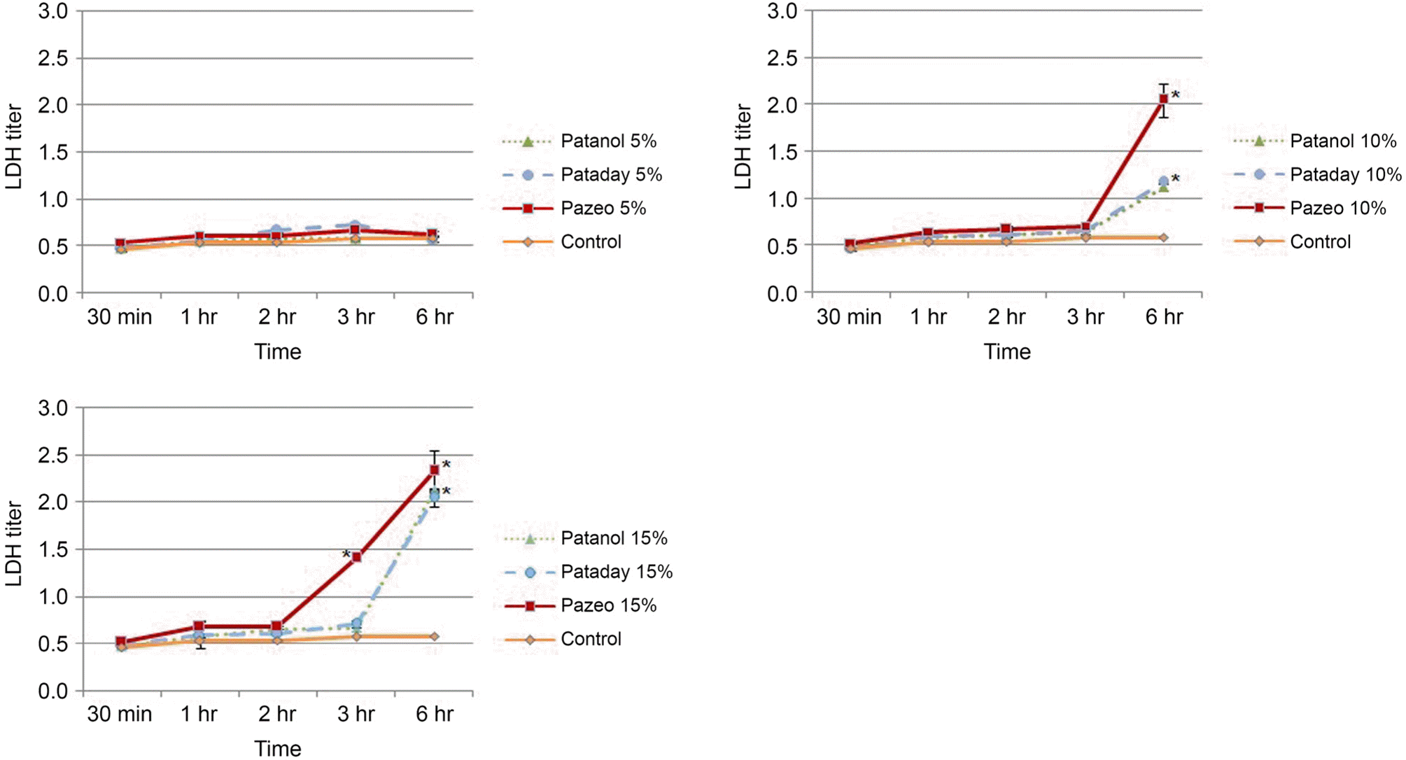
Figure 3.
Inverted light microscopy of rabbit conjunctival cells taken after 3 hours of exposure to three agents at 10% concentration (×200). (A) Control, (B) 10% Patanol® (Alcon, Fort Worth, TX, USA), (C) 10% Pataday® (Alcon), (D) 10% Pazeo® (Alcon). Conjunctival cells of control maintained elongated, spindle-shaped appearance. But the number of cells in (B) and (C) was decreased, and (D) showed even less.
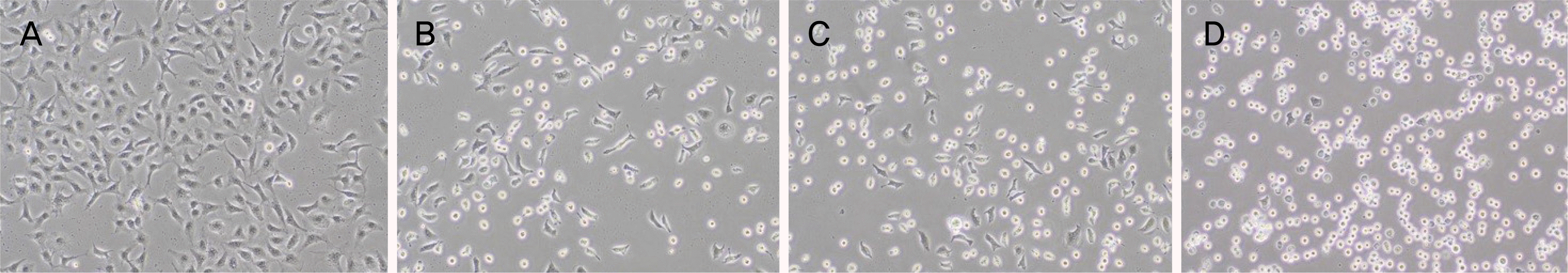
Figure 4.
Transmission electron micrography of rabbit conjunctival cells taken after 3 hours of exposure to three agents at 10% concentration (bar length 2 μ m, origianl magnification, ×8,000). (A) Control, (B) 10% Patanol® (Alcon, Fort Worth, TX, USA), (C) 10% Pataday® (Alcon), (D) 10% Pazeo® (Alcon). Conjunctival cells exposed to olopatadine showed loss of microvilli, vacuole formation, and disrupted nuclear membrane.
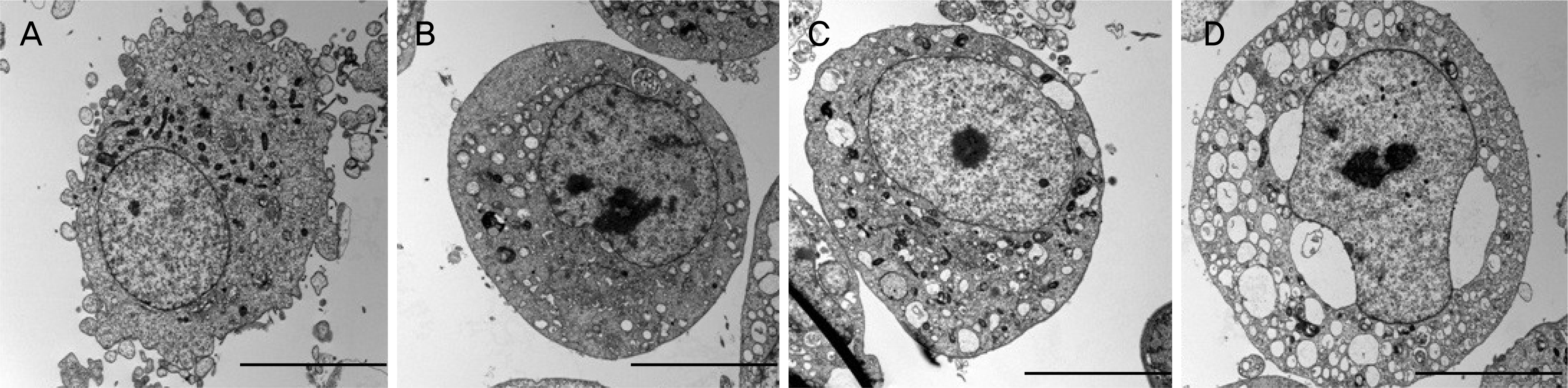
Table 1.
The ingredient, electrolyte composition, pH, osmolality and preservatives of the three olopatadine agents
 XML Download