

 PDF
PDF ePub
ePub Citation
 Print
Print


INTRODUCTION
Gastric cancer is one of the most common malignancies in Asian countries, including Korea. Gastric cancer patients in Korea now benefit from better prognoses due to early diagnosis, improved surgical techniques, and perioperative management [123]. Hence, the quality of life has gained importance due to the extended life expectancies in patients undergoing gastrectomy for gastric cancer. However, some patients further experience gallbladder (GB) stones or combined cholecystitis, necessitating additional surgical management during the follow-up period [45678]. The etiology or precise mechanism of GB stone formation after gastrectomy for gastric cancer is yet to be discovered, with few studies investigating this issue, even in Korea with its high prevalence of gastric cancer. Previously reported risk factors for GB stone formation after gastrectomy included patient sex, age, decreased body mass index (BMI), or type of gastrectomy in accordance with Paik et al. [9] and Park et al. [10], and included variables regarding the implementation of duodenal exclusion during reconstruction or pre-existing diabetes mellitus (DM). Other studies have shown that extensive lymph node dissection is associated with an increased incidence of GB stone formation after gastric cancer surgery [11121314]. Further information and evidence are crucial to establish prophylactic management techniques to mitigate or prevent additional abdominal interventions such as cholecystectomy. The goal of this study was to evaluate the incidence of GB stone formation and investigate the risk factors leading to cholecystectomy after gastrectomy in gastric cancer.
MATERIALS AND METHODS
Patients
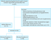
A total of 917 patients underwent gastrectomy for gastric cancer performed by a single surgeon at the Division of Stomach Surgery, Asan Medical Center between August 2012 and December 2015. The exclusion criteria included a history of cholecystectomy (n=20), concomitant cholecystectomy during gastrectomy (n=82), segmental resection of stomach (n=47), palliative gastrectomy (n=13), neoadjuvant chemotherapy performed prior to gastrectomy (n=18), combined resection of intraabdominal organs due to multi-primary cancer (n=19), and body weight not checked at routine follow-up (n=157) (Fig. 1). Among the 561 patients without any of the exclusion criteria, 36 presented with GB stone formation after gastrectomy for gastric cancer. GB stones were detected by evaluation via perioperative scans using ultrasonography (USG) or abdominal computed tomography (CT). The characteristics evaluated were patient sex, age, preoperative height, weight, BMI, body weight change in the first 6 months after gastrectomy, history of previous abdominal operation, previous diagnosis with liver cirrhosis or DM, location of tumor, operation duration, type of surgery (open or laparoscopic), type of gastrectomy and reconstruction (with or without excluded duodenum), extent of lymph nodes dissection, excision of other adjacent organs, presence of postoperative complications, T/N/M staging using the American Joint Committee on Cancer tumor, node, and metastasis (TNM) classification and staging system for gastric cancer, 7th Edition (2010), implementation of adjuvant chemotherapy, preoperative serum cholesterol level, serum alkaline phosphatase (ALP) level, and serum total bilirubin level.
Fig. 1
Algorithm of enrolled patients and each outcome. The figure shows the algorithm of enrolled patients and each outcome including the exclusion criteria of the study.
GB = gallbladder.
All included patients underwent either open or laparoscopic surgery for distal gastrectomy (DG), total gastrectomy (TG), or pylorus-preserving gastrectomy (PPG) with regional and/or systemic lymph nodes dissection. After gastrectomy, patients also underwent reconstruction with Billroth I (gastroduodenostomy) or Billroth II (gastrojejunostomy) anastomosis, Roux-en-Y or uncut Roux-en-Y for DG, Roux-en-Y esophagojejunostomy for TG, or gastrogastrostomy for PPG. USG and CT scans were used to detect GB stones and related cholecystitis during the follow-up period. Additionally, these imaging studies, mainly used to monitor recurrence or metastasis after gastrectomy, were also useful for detecting GB stones.
Statistics
The cumulative incidence of GB stone formation after gastrectomy was assayed by the Kaplan-Meier method, with differences between each group evaluated by the log-rank test. Categorical variables were compared using the Fisher's exact test and Pearson χ2 test. Continuous variables were examined using the Student's t-test, and the mean values were assessed using the Mann-Whitney U test. To evaluate risk factors for GB stone formation after gastrectomy in gastric cancer, univariate and multivariate analyses were conducted using the Cox proportional hazards model. P-values less than 0.05 were considered statistically significant. The receiver operating characteristic analysis and Youden's index were used to find the optimal cut-off value. All statistical calculations were performed using IBM SPSS ver. 25 for Windows (IBM Corporation, Armonk, NY, USA).
Ethics statement
The study was approved by the Institutional Review Board (IRB) of the Asan Medical Center and the University of Ulsan College of Medicine. (No. 2019-0465). All procedures were followed in accordance with the ethical standards of the responsible committee on human experimentation (institutional and national) and with the Helsinki Declaration of 1964 and later versions. Informed consents from patients for this retrospective study were waived by the IRB.
RESULTS
The characteristics of the 561 patients are shown in Table 1. The median follow-up period was 32.7 months. Among the 561 patients enrolled, 357 were men (63.6%), and 204 were women (26.4%). Thirty-six patients presented with the formation of GB stones, presenting an incidence rate of 6.4% for GB stone formation after gastrectomy for gastric cancer (Fig. 2A). The mean interval between gastrectomy and diagnosis of GB stone formation was 21.9 months. The incidence rate of GB stone formation was significantly higher in patients over 63 years of age. (15.7% vs. 6.7%, P=0.003; Fig. 2B).
Table 1
Characteristics of patients
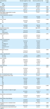
Values are presented as mean±standard deviation or number (%).
GB = gallbladder; BMI = body mass index; DM = diabetes mellitus; DG = distal gastrectomy; TG = total gastrectomy; PPG = pylorus-preserving gastrectomy; GD = gastroduodenostomy; GG = gastrogastrostomy; GJ = gastrojejunostomy; EJ = esophagojejunostomy; ALP = alkaline phosphatase; TNM = tumor, node, and metastasis. *According to the 7th Union for International Cancer Control/American Joint Committee on Cancer TNM system.
Fig. 2
Graph showing the incidence rate of GB stone formation after gastrectomy for gastric cancer.
(A) Kaplan-Meier analysis of the cumulative incidence of GB stone formation after gastrectomy for gastric cancer. (B) Incidence of GB stone formation after gastrectomy in patients over and under 63 years old (P=0.003). (C) Incidence of GB stone formation after gastrectomy in patients who lost under or over 6.2 kg in the 6 months after the surgery (P=0.002). (D) Incidence of GB stone formation after gastrectomy in patients who did or did not undergo adjuvant chemotherapy and (P=0.002). (E) Incidence of GB stone formation after gastrectomy in patients with relatively higher (≥0.5 mg/dL) or lower serum total bilirubin levels (<0.5 mg/dL) (P=0.003).
GB = gallbladder.
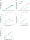
Initially, the effect of the degree of obesity on GB stone formation after gastrectomy was evaluated by comparing height, weight, BMI, and changes in body weight in the first 6 months after gastrectomy. No significant differences in height, weight, or BMI were uncovered. However, weight loss after gastrectomy was significantly associated with an increased risk of GB stone formation, with the incidence rate significantly higher in patients who lost more than 6.2 kg (15.5% vs. 7.4%, P=0.002; Fig. 2C). Patients who did not undergo postoperative chemotherapy also had a significantly higher incidence of GB stone formation than those who did (13.1% vs. 4.1%, P=0.002; Fig. 2D). The incidence of GB stone formation was significantly higher in patients with higher preoperative serum total bilirubin levels (13.4% vs. 3.5%, P=0.003; Fig. 2E); the cut-off value for serum total bilirubin level was 0.5 mg/dL.
No significant statistical differences in GB stone formation were observed for the other variables, with the results as follows: sex (P=0.100), height (P=0.188), weight (P=0.068), BMI (P=0.167), previous abdominal operation (P=0.779), liver cirrhosis (P>0.999), DM (P=0.214), operation time (P=0.275), open versus laparoscopic procedure (P=0.851), type of gastrectomy (P=0.410), type of reconstruction after gastrectomy (P=0.571), whether or not the duodenum was excluded (P=0.844), tumor location (P=0.206), N stage and M stage pathology (P=0.429 and P>0.999, respectively), combined resection of adjacent organs or not (P=0.069), postoperative complications (P=0.768), and preoperative serum cholesterol level and ALP levels (P=0.280 and P=0.637, respectively).
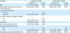
Variables associated with significant differences, revealed using univariate analyses, were then evaluated with multivariate analyses using the Cox proportional hazard model, which showed that patient age, weight loss in the first 6 months after the procedure, lack of postoperative chemotherapy, and elevated serum total bilirubin levels were significant risk factors for GB stone formation after gastrectomy for gastric cancer (Table 2).
Table 2
Univariate and multivariate analysis of risk factors for GB stone formation after gastrectomy
DISCUSSION
Gastric cancer is one of the most prevalent malignancies in Asia, and particularly in Korea. The quality of life in patients undergoing gastrectomy for gastric cancer has gained significance owing to extended life expectancies associated with early diagnosis and advanced management. Therefore, we retrospectively inspected patient data to discover potential risk factors for GB stone formation in patients after gastrectomy for gastric cancer in order to minimize the likelihood of patients needing additional interventions such as cholecystectomy in the future.
Unlike in previous studies, we observed that risk factors such as obesity, male gender, and exclusion of the duodenum during reconstruction after gastrectomy did not affect the incidence of GB stone formation [9]. Furthermore, no correlation was reported between the incidence of GB stone formation and patient history of DM with Roux-en-Y reconstruction after gastrectomy [11]. Some studies included TG, rather than partial gastrectomy, as a significant factor resulting in the higher incidence of GB stone formation [15]. Conversely, the current study revealed that certain other factors influenced the incidence of GB stone formation after gastrectomy for gastric cancer.
As shown in previous studies, this study confirmed that the incidence of GB stone formation was significantly higher in older patients regardless of whether they underwent gastrectomy or not. The rate of GB stone formation increases with each decade after 20 years of age due to an increased amount of cholesterol in the bile. Moreover, hemoperfusion of the GB wall decreases with age due to sclerotic changes, contributing to GB dysfunction [161718]. The same is probably true in case of patients undergoing gastrectomy for gastric cancer.
Furthermore, patients who demonstrated greater body weight loss in the 6 months after gastrectomy reported a higher incidence of GB stone formation. Other studies have mentioned a correlation between obesity, as measured by BMI, and showed an association between high BMI and GB stone formation. However, we observed a significant effect associated with weight loss in the 6 months after gastrectomy rather than a particular BMI value. One of the major pathophysiologic mechanisms of GB stone formation is the hypo-motility of the gallbladder, resulting in slower gallbladder emptying, and leading to the precipitation of cholesterol crystals via unstable vesicles [16192021]. This mechanism can be observed in patients who lose a great deal of body weight in a shorter period of time. As many obese patients lost a significant amount of weight after gastrectomy for gastric cancer, this study suggests a correlation between significant fluctuations in body weight and a higher incidence of GB stone formation [22].
Interestingly, this study revealed that patients with relatively high levels of preoperative serum total bilirubin presented a higher incidence of GB stone formation after gastrectomy. We propose that the high levels of bilirubin, a major source of GB stones, easily crystallize, especially into brown or black pigmented stones regardless of antecedent gastrectomy. This same principle of GB stone formation might account for the association between high preoperative serum bilirubin levels and higher incidence of GB stone formation after gastrectomy for gastric cancer. To avoid any redundant management such as cholecystectomy following GB stone formation after gastrectomy, it is recommended that patients reduce their serum bilirubin level with ursodeoxycholic acid prior to surgery [23].
There are some limitations to this study. Firstly, this was a retrospectively designed study based on data from a single center. To confirm these outcomes, a prospectively designed study using multicenter data is necessary. Also, the critical serum total bilirubin level resulting in a higher incidence of GB stone formation after gastrectomy was 0.5 mg/dL, which is of little practical significance as the normal serum bilirubin levels ranges from 0.2 to 1.2 mg/dL. In our study, while patients with GB stone formation demonstrated relatively higher serum total bilirubin levels, several patients were within the normal range. We also lacked an adequate population to evaluate potential correlations between postoperative chemotherapy and the incidence of GB stone formation after gastrectomy for gastric cancer. In the current study, significant differences with regard to the incidence of GB stone formation after gastrectomy and adjuvant chemotherapy were uncovered. Thirty-six patients reported GB stone formation; however only 3 out of the total 561 enrolled patients underwent postoperative chemotherapy. Previous studies have suggested that there is no correlation between these 2 factors. However, our results conflicted with previous reports [24]. Next, we will attempt to resolve these differences in an upcoming investigation using a larger population.
In conclusion, multivariate analyses in this study revealed that older patients, patients demonstrating a significant loss in body weight during the first 6 months after gastrectomy, patients receiving no postoperative adjuvant chemotherapy, and patients with relatively high preoperative serum total bilirubin levels present a greater risk for GB stone formation after gastrectomy for gastric cancer. Therefore, special attention should be paid to patients with these indicating risk factors.
 XML Download