

 PDF
PDF ePub
ePub Citation
 Print
Print


INTRODUCTION
Gastric cancer constitutes a major global health problem, as it remains the fifth most frequently diagnosed cancer worldwide and the third leading cause of cancer-related deaths [1]. Surgery is the only treatment option with a potential to cure patients with early-stage gastric cancer. However, only half of these patients undergo complete resection of their primary tumor and, in most cases, relapse with metastatic disease occurs few years after surgery [2]. In this regard, combined modality therapies have been shown to significantly increase survival in gastric cancer patients in this setting.
There are some differences in the therapeutic management of locally advanced gastric and esophagogastric junction cancer between Western and Eastern countries. Currently, in Western countries, perioperative systemic therapy with 5-fluorouracil, leucovorin, oxaliplatin, and docetaxel (FLOT) is new standard of care for gastric cancer andthe CROSS regimen is the standard treatment for neoadjuvant chemoradiotherapy in esophagogastric junction cancer [34]. This discrepancy is the basis for the ongoing ESOPEC study comparing the CROSS and FLOT regimens in non-metastatic esophageal or esophagogastric junction adenocarcinoma [5].
In Eastern countries, adjuvant chemotherapy (S-1 or capecitabine with oxaliplatin) is the standard of care for patients with pathological stage II or III after curative sub-total or total gastrectomy with D2 lymphadenectomy [6]. Indeed, 2 randomized phase III trials conducted in Asian population showed an overall survival (OS) increase in patients with resectable gastric cancer who received adjuvant chemotherapy after curative surgery with D2 lymphadenectomy [78].
In Western countries, perioperative chemotherapy and preoperative chemoradiotherapy increase the OS in patients with locally advanced gastric and esophagogastric junction adenocarcinoma and are currently regarded as the standard treatment [49].
In order to further improve the advantage given by this strategy, a number of clinical studies are currently exploring the combination of systemic therapies with targeted and immunotherapeutic agents already approved for the treatment of patients with metastatic disease.
These clinical trials constitute a modern approach for improving the prognosis of gastric cancer patients, treating the potential metastatic disease by taking advantage of the patients' better performance status prior to surgery. Most importantly, these randomized clinical studies represent a unique opportunity for developing predictive biomarkers that could be useful for selecting patients who would benefit the most from a given treatment, reducing the risk of unnecessary toxic therapy and postponing a potentially curative surgery.
It is of utmost importance to incorporate the evaluation of potential biomarkers in these trials, corroborated by extensive preclinical and translational research evidence. The aim of this review article is to present the most promising biomarkers of response to classic chemotherapeutic, targeted, and immunotherapeutic agents that can be potentially useful for personalized preoperative systemic therapies in gastric cancer patients (Table 1).
Table 1
Potential novel biomarkers for the prediction of response to preoperative systemic therapies
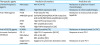
| Therapeutic agents | Predictive biomarkers | Predictive role | |
|---|---|---|---|
| Chemotherapeutic agents | MSI status | MSI-H [14151617] | Resistance to platinum-based chemotherapy |
| BIRC3 | High BRIC3 expression [27] | Resistance to chemoradiotherapy | |
| Anti-HER2 agents | PTEN | PTEN loss [464748] | Resistance to trastuzumab and/or lapatinib |
| AMNESIA panel | EGFR/MET/KRAS/PI3K/PTEN mutations and EGFR/MET/KRAS amplifications [49] | ||
| NRF2 | High NRF2 expression [54] | ||
| MET | MET amplification [55] | ||
| FGFR3 | High FGFR3 expression [58] | ||
| Anti-VEGF(R) agents | HOXB9 | HOXB9-positive [74] | Resistance to bevacizumab (in CRC) |
| Immune checkpoint inhibitors | PD-L1 | High PD-L1 expression [9091] | Response to anti-PD-1 |
| MSI-status | MSI-H [8490] | ||
| EBV | EBV-positive [90] | ||
| Epigenomic promoter | Epigenomic promoter alterations [93] | Resistance to anti-PD-1 | |
MSI = microsatellite instability; MSI-H = microsatellite instability-high; BIRC = baculoviral inhibitor of apoptosis repeat containing; PTEN = phosphatase and tensin homolog; EGFR = epidermal growth factor receptor; PI3K = phosphoinositide 3-kinases; NRF2 = nuclear factor erythroid 2-related factor 2; FGFR = fibroblast growth factor receptor; HOXB9 = homeobox B9; VEGF(R) = vascular endothelial growth factor (receptor); CRC = colorectal cancer; PD-L1 = programmed death-ligand 1; EBV = Epstein–Barr virus; PD-1 = programmed death-1.
PREDICTIVE BIOMARKERS OF RESPONSE TO CLASSICAL CHEMOTHERAPEUTIC AGENTS
Currently, no predictor of response to classical chemotherapeutic agents has been prospectively validated. In the largest effort to determine genetic predictors of response to systemic therapies in esophageal and gastric cancer, tumor tissue from 187 patients affected with HER2-negative esophageal cancer and receiving first-line treatment with a fluoropyrimidine plus platinum combination was analyzed by using MSK-IMPACT, a capture-based, new generation sequencing platform that can detect mutations, copy-number alterations, and rearrangements. The analysis showed that no single mutant allele or gene, including those with a role in DNA repair pathways such as BRCA1/2, was significantly associated with treatment response. In particular, not even large-scale state transition—a surrogate marker for defects in homologous recombination—was associated with response to platinum-based chemotherapy in this patients' cohort [10].
More recently, compelling evidence has pointed to microsatellite instability (MSI) as a potential predictive factor of response to chemotherapy in gastric cancer [111213]. In this regard, a retrospective analysis of the MAGIC trial evaluated the association between mismatch repair (MMR) deficiency (MMR-D) and MSI with survival in patients with resectable esophageal and gastric cancer randomly allocated to receive surgery alone or perioperative combination polichemotherapy with epirubicin, cisplatin and fluoropyrimidine. The analysis was conducted only on tumor tissues from patients undergoing surgery. Only 6.7% of the cases had MSI-high (MSI-H) tumors and all of them were gastric cancer. Interestingly, none of the patients with an MSI-H tumor treated with chemotherapy demonstrated a significant response as a tumor regression grade of 1 or 2 in the resection specimen. Conversely 16.3% of patients with microsatellite stable (MSS) or MSI-low (MSI-L) tumors treated with chemotherapy had a tumor regression grade of 1 or 2. Among patients allocated to receive upfront surgery, those with MSI-H tumors had a longer survival than those with MSS/MSI-L or MMR proficient (MMR-P) tumors. On the other hand, patients with MSI-H had a shorter OS than patients with MSS/MSI-L or MMR-P tumors when treated with perioperative chemotherapy plus surgery [14].
A potential predictive role of MSI status for resistance to chemotherapeutic agents is emerging also in the adjuvant setting. A recent post hoc analysis of the CLASSIC trial, an open-label, phase III, randomized controlled trial that demonstrated the benefit of adjuvant capecitabine plus oxaliplatin after D2 gastrectomy over D2 gastrectomy alone for stage II/III gastric cancer [15], showed that adjuvant chemotherapy improved disease-free survival (DFS) in the MSS group (5-year DFS: 66.8% vs. 54.1%; P=0.002), but no benefit was observed in the MSI-H group (5-year DFS: 83.9% vs. 85.7%; P=0.931) [16]. Consistently, MSI was independently associated with DFS and OS in a recent individual patient data meta-analysis of resectable gastric cancer patients enrolled in MAGIC, CLASSIC, ARTIST, and ITACA-S randomized clinical trials [17]. Collectively, these evidences strongly highlight MSI-H status as a predictive biomarker for resistance to chemotherapeutic agents in gastric cancer; thus, perioperative systemic therapies might be spared in this patient population. Omission of perioperative chemotherapy should be prospectively investigated in MSI-H gastric cancer patients.
Our research group recently contributed to this field by exploring the role of the transforming growth factor (TGF)-β activated kinase 1 (TAK1, also called MAP3K7) pathway—a unique Smad-independent TGF-β intracellular signaling pathway—in the resistance of esophagogastric junction carcinoma to preoperative chemoradiotherapy. TAK1 is a serine/threonine kinase acting as a central hub for the regulation of apoptosis by integrating the most relevant signals from various cytokines—including interleukin (IL)-1, TGF-β, and tumor necrosis factor α [1819]—and controlling, in turn, the activation of different transcription factors, including activator protein 1 (AP-1) and nuclear factor kappa B (NF-κB) [2021]. Activation of the transcriptional activities of NF-κB and AP-1 sustains a potent resistance to chemotherapeutic treatments through the expression of several genes, including baculoviral inhibitor of apoptosis (IAP) repeat containing (BIRC) 3 [22]. BIRC3 encodes for the cellular IAP 2 protein [23], a member of the IAP family that inhibits apoptosis by directly inhibiting the caspases cascade [2425]. Initially, we demonstrated that targeting the kinase activity of TAK1 dramatically led to a proapoptotic phenotype and, in turn, to a significantly higher sensitivity to chemotherapy in models of pancreatic cancer [26]. More recently, we demonstrated that the TAK1-regulated expression of BIRC3 is responsible for the resistance of distal esophageal and esophagogastric junction carcinoma to the proapoptotic effect of chemoradiotherapeutic treatments [27]. We measured an elevated expression of BIRC3 in distal esophageal adenocarcinoma cell lines that was regulated by TAK1 kinase activity. Downregulating BIRC3 expression through the inhibition of TAK1 sensitized esophageal adenocarcinoma cell lines to the proapoptotic activity of radiotherapy and classic chemotherapeutic agents. In order to translate our findings into the clinical setting and evaluate the potential of BIRC3 expression to predict chemoradioresistance in esophageal and esophagogastric junction cancers, we measured the expression levels of BIRC3 mRNA in pretreatment biopsies from 32 patients with adenocarcinoma and 33 patients with squamous cell carcinoma treated with a preoperative schedule including weekly docetaxel and cisplatin, continuous infusion of 5-fluorouracil (5-FU) and concomitant radiotherapy. Initially, we demonstrated a significantly lower expression of BIRC3 in the more sensitive population of patients affected by squamous cell carcinoma than in those affected by adenocarcinoma. Most importantly, tumor expression levels of BIRC3 could not distinguish between sensitive or resistant esophageal squamous cell carcinoma, but it significantly discriminated patients with sensitive or resistant adenocarcinoma. In particular, pathologic complete response was achieved in 6 out of 11 (54.5%) patients with a BIRC3 expression lower than the cutoff threshold, and only in 4 out of 21 (19.0%) patients BIRC3 expression was higher than the cutoff. Thus, we demonstrated BIRC3 gene expression is a potential biomarker for identifying patients with adenocarcinoma of the distal esophagus and of the esophagogastric junction that could most likely benefit from preoperative chemoradiotherapy [27].
The results of this study led to the design of BoRgEs trial. This is an investigator-initiated, randomized multicenter phase II trial of activity and safety of radiochemotherapy treatment guided by the expression of BIRC3 in patients with resectable adenocarcinoma of the distal esophagus or esophagogastric junction (cT2-4 N0 or any T N +). In the experimental arm, patients with BIRC3 expression on tumor tissues higher than the cutoff for resistance were selected for upfront surgery, while patients with BIRC3 expression lower than the cutoff were selected for standard neoadjuvant chemoradiotherapy according to the CROSS regimen [4]. Patients in the calibration arm will not be selected based on the biomarker expression, but will receive standard neoadjuvant chemoradiotherapy followed by surgery. The primary objective of the study is the R0 resection rate. The secondary objectives are DFS, OS, and tumor response rate.
PREDICTIVE MARKERS TO ANTI HER2 AGENTS
HER2 is a tyrosine kinase receptor belonging to the epidermal growth factor receptor (EGFR) family, and is encoded by the pro-oncogene ERBB2 (avian erythroblastosis oncogene B) located at chromosome 17q2. Activation of HER2 predominantly occurs through the binding to its ligand involving hetero- or homo-dimerization with another member of the HER family [28]. The oncogenic activity of HER2 is mainly due to gene amplification and less frequently to activating mutations. When HER2 is mutated or amplified, its activation also occurs in a ligand-independent manner [29]. The transduction of the signal of HER2 occurs through 2 main pathways: the RAS-mitogen activated protein kinase and the phosphoinositide 3-kinases (PI3K)-AKT pathway [30]. The amplification of ERBB2 leads to an increased expression of HER2 protein, which plays a pivotal role in cell proliferation and survival in several types of cancer, including gastric cancer [31]. In gastric cancer, HER2 amplification is more often associated with proximal tumors (34%) compared to distal forms (20%) [32]. Similarly, the expression of HER2 is more frequent in intestinal subtypes (about 34%) than in diffuse subtypes (approximately 6%) [33]. In the TCGA classification, tumors with chromosomal instability more often express HER2 as consequence of gene amplification (24%) [34]. In clinical practice, there are several drugs targeting HER2, which can be distinguished into monoclonal antibodies and tyrosine kinase inhibitors.
Trastuzumab is a monoclonal antibody directed against the extracellular domain (IV) of HER2 [35]. In first line of therapy, treatment of metastatic gastric cancer patients with high expression of HER2 (IHC3+ or IHC2+/FISH+) with trastuzumab in addition to chemotherapy of a fluoropyrimidine (capecitabine or 5-FU) plus cisplatin has shown an advantage in OS compared to chemotherapy alone [36].
Pertuzumab is a monoclonal antibody which binds to the extracellular domain of HER2 (domain II) preventing its heterodimerization with other members of the HER family (HER1, HER3, and HER4). Pertuzumab and trastuzumab bind to a distinct extracellular domain of HER2, resulting in synergistic antiproliferative activity in vitro and in vivo [37]. Unfortunately, in patients with metastatic gastric cancer, the addition of pertuzumab to a first-line therapy containing trastuzumab, cisplatin, and fluoropyrimidine did not result in a statistically significant increase of OS [38].
Trastuzumab emtansine (TDM-1) is a HER2-targeted antibody-drug conjugate. The small molecule cytotoxin, DM1, is a microtubule inhibitor. This molecule has been studied as a second line treatment in metastatic gastric cancer patients, but TDM-1 therapy was not superior to standard taxane therapy [39]. Lapatinib is a multi-kinase inhibitor with an activity against EGFR, including HER2. In patients with metastatic gastric cancer, the addition of lapatinib to chemotherapy compared to chemotherapy alone has not shown an advantage in OS neither in first nor in second line therapy [4041].
While the role for anti-HER2 therapy for advanced gastric cancer is well established, its value is undetermined in locally advanced gastric cancer. Currently, at least 2 clinical trials are exploring the association of peri-operative chemotherapy in association with anti-HER2 agents in gastric and esophagogastric junction cancer: the EORTC INNOVATION (NCT02205047) and the AIO-FLOT6 PETRARCA (NCT02581462) studies (Table 2). The INNOVATION trial is a randomized, multicenter, open-label, phase II trial. This study is investigating whether adding trastuzumab or trastuzumab plus pertuzumab to a chemotherapy regimen containing fluoropyrimidine and cisplatin in a perioperative setting with HER2 positive gastric and esophagogastric junction carcinoma is able to increase the complete pathological response rate, compared to fluoropyrimidine and cisplatin chemotherapy alone (NCT02205047). The PETRARCA/FLOT6 trial is a multicenter, randomized, phase II/III trial. Patients with HER2-positive locally advanced gastric and esophagogastric junction carcinoma (≥cT2 any N or any T N-positive) can be enrolled. This trial is investigating whether adding trastuzumab plus pertuzumab to the FLOT chemotherapy regimen is able to increase the complete pathological response rate and progression-free survival (PFS) compared with FLOT chemotherapy alone (NCT02581462). Although pertuzumab did not increase OS in patients with HER2 amplified metastatic gastric cancer, the combination of trastuzumab with pertuzumab was associated with a higher rate of objective response [38]. The rationale of using pertuzumab and trastuzumab in the perioperative setting is based on the probability of increasing the rate of pathological response in patients with localized gastric and gastroesophageal cancers. These increase of tumor response rates could also translate into better surgical outcomes with improved R0 resection rates in patients with operable disease.
Table 2
Randomized controlled trial with perioperative anti-HER2 agents in HER2+ gastric cancer and EGJ carcinoma

| NCT number | Study name | Phase | Setting | Treatment | Primary endpoint |
|---|---|---|---|---|---|
| NCT02581462 | PETRARCA | II/III | Untreated GC and EGJ HER2+ ≥cT2 or cN+ | Perioperative 5-fluorouracil+leucovorin+docetaxel+oxaliplatin+trastuzumab/pertuzumab | pCR (phase II), PFS (phase III) |
| NCT02205047 | INNOVATION | II | Untreated GC and EGJ HER2+ stage Ib–III | Perioperative cisplatin+capecitabine/5-fluorouracil with or without trastuzumab or trastuzumab+pertuzumab | pCR |
In this regard, the identification of molecular biomarkers useful for selecting patients eligible for an anti HER2 strategy would be of particular interest. Several studies have shown that selection of non-amplified HER2 clones is a recurrent mechanism of trastuzumab resistance. The loss of amplified HER2 clones seems to be more frequent especially in patients with uncertain immunohistochemistry positivity (IHC2+) [42]. Furthermore, the loss of amplified HER2 clones may be due to tumor progression or tumor heterogeneity between the metastatic lesions and the primary tumor in the same patients [43].
Several studies investigated the association between PI3K and phosphatase and tensin homolog (PTEN) pathway aberrations and clinical outcomes in HER2 positive gastric cancer patients treated with anti-HER2 drugs. PTEN is a tumor suppressor gene coding for a protein member of the tyrosine phosphatase family, and is frequently mutated in human cancer [44]. PTEN gene is a negative regulator of PI3K signaling that, through phosphatidylinositol-3,4,5-triphosphate production, leads to the activation of the AKT pathway regulating cell proliferation, metabolism, and survival [45]. PTEN loss is frequently detected in HER2 amplified gastric cancer, and it is associated with a poor response to anti-HER2 therapy. In these patients, PTEN loss is associated with poor clinical outcomes, and shorter PFS and OS compared to those of patients with HER2 positive gastric cancer without a PTEN deficiency [4647]. In order to identify the correlation between PTEN and the PI3K pathway in HER2 positive gastroesophageal cancer, a recent study showed that PTEN loss, through the constitutive activation of AKT, leads to inhibition of apoptosis and G1 cell cycle arrest, resulting in trastuzumab resistance [48].
The AMNESIA study is a multicenter, prospective, case-control study, which has shown the negative predictive impact of a panel of genomic aberrations, including EGFR/MET/KRAS/PI3K/PTEN mutations and EGFR/MET/KRAS amplifications in HER2 positive metastatic gastric cancer patients eligible for first line therapy whit trastuzumab plus cisplatin and fluoropyrimidine chemotherapy. In this study, patients who experienced progression or stable disease within 4 months of starting treatment were defined as tumor-resistant, while patients who had a complete or partial response to treatment and subsequently experienced disease progression at least 3 months after the end of chemotherapy were defined as tumor-sensitive. The AMNESIA panel of alterations was statistically significantly more frequently in treatment of resistant patients (55%) than in sensitive (0%) ones (P<0.001). In addition, patients without panel alterations had longer PFS and OS than patients with panel alterations. The AMNESIA panel has a 76% predictive accuracy to identify patients with primary molecular resistance to trastuzumab [49].
Nuclear factor erythroid 2-related factor 2 (NRF2) is a transcription factor that, in physiological conditions, is involved in resistance to oxidative stress [50]. Previous studies showed that, in many cancer types, overexpression of NRF2 is associated with chemo- and radioresistance, a more aggressive tumor phenotype and positive correlations between level of expression of ribosomal protein S6 (RPS6) and PI3K/AKT/mTOR [515253]. In this scenario, a recent study reported that NRF2 through RPS6 is a primary resistance factor to trastuzumab and lapatinib. NRF2 and RPS6 were overexpressed in HER2-amplified gastric cancer cells selected for lapatinib and/or trastuzumab resistance. Silencing of RPS6 expression or pharmacological inhibition of PI3K/AKT/mTOR was associated with reduced cellular viability and expression level of NRF2, suggesting a pivotal role of this pathway in anti-HER2 drug resistance. Moreover, this study retrospectively analyzed a cohort of patients with HER2 amplified locally advanced or metastatic gastric cancer treated with trastuzumab in combination with platinum-based chemotherapy. In this analysis, patients with high NRF2 expression had worse prognosis than patients with low NRF2 expression [54]. These data point to NRF2 as one of the molecules implicated in resistance to anti-HER2 therapy and open a new scenario for identifying biomarkers.
Co-alterations in genes coding for proteins members of different tyrosine-kinase receptor intracellular pathways are emerging as relevant mechanisms of intrinsic and acquired resistance to anti-HER2 therapeutic strategies. Patients with these co-alterations have a lower benefit from trastuzumab therapy and a shorter PFS [10]. These patients can benefit from an inhibition of 2 driver targets. A recent study showed that 50% of esophagogastric junction cancer with MET amplifications were associated with HER2/EGFR amplifications in the same tumor cells. When the amplification of MET and HER2 were simultaneously present in the same tumor cells, no disease response was obtained with the single inhibition of HER2 or MET, while a considerable response was obtained with dual inhibition of MET and HER2 [55].
Fibroblast growth factor (FGF) receptor (FGFR) 3 is a member of a family of 4 tyrosine kinase receptors that is implicated in cellular growth, survival, and tumor angiogenesis [56]. FGF9 is one of the members of the heparin binding polypeptide ligands of the FGF family that has unique affinity for FGFR3 [57]. Our group recently identified that, in gastric cancer, the overexpression of FGFR3 and FGF9 sustains the acquired resistance to trastuzumab through the activation of the PI3K/AKT/mTOR signaling pathway. In this study, we found that gastric cancer models selected for resistance to trastuzumab, in addition to a loss of HER2 expression, were associated with overexpression of FGFR3, FGF9 and phosphorylated AKT. In in vivo murine models resistant to trastuzumab, treatment with dovitinib—a selective inhibitor of FGFR3—was associated with a greater reduction in tumor burden and an increase in OS compared to control. The clinical relevance of the overexpression of FGFR3 has also been confirmed in 3 patients of whom biopsies were available before treatment and after progression with trastuzumab. In all 3 patients, samples taken at progression with trastuzumab had significantly higher FGFR3 expression than their matched pretreatment samples [58].
These preclinical data led to the design of the FiGhTeR trial. This is an investigator-sponsored, phase II, single-arm, open-label trial that is currently assessing the safety, tolerability, and activity of Pemigatinib (INCB54828), an inhibitor of FGFR-1, -2 and -3, as a second-line treatment strategy in advanced esophagogastric junction and gastric cancer patients refractory to first-line trastuzumab-containing therapies.
PREDICTIVE MARKERS TO ANTIANGIOGENIC DRUGS
Angiogenesis is a necessary process for tumor progression and metastasis, and its inhibition is an important therapeutic strategy for many tumor types [59]. Vascular endothelial growth factor (VEGF) plays an important role in the development of gastric and esophagogastric junction cancers [6061]. Bevacizumab, a humanized monoclonal antibody targeting VEGF, failed to improve OS in first-line treatment of advanced gastric cancer [62]. Not even the combination of bevacizumab with perioperative combined chemotherapy of epirubicin, cisplatin, and capecitabine has demonstrated an improvement in OS compared with standard chemotherapy. On the contrary, the addition of bevacizumab was associated with a higher rate of anastomotic dehiscence [63].
Ramucirumab is a human IgG1 monoclonal antibody that binds to vascular endothelial growth factor receptor (VEGFR)-2 on the endothelial cells in blood vessels and limits angiogenesis by inhibiting VEGF ligand binding and receptor signaling [64]. In 2 studies of second-line treatment of metastatic gastric cancer, ramucirumab improved OS in monotherapy or in combination with paclitaxel [6566]. On the other hand, the combination of ramucirumab with platinum-based chemotherapy in first-line treatment of advanced gastric cancer has failed to determine a clinically significant advantage of OS [67].
Considering the clinical benefit of adding ramucirumab to second-line treatment, the ongoing RAMSES/FLOT7 study (NCT02581462) is investigating the addition of ramucirumab to FLOT chemotherapy in a perioperative setting (Table 3).
Table 3
Randomized controlled trial with perioperative antiangiogenic agent in HER2− gastric cancer and EGJ carcinoma

| NCT number | Study name | Phase | Setting | Treatment | Primary endpoint |
|---|---|---|---|---|---|
| NCT02661971 | RAMSES | II | Untreated GC and EGJ HER2− ≥cT2 or cN+ | Perioperative 5-fluorouracil+leucovorin+docetaxel+oxaliplatin with or without ramucirumab | OS |
A recently retrospective exploratory analysis of the REGARD study was aimed at identifying predictive biomarkers of ramucirumab response. In this study, none of the tested biomarkers—tumoral HER2 or VEGFR-2, serum VEGF-C and -D, and soluble VEGFR-1 and -3—were found to be predictive of response to ramucirumab. Although predictive biomarkers to select patients for ramucirumab treatment is still lacking, this study indicates that the benefit of ramucirumab may be more pronounced in patients with high VEGFR-2 tumor expression [68]. A similar analysis was performed in plasma of patients enrolled in the RAINBOW study. This study did not identify any predictive biomarkers to ramucirumab response [69].
In the last decade, our group contributed to the identification of mechanisms of resistance to anti-VEGF strategies in pancreatic and colon cancer. Initially, we demonstrated the overexpression of a signature of proinflammatory and proangiogenic factors—including angiopoietin-like protein (Angptl) 2 [70], ligands of CXC receptors (CXCR)1/2 such as CXCL1 and IL-8, IL-1α and β, and TGF-β—in bevacizumab-resistant preclinical models compared to their sensitive counterparts [71]. These factors indirectly sustained angiogenesis through the recruitment of CD11b+ myeloid cells and increased the aggressiveness of bevacizumab-resistant tumors by inducing an epithelial-to-mesenchymal transition [72]. The combined inhibition of IL-1, CXCR1/2, and TGF-β signaling pathways modulated in vivo bevacizumab resistance by reversing the epithelial-to-mesenchymal transition and inhibiting tumor infiltration of CD11b+ cells [73].
More recently, we focused our studies on homeobox (HOX) B9 (HOXB9), a member of the highly conserved HOX transcription factor gene family, which is critical for the regulation of the expression of Angptl2, CXCL1 and IL-8, IL-1, and TGF-β. We demonstrated HOXB9 is a crucial transcription factor in sustaining tumor resistance to bevacizumab, and that silencing its expression could be a promising approach to modulate this resistance. HOXB9-positive tumors were resistant to bevacizumab, whereas mice bearing HOXB9-negative tumors were cured by this agent. Silencing HOXB9 in bevacizumab-resistant models significantly reduced Angptl2, CXCL1, IL-8, and TGF-β levels, reverted their mesenchymal phenotype, and restored, in turn, sensitivity to bevacizumab. HOXB9 had no prognostic value in colon cancer patients treated with a first-line chemotherapeutic regimen not containing bevacizumab. However, patients affected by a HOXB9-negative tumor had a significantly longer PFS compared with those with an HOXB9-positive tumor if treated with a first-line regimen containing bevacizumab [74]. In this regard, we are currently exploring the potential role of HOXB9 in the resistance of gastric cancer to ramucirumab by sustaining the activation of the ANGPTL2–LILRB2 signaling pathway. Thus, targeting the HOXB9–ANGPTL2–LILRB2 axis could enhance the activity of ramucirumab. Moreover, the expression of HOXB9 might serve as potential biomarker for selecting patients with gastric cancer that are more likely to benefit from this therapeutic strategy.
PREDICTIVE MARKERS TO IMMUNOTHERAPEUTIC AGENTS
Programmed death (PD)-1 receptor is an inhibitory T-cell receptor that binds to programmed death-ligand 1 (PD-L1) and PD-L2 [75]. Their interaction determines the inhibition of the lymphocyte response and represents a known mechanism for tumoral immune escape [76]. In recent years, the use of these drugs has dramatically changed the prognosis of patients affected by some tumors, including melanoma and lung cancer [77]. Although the use of immunotherapy in gastric cancer is not present in clinical practice yet, there are preclinical and clinical data that suggest its potential beneficial role also in gastric and esophagogastric junction cancers [78].
Currently, there are 2 ongoing clinical trials of perioperative chemotherapy plus immunotherapy in gastric cancer and esophagogastric junction adenocarcinoma: KEYNOTE-585 (NCT03221426) and DANTE trail (NCT03421288). KEYNOTE-585 is a phase III, randomized, double-blind study that aims to investigate the added value of combining pembrolizumab with perioperative chemotherapy (cisplatin plus 5-FU) in previously untreated, resectable gastric and gastroesophageal adenocarcinoma. The DANTE trial is a randomized open-label, phase III study to evaluate the safety and efficacy of atezolizumab in combination with FLOT chemotherapy vs. FLOT alone for operable gastric and gastroesophageal adenocarcinoma (Table 4).
Table 4
Randomized controlled trial with perioperative immunotherapeutic agents in gastric cancer and EGJ carcinoma

| NCT number | Study name | Phase | Setting | Treatment | Primary endpoint |
|---|---|---|---|---|---|
| NCT03421288 | DANTE | III | Untreated GC and EGJ ≥cT2 or cN+ | Perioperative 5-fluorouracil+leucovorin+docetaxel+oxaliplatin with or without atezolizumab | PFS |
| NCT03221426 | KEYNOTE-585 | III | Untreated GC and EGJ ≥cT3 or cN+ | Perioperative cisplatin+capecitabine/5-fluorouracil with or without pembrolizumab (safety cohort: perioperative 5-fluorouracil+leucovorin+docetaxel+oxaliplatin with or without pembrolizumab | pCR |
In gastric cancer the main predictors of response to immunotherapy appear to be the tumor expression of PD-L1 and the MSI or positive Epstein–Barr virus (EBV) status. In fact, in gastric cancer, the MSI-H subtype is associated with a high tumor mutation burden [79], while the EBV subtype is characterized by a rich intra- or peritumoral immune cell infiltration and high PD-L1/PD-L2 expression [80]. These biological features are positive predictive factors of response to immunotherapy [818283].
Some retrospective analyses have evaluated PD-L1 expression and MSI and EBV status in gastric cancer samples. In a series of 41 resected gastric cancer, 7 patients were EBV+, 16 were MSI-H and 21 were MSS. This analysis showed that in EBV+ or MSI-H tumors, the overexpression of PD-L1 is much more frequent than in EBV- or MMS (7/7, 100%, 14/16, 87%, 11/21, 52% +; P=0.013). EBV+ or MSI-H gastric cancers with positive expression of PD-L1 were associated with a significantly higher number of CD8+ T cells at the tumor invasive front. In the same study, PD-L1 expression was not an independent prognostic factor, whereas MSI-H status was an independent predictor of better DFS [84]. Furthermore, a large multi-center study evaluating the frequency of MSI and EBV status in gastric and esophageal cancer demonstrated that none of the esophageal cancer were EBV+ and only 0.8% of tumors were MMR-D. On the contrary, 10.3% of gastric cancer were MMR-D and 4.8% was EBV+ [85]. These data indicate a different tumor pathogenesis between esophageal and gastric cancer and confirm the need for a greater selection of patients with gastric cancer based on the MMR and EBV status.
In this field, a recent study identified a positive correlation between PD-L1 membrane expression and CD8+ T cell infiltration. Expression of PD-L1 is regulated by interferon gamma through the activation of the JAK-STAT pathway in gastric cancer [86]. Indeed, the antitumor effect of interferon gamma is due to increased expression of human leukocyte antigen class I and PD-L1 in tumor cells [878889]. The main hypothesis is that interferon gamma is produced by CD8+ T cells present in the tumor microenvironment. Together, these data suggest that the gastric cancer microenvironment rich in CD8+ T cells and with high expression levels of interferon gamma may benefit from PD-1/PD-L1 antagonists.
More recently, a molecular characterization of tissues and circulating tumor DNA from 61 patients with metastatic gastric cancer enrolled in a prospective phase II clinical trial with pembrolizumab as a salvage treatment was conducted in order to identify determinants of response to this immunotherapeutic agent. In this study, 6 (9.8%) patients were EBV+ and 7 (11.5%) had a MSI-H tumor. EBV+ and MSI-H status were mutually exclusive. In patients with MSI-H and EBV+ tumors, significant responses to pembrolizumab were observed (overall response rate [ORR] of 85.7% in MSI-H and 100% in EBV+). In addition, a significantly higher ORR was observed in patients with PD-L1 ≥1% compared to patients with PD-L1 negative (50.0% vs. 0.0%, P<0.001). Furthermore, a decrease in ctDNA levels during treatment was associated with a significant improvement of disease control rate, ORR, and PFS [90]. These results parallel with those of a different study where tumor analysis with a next generation sequencing platform (MSK-IMPACT) showed that patients with MSI-H and EBV+ gastric cancer achieve durable responses to immunotherapy, whereas they are unlikely to respond to chemotherapy [10]. A strong evidence for the importance of MSI status as a biomarker for immunotherapy in gastric cancer was more recently provided by the results of the KEYNOTE-061 trial (29880231). In this trial, patients with advanced gastric or esophagogastric junction adenocarcinoma that progressed on first line chemotherapy containing a platinum and fluoropyrimidine were randomly assigned to receive either pembrolizumab 200 mg every 3 weeks for up to 2 years or standard-dose paclitaxel. Overall, pembrolizumab failed the primary endpoints of significantly improving OS and PFS in patients with a PD-L1 combined positive score of 1 or higher. However, data from post-hoc analyses suggested that pembrolizumab had antitumor activity in patients whose tumors had high levels of MSI irrespective of the combined positive score (hazard ratio, 0.42; 95% confidence interval [CI], 0.13–1.31; median OS not reached [95% CI, 5.6 months–not reached] vs. 8.1 months [2.0–16.7]). Responses were observed in 7 (47%) of 15 patients in the pembrolizumab group and in 2 (17%) of the 12 patients in the paclitaxel group [91]. These findings confirm the use of MMR-D and MSI as predictive biomarkers for pembrolizumab in metastatic gastric cancer.
Epigenomic promoter alterations have been described as a mechanism of immune escape [92] and as negative predictive biomarkers in gastric cancer patients who receive checkpoint inhibitors [93]. In fact, alternate promoters influence the expression of mRNA [94], the translation process, and they generate different protein isoforms [95]. The clinical relevance of this mechanism has been demonstrated by evaluating the intensity of use of alternate promoters in gastric cancer patients treated with nivolumab and pembrolizumab through transcriptomic analyses performed on fresh tumor tissue by RNAseq. Patients with high alternate promoter utilization had lower markers of T cell cytolytic activity, and lower response rate and survival than patients with low alternate promoter utilization. Additionally, no association has emerged between alternate promoter utilization and mainly immune-related markers such as PD-L1 expression and tumor mutational load [93]. These findings point to alternate promoter utilization as a potentially negative predictive biomarker in gastric cancer patients who receive immune checkpoint inhibitors.
CONCLUSION
This review article summarizes the novel biomarkers for prediction of response to preoperative systemic therapies: classic chemotherapeutic, anti-HER2, antiangiogenic, and immunotherapeutic agents (Table 1).
Gastric cancer is a highly heterogeneous disease [34]. In the current time of personalized tumor treatments, preoperative systemic therapies should be tailored to the individual tumor. The identification and validation of predictive biomarkers might make a better selection of patients possible, in order to propose the best therapeutic strategy based on the molecular features of their tumors.
In current international guidelines, HER2 overexpression and MSI status are the only biomarkers recommended to be evaluated in patients with advanced metastatic gastric cancer for whom is being considered a therapy with trastuzumab or pembrolizumab, respectively [6]. Of note, none of the biomarkers identified in metastatic patients were prospectively validated in randomized clinical investigations in localized gastric cancer. However, angiogenesis, HER2 amplification, MSI, and EBV+ status are early events in the development of gastric cancer [969899] and it is reasonable to conceive that their predictive value could be relevant also in the preoperative setting. The ongoing clinical trials of perioperative systemic therapies in localized gastric cancer will provide an answer to these questions.
The studies presented here indicate MSI and antiapoptotic genes such as BIRC3 are potential biomarker of resistance to chemotherapeutic agents. Co-alterations in tyrosine kinase receptors, including FGFR3, might contribute to intrinsic and acquired resistance to trastuzumab. Transcriptional factors crucial for inflammatory responses such as HOXB9 might be considered instead of classic VEGF family members in gastric cancer patient selection for perioperative systemic therapies, including antiangiogenic strategies.
This approach could ensure that an individual patient with a potentially resectable gastric cancer receives preoperative combination systemic therapies with a high chance of efficacy [100], thereby providing the best ratio between risk of developing adverse events and benefit by improving OS.
 XML Download