

 PDF
PDF ePub
ePub Citation
 Print
Print


Sung Hyun Lee, Young Yi*, Saintpee Kim, Hong Je Kang
Abstract
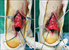
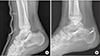
In clinical practice, recurrent Achilles ruptures have been noted to occurr at the original ruptured site. However, reports of new developed fresh rupture of the Achilles tendon in other sites are is extremely rare. Our report is about one uncommon case of a traumatic calcaneal tuberosity avulsion fracture following augmented repair, which was performed using the Krackow locking loop technique. We performed open reduction and intra-osseous fixation using a suture anchor. This procedure was done through the primary longitudinal incision for the calcaneal avulsion fracture fragment. After 6 months of follow-up, our patient has achieved a complete functional recovery and he can normally perform daily and work-related tasks without pain.
Figures and Tables
Figure 1
Clinical photo of initial injury treated using Krackow locking loop technique. (A) Laceration wound on Achilles tendon. (B) Initial repair using Krakow method.
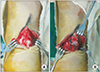
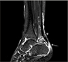
Figure 2
Magnetic resonance image before 2nd surgery (asterisk: primary repair site with near complete healing, arrow: avulsion fracture of calcaneal tuberosity).
References
1. Ajis A, Maffulli N. Management of acute tendo Achillis ruptures. Foot Ankle Surg. 2007; 13:132–135. DOI: 10.1016/j.fas.2007.02.002.

2. Cetti R, Christensen SE, Ejsted R, Jensen NM, Jorgensen U. Operative versus nonoperative treatment of Achilles tendon rupture A prospective randomized study and review of the literature. Am J Sports Med. 1993; 21:791–799. DOI: 10.1177/036354659302100606.
3. Rushton PR, Singh AK, Deshmukh RG. A case of 'second rupture' following open repair of a ruptured Achilles tendon. Foot Ankle Surg. 2011; 17:e17–e19. DOI: 10.1016/j.fas.2010.08.005.
4. García-Germán D, Rubio-Quevedo R, Lopez-Goenaga J, Martin-Guinea J. Achilles tendon recurrent rupture following surgical repair: report on two cases. Foot Ankle Surg. 2009; 15:152–154. DOI: 10.1016/j.fas.2008.09.001.
5. Pierre-Jerome C, Moncayo V, Terk MR. MRI of the Achilles tendon: a comprehensive review of the anatomy, biomechanics, and imaging of overuse tendinopathies. Acta Radiol. 2010; 51:438–454. DOI: 10.3109/02841851003627809.
6. Beavis RC, Rourke K, Court-Brown C. Avulsion fracture of the calcaneal tuberosity: a case report and literature review. Foot Ankle Int. 2008; 29:863–866. DOI: 10.3113/FAI.2008.0000.
7. Pramod J, Nitin S, Vasant G, Badole CM, Pritish S. Avulsion fracture of calcaneal tuberosity and heel pad avulsion : tension band fixation as novel fixation. J Clin Diagn Res. 2012; 6:738–739.
8. Greenhagen RM, Highlander PD, Burns PR. Double row anchor fixation: a novel technique for a diabetic calanceal insufficiency avulsion fracture. J Foot Ankle Surg. 2012; 51:123–127. DOI: 10.1053/j.jfas.2011.09.006.
9. Lui TH. Fixation of tendo Achilles avulsion fracture. Foot Ankle Surg. 2009; 15:58–61. DOI: 10.1016/j.fas.2008.06.004.
 XML Download