

 PDF
PDF ePub
ePub Citation
 Print
Print


Abstract
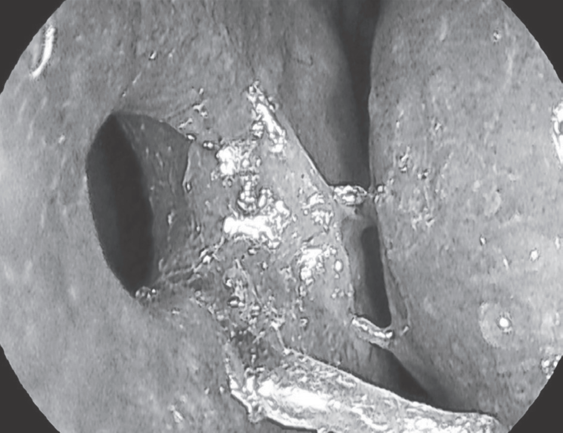
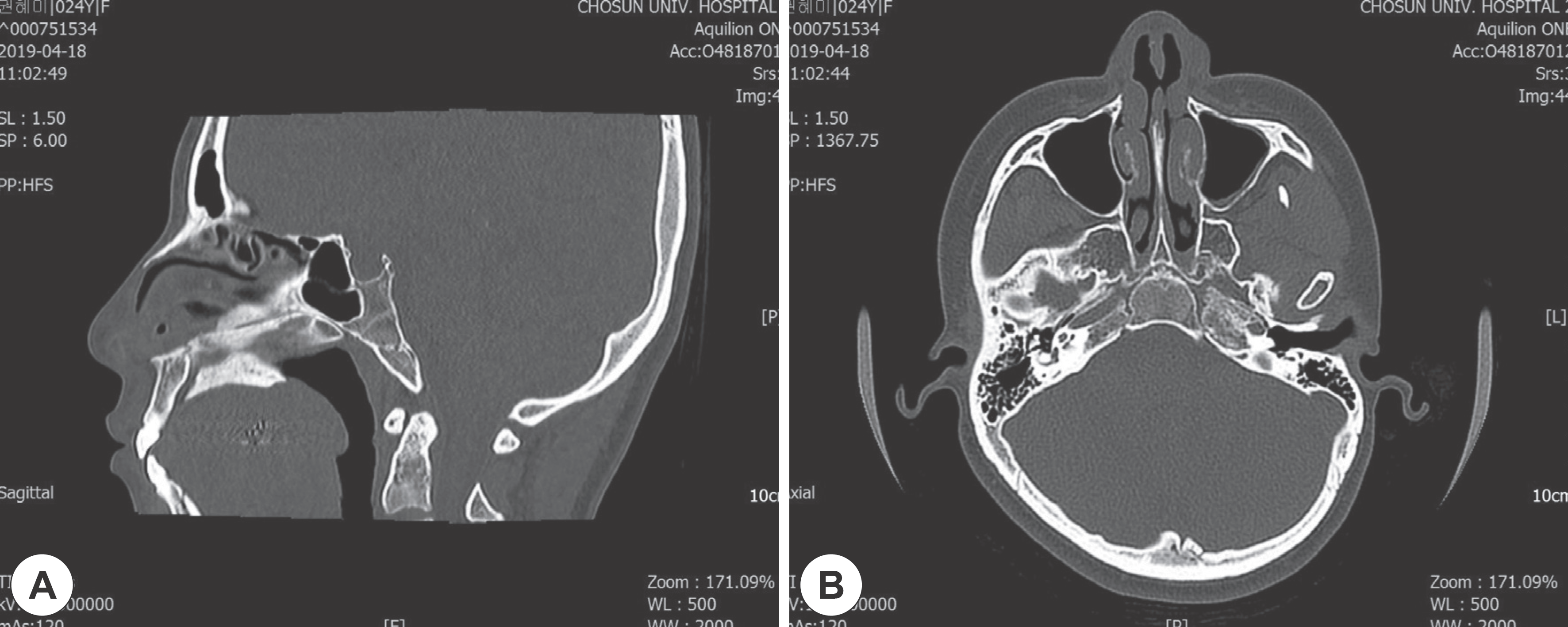
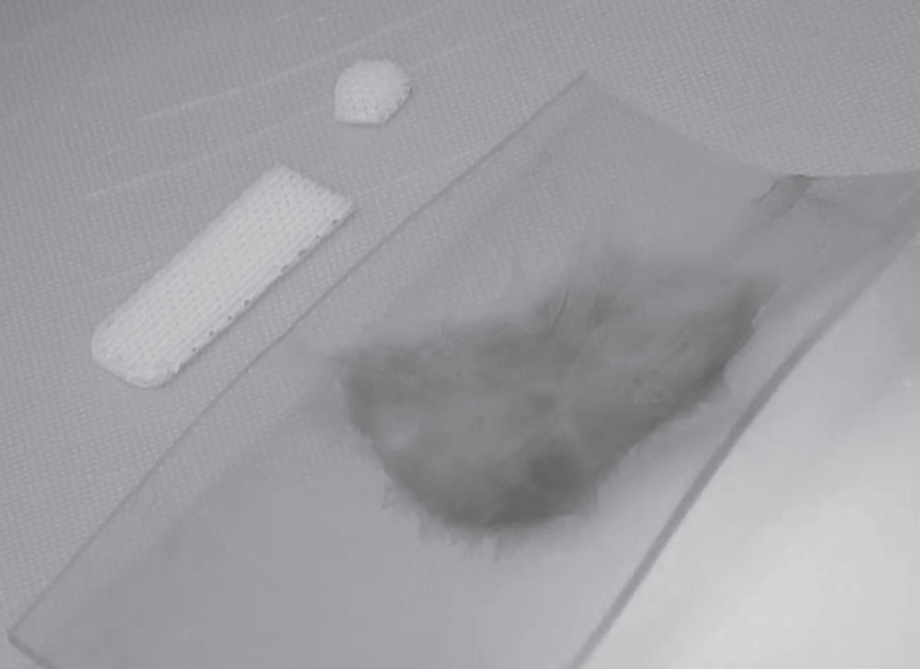
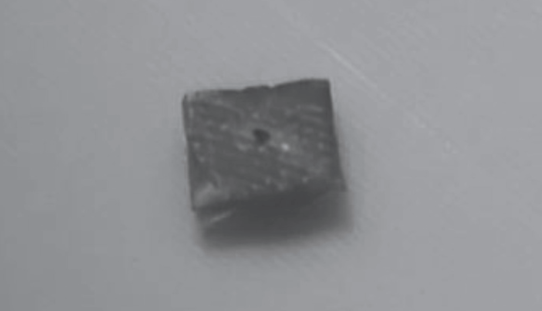
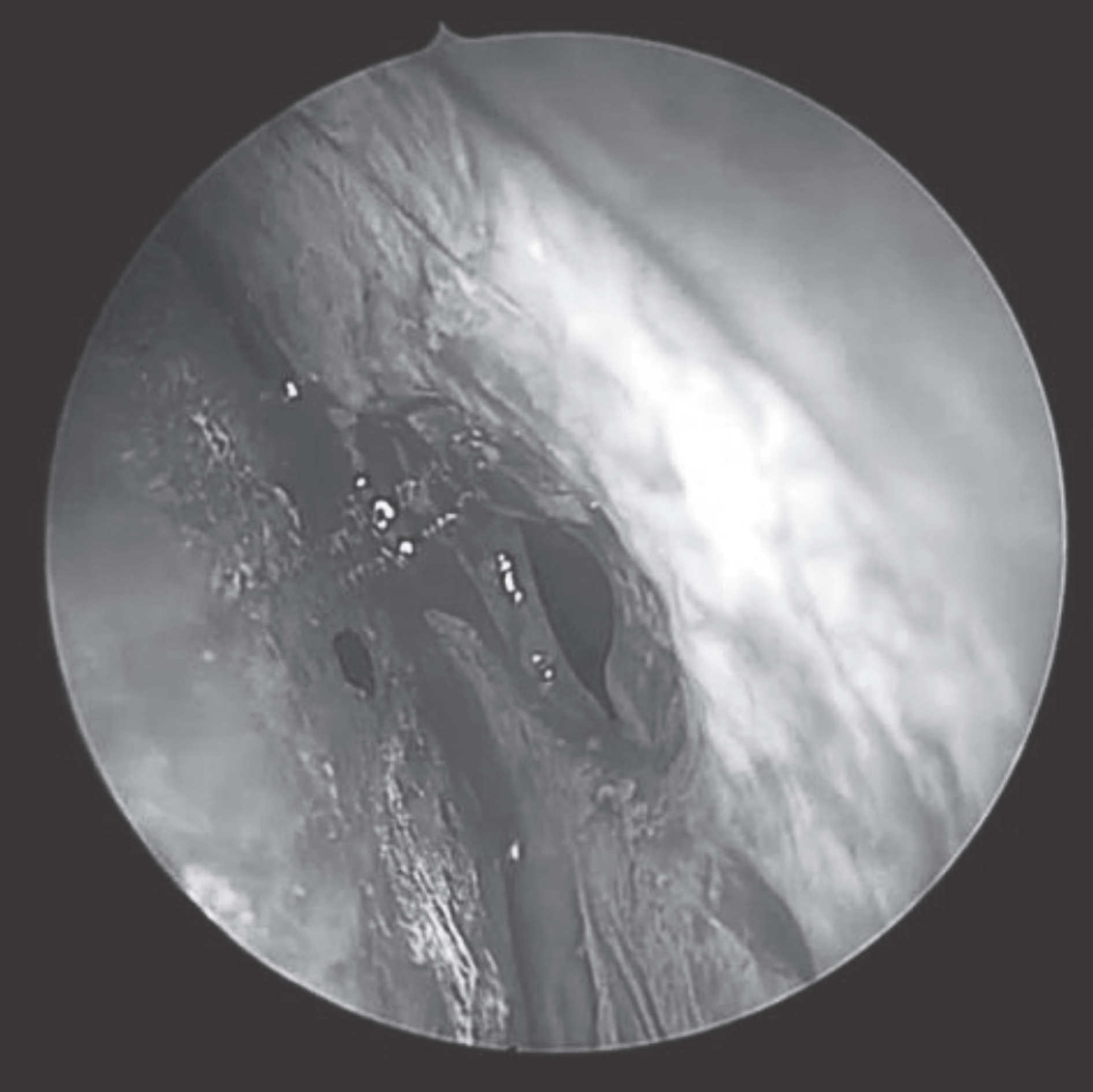
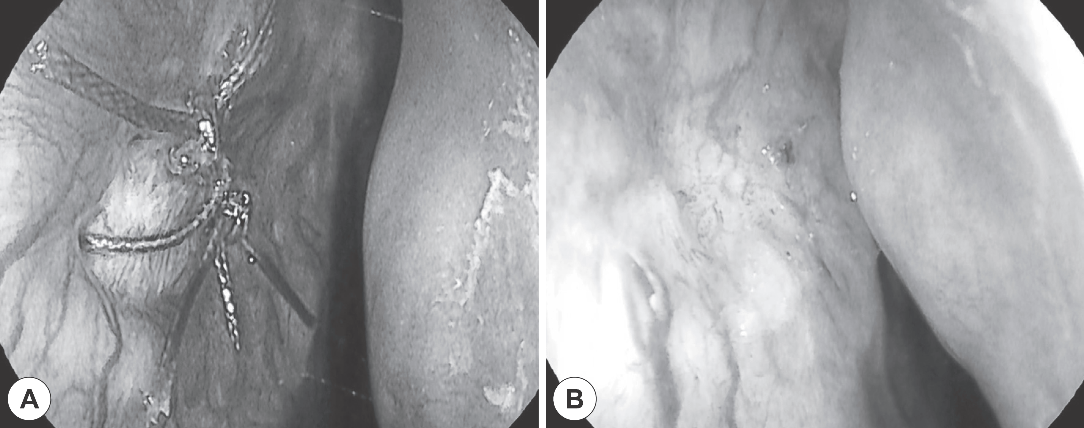
Trauma is the predominant cause of septal perforation resulting from surgical operation for nasal septum including submucosal resection. A 24-year-old female patient was diagnosed with nasal septal perforation after septoplasty. The patient manifested no specific symptoms except for occasional nasal bleeding, whistling and stuffy nose. Nasal septal perforation measuring 5×5 mm2 in size was observed at the anterior portion of nasal septum. The present study involves the repair of nasal septal perforation employing a polycaprolactone (PCL) plate and temporalis fascia graft, and discusses the consequences of complete closure of perforation without complications.
References
1. Yang SC, Lee KB, Lee JH, Kim CH. Repair of the septal perforation by temporalis fascia autografting and mucosal advancement fap. Korean Journal of Otorhinolaryngology-Head and Neck Surgery. 1996; 39(9):1492–96.
2. Mocella S, Muia F, Giacomini PG, Bertossi D, Residori E, Sgroi S. Innovative technique for large septal perforation repair and radiological evaluation. Acta OtorhinolaryngologicaItalica. 2013; 33(3):202.
3. Lee CU, Kang SH, Lee HS, Ahn KS. Repair of large septal perforation by external rhinoplasty approach. Korean Journal of Otorhinolaryngology-Head and Neck Surgery. 1991; 34(6):1232–36.
4. Morse J, Harris J, Owen S, Sowder J, Stephan S. Outcomes of nasal septal perforation repair using combined temporoparietal fascia graft and Polydioxanone plate construct. JAMA Facial Plast Surg. 2019; 21(4):319–26.

5. Delaney SW, Kridel RWH. Contemporary trends in the surgical management of nasal septal perforations: A community survey. Facial Plast Surg. 2019; 35(1):78–84.
6. Cassano M. Endoscopic repair of nasal septal perforation. Acta Otorhinolaryngol Ital. 2017; 37(6):486–92.
7. Re M, Paolucci L, Romeo R, Mallardi V. Surgical treatment of nasal septal perforations. Our experience. Acta Otorhinolaryngol Ital. 2006; 26(2):102–9.
8. Hong SK, Min YG. Repair of Nasal septal perforation by intranasal approach using a free composite graft of the auricular cartilage and the temporalis muscle fascia. Korean Journal of Otorhinolaryngolo-gy-Head and Neck Surgery. 2002; 45(10):969–74.
9. Park YJ, Cha JH, Bang SI, Kim SY. Clinical application of three-di-mensionally printed biomaterial Polycaprolactone (PCL) in augmentation rhinoplasty. Aesthetic Plast Surg. 2019; 43(2):437–46.
10. Virkkula P, Makitie AA, Vento SI. Surgical outcome and complications of nasal septal perforation repair with temporal fascia and periosteal grafts. Clin Med Insights Ear Nose Throat. 2015; 8:7–11.
 XML Download