Journal List > J Periodontal Implant Sci > v.49(6) > 1139189

Ahn, Kim, Joo, and Lee: Prevalence and risk factors of peri-implant mucositis and peri-implantitis after at least 7 years of loading

Abstract

Purpose

This study examined the prevalence and risk factors of peri-implant disease after at least 7 years of dental implant loading.

Methods

A total of 111 patients with 218 dental implants were treated. The follow-up period for all implants was at least 7 years. The patients' dental records were collected and risk factors of peri-implant disease were investigated through logistic regression analysis.

Results

The overall implant survival rate was 95.87%, because 9 of the 218 implants failed. The prevalence of peri-implant mucositis and peri-implantitis was 39.7% and 16.7%, respectively. As risk factors, smoking and prosthetic splinting showed significant associations with peri-implantitis (P<0.05).

Conclusions

Within the limits of this study, no significant correlations were found between any risk factors and peri-implant mucositis, but a significantly elevated risk of peri-implantitis was observed in patients who smoked or had splinted prostheses in 2 or more implants.

Graphical Abstract

jpis-49-397-abf001

INTRODUCTION

Owing to advances in surface treatments and the surgical and prosthodontic techniques used with dental implants, survival rates of more than 96% have been reported in implants that have functioned for a long-term period [12]. Nonetheless, many researchers have not distinguished between the “survival rate” and the “success rate”.
The term “success” and “survival” can be interpreted differently depending on the criteria used. In 1997, Roos et al. [3] proposed criteria for the success of implants that are now widely accepted, according to which the success of an implant is determined based on an evaluation of its status, that of the peri-implant tissue, and patients' satisfaction, whereas the survival of an implant is interpreted only as whether it is present or not. For example, an implant with 50% bone loss may continue to exist in the alveolar ridge, while causing many problems, such as swelling, pain, and halitosis. This would be categorized as “survival”, but not as “success”. Misch et al. [4] proposed a health scale for dental implants. According to this scale, an implant showing mobility or more than 50% radiographic bone loss is categorized as a failure. Numerous implants are placed in patients every year and the prevalence of implant complications has been reported to be 45% [5]. Hence, surviving implants that do not have ideal peri-implant tissue conditions are a growing problem.
Peri-implant disease is an inflammatory process that occurs in the tissues surrounding an implant, and includes peri-implant mucositis and peri-implantitis [6]. Peri-implant mucositis refers to the presence of an inflammatory lesion in the mucosa only, without additional bone loss after initial bone remodeling, whereas peri-implantitis is a condition characterized by gradual loss of the supporting bone [7]. In a clinical examination, the condition of peri-implant tissue can be categorized as follows (all cases of this study were classified using this categorization system) [89].
  • 1. Healthy peri-implant tissue: bleeding on probing (BOP) (−), marginal bone loss <2 mm

  • 2. Peri-implant mucositis: BOP (+), probing pocket depth (PPD) >5 mm, marginal bone loss ≤2 mm

  • 3. Peri-implantitis: BOP (+), PPD >5 mm, marginal bone loss >2 mm

Treatment protocols for peri-implant disease have been reported [1011]. Nonetheless, the disease is often not well controlled and frequently recurs. Despite the high prevalence and aggressive character of peri-implant disease, an ideal treatment protocol has not been established. Therefore, to better understand the characteristics of peri-implant disease, we investigated the prevalence and risk factors of peri-implant mucositis and peri-implantitis in dental implants that had functioned for at least 7 years.

MATERIALS AND METHODS

The implants in this study were placed at our dental hospital and had functioned for at least 7 years. In all patients, implant surgery was performed after the completion of periodontal treatment suitable for each individual patient's periodontal status. A total of 218 implants were placed in 111 patients, and the research protocol was approved by the Pusan National University Dental Hospital Institutional Review Board (PNUDH-2017-005).

Analysis of patients' files

The following data were extracted from the patients' dental records:
  • 1. Patient factors: sex, age, smoking, plaque index, maintenance visit, gingival biotype, and keratinized tissue

  • 2. Implant factors: loading period, implant length, diameter, number of implants, position of placement (maxilla or mandible; anterior, canine, posterior), design of the supraconstruction (splinting or not), implant surface (anodized type or resorbable blast media type)

  • 3. Surgical factors: regenerative procedure (bone graft, guided bone regeneration), surgeon's experience (first-year resident, second-year resident, third resident-year, or professor)

Clinical examination

The clinical examinations, which were carried out by a single periodontist (D.H.A), included the modified plaque index, PPD, and BOP. All clinical parameters were measured manually at each implant to the nearest millimeter using a periodontal probe (PGF-W, Osung, Gwangmyeong, Korea) [12]. The above variables were recorded at 6 surfaces (mesio-buccal, mid-buccal, disto-buccal, mesio-lingual, mid-lingual, and disto-lingual) of each implant. The presence of bleeding at 3 or more aspects was considered to indicate BOP. The gingival biotype was classified as thin or thick according to the transparency of the probe located in the gingival sulcus. Keratinized tissue around the implant was recorded as present or absent.

Radiographic examination

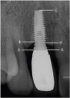
Standard periapical radiographs of each implant were taken using the parallel technique (Figure 1). Radiographic measurements were recorded using image analysis software (AxioVision, Carl Zeiss MicroImaging GmbH, Jena, Germany). Calibration was performed based on the length of each implant. The distances between the shoulder of the implant and the first bone-to-implant contact, mesially and distally, were noted in millimeters. The mean value was recorded.
Figure 1

Distances between the shoulder of the implant and the first bone-to-implant contact.

X=(A−B)×Implant Length/(A−C)
X′=(A−B′)×Implant Length/(A−C)
Mean Marginal Bone Level=(X+X′)/2
A–B: distance from the implant shoulder to the marginal bone level (distal), A–B′: distance from the implant shoulder to the marginal bone level (mesial), A–C: length of the implant (implant-abutment junction to apex).
jpis-49-397-g001

Data analysis

The prevalence of health and peri-implant disease was assessed at the implant level. Binary logistic regression analysis was performed to examine the effects of the patient, implant, and surgical factors. Subsequently, multiple logistic regression analysis was performed to analyze the statistical significance of various factors. The statistical analysis was performed using SPSS version 21.0 (IBM Corp., Armonk, NY, USA). P values less than 0.05 were considered to indicate statistical significance. The odds ratios and predicted probabilities, including 95% confidence intervals, were analyzed. The overall fit (calibration) of the predictive scale was assessed using the Hosmer–Lemeshow statistic.

RESULTS

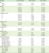
Nine of the 218 implants showed mobility or more than 50% of radiographic bone loss, and were categorized as failed implants. The overall implant survival rate was 95.87%. Table 1 presents the distribution of implants according to various factors.
Table 1

Distribution of implants according to various factors

jpis-49-397-i001
Variables Survived implants Failed implants Total implants
Sex
Male 72 (33.0) 5 (2.3) 77 (35.3)
Female 137 (62.8) 4 (1.8) 141 (64.7)
Age (yr)
≤49 32 (14.7) 0 (0) 32 (14.7)
50–59 67 (30.7) 6 (2.7) 73 (33.5)
60–69 76 (34.9) 3 (1.4) 79 (36.2)
≥70 34 (15.6) 0 (0) 34 (15.6)
Loading period (yr)
≥11 29 (13.3) 0 (0) 29 (13.3)
10 39 (17.9) 5 (2.3) 44 (20.2)
9 49 (22.5) 2 (0.9) 51 (23.4)
8 29 (13.3) 1 (0.5) 30 (13.8)
7 63 (28.9) 1 (0.5) 64 (29.4)
Implant surface
RBM 174 (79.8) 4 (1.8) 178 (81.7)
Anodized 33 (15.1) 5 (2.3) 38 (17.4)
Missing data 2 (0.9)
Length (mm)
<10 85 (39.0) 7 (3.2) 92 (42.2)
≥10 124 (56.9) 2 (0.9) 126 (57.8)
Diameter (mm)
Narrow (≤3.75) 13 (6.0) 0 (0) 13 (6.0)
Regular (4) 70 (32.1) 3 (1.4) 73 (33.5)
Wide (≥5) 126 (57.8) 6 (2.8) 132 (60.6)
Position of placement
Maxillary anterior 6 (2.8) 0 (0) 6 (2.8)
Maxillary canine 7 (3.2) 0 (0) 7 (3.2)
Maxillary posterior 79 (36.2) 4 (1.8) 83 (38.1)
Mandibular anterior 5 (2.3) 0 (0) 5 (2.3)
Mandibular canine 0 (0) 0 (0) 0 (0)
Mandibular posterior 112 (51.4) 5 (2.3) 117 (53.7)
Surgeon's experience
1st-yr resident 22 (10.1) 0 (0) 22 (10.1)
2nd-yr resident 73 (33.5) 5 (2.3) 78 (35.8)
3rd-yr resident 57 (26.1) 4 (1.8) 61 (28.0)
Professor 57 (26.1) 0 (0) 57 (26.1)
Values are presented as number (%).
RBM: resorbable blast media.

Prevalence of health, peri-implant mucositis, and peri-implantitis

A total of 209 implants, excluding failed implants, were investigated, of which 83 (39.7%) presented with peri-implant mucositis, whereas 35 (16.7%) exhibited peri-implantitis (Table 2).
Table 2

Prevalence of health and peri-implant disease in dental implants after at least 7 years of loading

jpis-49-397-i002
Variables Implants (total n=209)
Peri-implant healtha) 91 (43.5)
Peri-implant mucositisb) 83 (39.7)
Peri-implantitisc) (bone loss, mm)
2.0< bone loss ≤3.0 21 (60.0)
3.0< bone loss ≤4.0 9 (25.7)
4.0< bone loss ≤5.0 4 (11.4)
5.0< bone loss 1 (2.9)
Values are presented as number (%).
BOP: bleeding on probing, PPD: probing pocket depth.
a)No BOP and no bone loss >2 mm; b)BOP and PPD >5 mm, but no bone loss >2 mm; c)BOP, PPD >5 mm, and bone loss >2 mm.

Factors associated with peri-implant mucositis and peri-implantitis

No statistically significant associations were found between any of the investigated parameters and peri-implant mucositis (Table 3).
Table 3

Factors associated with peri-implant mucositis and peri-implantitis

jpis-49-397-i003
Factors Peri-implant mucositis Peri-implantitis
OR CI P OR CI P
Patient factors
Sex
Male (72) 1.000 1.000
Female (137) 1.513 0.834–2.744 0.173 1.009 0.470–2.167 0.982
Smoking
No (192) 1.000 1.000
Yes (17) 0.440 0.138–1.400 0.164 4.100 1.441–11.666 0.008a)
Plaque index
0 (135) 1.000 1.000
1 (63) 1.511 0.945–2.414 0.084 1.417 0.793–2.530 0.239
2 (11)
Maintenance visits (mon)
≤6 (111) 1.000 1.000
>6 (98) 0.929 0.533–1.619 0.795 0.714 0.341–1.495 0.372
Biotype
Thin (20) 1.000 1.000
Thick (189) 0.786 0.311–1.990 0.612 4.168 0.539–32.208 0.171
Keratinized tissue
No (36) 1.000 1.000
Yes (173) 1.392 0.653–2.966 0.391 1.007 0.384–2.638 0.989
Implant factors
Length (mm)
<10 (85) 1.000 1.000
≥10 (124) 0.897 0.345–2.333 0.823 1.787 0.394–8.108 0.452
Diameter (mm)
≤4 (83) 1.000 1.000
≥5 (126) 0.997 0.566–1.756 0.991 0.235 0.235–1.020 0.056
No. of implants
<4 (109) 1.000 1.000
≥4 (100) 1.585 0.906–2.775 0.107 0.638 0.307–1.328 0.230
Position of placement
Maxilla (92) 1.000 1.000
Mandible (117) 0.592 0.338–1.036 0.067 1.631 0.764–3.484 0.206
Design of supraconstruction
No splinting (81) 1.000 1.000
Splinting (128) 0.725 0.412–1.278 0.267 2.439 1.048–5.676 0.038b)
Implant surface
RBM (174) 1.000 1.000
Anodized (33) 1.782 0.843–3.767 0.130 0.636 0.209–1.941 0.427
Missing data (2)
Terminal placement
No (131) 1.000 1.000
Yes (78) 1.293 0.731–2.288 0.377 0.732 0.337–1.591 0.431
Surgical factors
Regenerative surgery
No (116) 1.000 1.000
Yes (93) 1.281 0.734–2.237 0.383 1.398 0.675–2.893 0.367
OR: odds ratio, CI: confidence interval, RBM: resorbable blast media.
a)P<0.01; b)P<0.05.
For peri-implantitis, smoking (as a patient factor) and prosthesis splinting (as an implant factor) were significantly associated with the development of peri-implantitis (P<0.01 and P<0.05, respectively), while no surgical factors showed a statistically significant relationship (Table 3).
The multiple logistic regression analysis showed that smoking was significantly correlated with peri-implantitis (P<0.01) (Table 4). Smokers showed a higher incidence of peri-implantitis than non-smokers. Prosthesis splinting was also associated significantly with peri-implantitis (P<0.05) (Table 4). The use of bridges as a prosthesis for 2 or more implants was associated with a significantly higher risk of peri-implantitis than was observed for single crown implants.
Table 4

Association of peri-implantitis with various factors according to multiple regression analysis

jpis-49-397-i004
Factors B±SE β t P value
Smoking 0.269±0.092 0.197 2.915 0.004a)
Prosthesis splinting 0.114±0.052 0.149 2.210 0.028b)
SE: standard error.
a)P<0.01; b)P<0.05.

DISCUSSION

Many researchers have reported high survival rates, exceeding 90%, in implants that have functioned for a long-term period, although the survival rate tends to decrease with longer functional periods [131415]. In this study, the 7-year and 10-year survival rates were 95.87% and 92.6%, respectively, showing a similar pattern to those reported in previous studies.
We adopted strict diagnostic criteria for peri-implant mucositis and peri-implantitis when evaluating the status of peri-implant tissue health. Peri-implant mucositis and peri-implantitis were observed in 39.7% and 16.7% of patients, respectively. In previous studies, the prevalence of these conditions has been reported to range very widely [51617]. This diversity in previous findings can be explained by differences in the sample size, the definition of disease, and the follow-up period.
No factors showing a statistically significant relationship with peri-mucositis were found in our study. Although not statistically significant, the position of the implant (P=0.067) and the plaque index (P=0.084) could be potential risk factors for peri-implant mucositis. The accumulation of plaque could induce peri-implant inflammation and affect peri-implant tissue health [181920]. In this study, the maxilla was found to be more closely associated with the incidence of peri-implant mucositis than the mandible. All dentists will agree that both the quantity and quality of residual bone for dental implant surgery are worse in the maxilla than in the mandible. For this reason, a lower success rate and more implant failures have been reported in the maxilla than in the mandible [1521].
The present study showed that smoking (P<0.01) was a significant factor affecting the risk of peri-implantitis. Smoking has been identified as a risk factor for peri-implant diseases [222324]. and smoking increases peri-implant marginal bone resorption during the initial healing period [25]. According to a systematic review, the annual rate of marginal bone loss was found to be 0.16 mm per year higher in smokers than in non-smokers [26]. Recently, Duan et al. [27] reported that the oral microbiome of smokers was more harmful than that of non-smokers. In particular, Porphyromonas gingivalis, which was present in significantly greater quantities in smokers, affected marginal bone loss during bone healing.
Prosthetic splinting was identified as another significant factor affecting the incidence of peri-implantitis in the present study (P<0.05). The inter-implant portion of the prosthetic splint or the area beneath a pontic may be more difficult to clean than a single crown due to the non-ideal emergence profile or inadequate space of the prosthesis around the implant. Poor oral hygiene has been reported to be associated with the risk of peri-implantitis [16]. Similarly, Serino and Ström [28] reported improper plaque control in 74% of implants. Therefore, the importance of effective plaque control has been emphasized as a way to increase the survival rate of implants [29].
A previous study reported that 5.0-mm-diameter implants exhibited higher failure rate than 3.75-mm-diameter or 4.0-mm-diameter implants [30]. Canullo et al. [31] also reported that implants with a wider diameter had a greater likelihood of peri-implantitis. This is because additional bone grafting, which is often required to facilitate the placement of implants with wider diameters, may be another factor that causes peri-implantitis. This may also be due to the higher compression force generated during the drilling process for wider implants and the presence of less bone surrounding the wider implant. In contrast, several studies reported no relationship between the implant diameter and the survival rate [3233]. In our study, peri-implantitis was more likely to occur in narrow-diameter implants (≤4 mm) than in wide-diameter implants (≥5 mm) (P=0.056). This result could be interpreted as implying that choosing a narrow diameter could result in a lack of residual bone quantity. Therefore, this topic is still open to debate. Most of the factors that have been considered to be predictors of peri-implant disease showed no statistical significance in the present study. Considering the details of this study, future research including a larger sample and a longer follow-up period will be needed.

Notes

Funding This work was supported by the National Research Foundation of Korea (NRF) grant NRF-2017M3A9B6062026, funded by the Korean government.

Author Contributions

  • Conceptualization: Ju-Youn Lee, Dae-Hee Ahn.

  • Data curation: Hyun-Joo Kim.

  • Formal analysis: Dae-Hee Ahn.

  • Funding acquisition: Ju-Youn Lee.

  • Investigation: Ju-Youn Lee, Dae-Hee Ahn, Hyun-Joo Kim.

  • Methodology: Ju-Youn Lee, Ji-Young Joo.

  • Project administration: Ju-Youn Lee, Hyun-Joo Kim.

  • Writing - original draft: Ju-Youn Lee, Dae-Hee Ahn, Hyun-Joo Kim.

  • Writing - review & editing: Ju-Youn Lee, Ji-Young Joo, Hyun-Joo Kim.

Conflict of Interest No potential conflict of interest relevant to this article is reported.

References

1. Niedermaier R, Stelzle F, Riemann M, Bolz W, Schuh P, Wachtel H. Implant-supported immediately loaded fixed full-arch dentures: evaluation of implant survival rates in a case cohort of up to 7 years. Clin Implant Dent Relat Res. 2017; 19:4–19.
crossref
2. Hasegawa T, Kawabata S, Takeda D, Iwata E, Saito I, Arimoto S, et al. Survival of Brånemark System Mk III implants and analysis of risk factors associated with implant failure. Int J Oral Maxillofac Surg. 2017; 46:267–273.
crossref
3. Roos J, Sennerby L, Lekholm U, Jemt T, Gröndahl K, Albrektsson T. A qualitative and quantitative method for evaluating implant success: a 5-year retrospective analysis of the Brånemark implant. Int J Oral Maxillofac Implants. 1997; 12:504–514.
4. Misch CE, Perel ML, Wang HL, Sammartino G, Galindo-Moreno P, Trisi P, et al. Implant success, survival, and failure: the International Congress of Oral Implantologists (ICOI) Pisa Consensus Conference. Implant Dent. 2008; 17:5–15.
crossref
5. Derks J, Schaller D, Håkansson J, Wennström JL, Tomasi C, Berglundh T. Effectiveness of implant therapy analyzed in a Swedish population: prevalence of peri-implantitis. J Dent Res. 2016; 95:43–49.
crossref
6. Albrektsson T, Isidor F. Consensus report of session IV. In : In : Lang NP, Karring T, editors. Proceedings of the First European Workshop on Periodontology; London; Quintessence. 1994. p. 365–369.
7. Renvert S, Persson GR, Pirih FQ, Camargo PM. Peri-implant health, peri-implant mucositis, and peri-implantitis: case definitions and diagnostic considerations. J Clin Periodontol. 2018; 45:Suppl 20. S278–S285.
crossref
8. Marrone A, Lasserre J, Bercy P, Brecx MC. Prevalence and risk factors for peri-implant disease in Belgian adults. Clin Oral Implants Res. 2013; 24:934–940.
crossref
9. Lang NP, Berglundh T. Working Group 4 of Seventh European Workshop on Periodontology. Periimplant diseases: where are we now?--Consensus of the Seventh European Workshop on Periodontology. J Clin Periodontol. 2011; 38:Suppl 11. 178–181.
crossref
10. Lang NP, Mombelli A, Tonetti MS, Brägger U, Hämmerle CH. Clinical trials on therapies for peri-implant infections. Ann Periodontol. 1997; 2:343–356.
crossref
11. Jovanovic SA. The management of peri-implant breakdown around functioning osseointegrated dental implants. J Periodontol. 1993; 64:1176–1183.
crossref
12. Mombelli A, van Oosten MA, Schürch E Jr, Land NP. The microbiota associated with successful or failing osseointegrated titanium implants. Oral Microbiol Immunol. 1987; 2:145–151.
crossref
13. Jung RE, Zembic A, Pjetursson BE, Zwahlen M, Thoma DS. Systematic review of the survival rate and the incidence of biological, technical, and aesthetic complications of single crowns on implants reported in longitudinal studies with a mean follow-up of 5 years. Clin Oral Implants Res. 2012; 23:Suppl 6. 2–21.
14. Simonis P, Dufour T, Tenenbaum H. Long-term implant survival and success: a 10–16-year follow-up of non-submerged dental implants. Clin Oral Implants Res. 2010; 21:772–777.
crossref
15. French D, Larjava H, Ofec R. Retrospective cohort study of 4591 Straumann implants in private practice setting, with up to 10-year follow-up. Part 1: multivariate survival analysis. Clin Oral Implants Res. 2015; 26:1345–1354.
crossref
16. Ferreira SD, Silva GL, Cortelli JR, Costa JE, Costa FO. Prevalence and risk variables for peri-implant disease in Brazilian subjects. J Clin Periodontol. 2006; 33:929–935.
crossref
17. Lindhe J, Meyle J. Group D of European Workshop on Periodontology. Peri-implant diseases: consensus report of the Sixth European Workshop on Periodontology. J Clin Periodontol. 2008; 35:282–285.
crossref
18. Zitzmann NU, Berglundh T, Marinello CP, Lindhe J. Experimental peri-implant mucositis in man. J Clin Periodontol. 2001; 28:517–523.
crossref
19. Costa FO, Takenaka-Martinez S, Cota LO, Ferreira SD, Silva GL, Costa JE. Peri-implant disease in subjects with and without preventive maintenance: a 5-year follow-up. J Clin Periodontol. 2012; 39:173–181.
crossref
20. Mombelli A, Décaillet F. The characteristics of biofilms in peri-implant disease. J Clin Periodontol. 2011; 38:Suppl 11. 203–213.
crossref
21. Buser D, Mericske-Stern R, Bernard JP, Behneke A, Behneke N, Hirt HP, et al. Long-term evaluation of non-submerged ITI implants. Part 1: 8-year life table analysis of a prospective multi-center study with 2359 implants. Clin Oral Implants Res. 1997; 8:161–172.
crossref
22. Haas R, Haimböck W, Mailath G, Watzek G. The relationship of smoking on peri-implant tissue: a retrospective study. J Prosthet Dent. 1996; 76:592–596.
crossref
23. Lindquist LW, Carlsson GE, Jemt T. Association between marginal bone loss around osseointegrated mandibular implants and smoking habits: a 10-year follow-up study. J Dent Res. 1997; 76:1667–1674.
crossref
24. Bain CA, Moy PK. The association between the failure of dental implants and cigarette smoking. Int J Oral Maxillofac Implants. 1993; 8:609–615.
25. Shibli JA, Piattelli A, Iezzi G, Cardoso LA, Onuma T, de Carvalho PS, et al. Effect of smoking on early bone healing around oxidized surfaces: a prospective, controlled study in human jaws. J Periodontol. 2010; 81:575–583.
crossref
26. Clementini M, Rossetti PH, Penarrocha D, Micarelli C, Bonachela WC, Canullo L. Systemic risk factors for peri-implant bone loss: a systematic review and meta-analysis. Int J Oral Maxillofac Surg. 2014; 43:323–334.
crossref
27. Duan X, Wu T, Xu X, Chen D, Mo A, Lei Y, et al. Smoking may lead to marginal bone loss around non-submerged implants during bone healing by altering salivary microbiome: a prospective study. J Periodontol. 2017; 88:1297–1308.
crossref
28. Serino G, Ström C. Peri-implantitis in partially edentulous patients: association with inadequate plaque control. Clin Oral Implants Res. 2009; 20:169–174.
crossref
29. Renvert S, Persson GR. Supportive periodontal therapy. Periodontol 2000. 2004; 36:179–195.
crossref
30. Ivanoff CJ, Gröndahl K, Sennerby L, Bergström C, Lekholm U. Influence of variations in implant diameters: a 3- to 5-year retrospective clinical report. Int J Oral Maxillofac Implants. 1999; 14:173–180.
31. Canullo L, Peñarrocha M, Monje A, Catena A, Wang HL, Peñarrocha D. Association between clinical and microbiologic cluster profiles and peri-implantitis. Int J Oral Maxillofac Implants. 2017; 32:1054–1064.
crossref
32. Romeo E, Lops D, Margutti E, Ghisolfi M, Chiapasco M, Vogel G. Long-term survival and success of oral implants in the treatment of full and partial arches: a 7-year prospective study with the ITI dental implant system. Int J Oral Maxillofac Implants. 2004; 19:247–259.
33. Lemmerman KJ, Lemmerman NE. Osseointegrated dental implants in private practice: a long-term case series study. J Periodontol. 2005; 76:310–319.
crossref
TOOLS
ORCID iDs

Dae-Hee Ahn
https://orcid.org/0000-0001-6746-0931

Hyun-Joo Kim
https://orcid.org/0000-0001-7553-6289

Ji-Young Joo
https://orcid.org/0000-0002-4050-5797

Ju-Youn Lee
https://orcid.org/0000-0002-0772-033X

Similar articles