

 PDF
PDF ePub
ePub Citation
 Print
Print


Jung Hoei Ku , Hyung Lae Cho, Jong Min Kim
, Hyung Lae Cho, Jong Min Kim
Abstract
Purpose
This study aimed to evaluate the diagnostic characteristics and clinical results after surgical repair of traumatic superimposed posterior rotator cuff tear in the setting of preexisting retracted supraspinatus tendon tear.
Methods
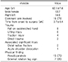
A total of 20 patients (mean age, 62.1 years) were included and all patients had significant traumatic events mean 3.7 weeks prior to the surgery. Preoperative acromiohumeral distance (AHD, mean 3.2 mm) and arthritis change were analyzed on plain radiograph and magnetic resonance imaging was evaluated for the nature and extent of torn tendon, and fatty degeneration (FD) of all cuff muscles to validate if the tears were traumatic or chronic.
Results
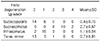
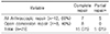
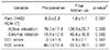
Complete repairs were achieved in 15 patients and partial repair including posterior cuff in five. Functional and radiographic results were statistically evaluated and repair integrities were assessed with ultrasound at average 17.3 months. Overall functional outcome scores were significantly improved and 17 patients (85%) were satisfied with their symptoms. AHD was significantly recovered (mean, 6.7 mm), but two patients showed progression of arthritic change. Retears after the complete repair were three patients (20%), who showed poor outcome, with advanced preoperative FD of posterior cuff muscles. Five patients with partial repair of posterior cuff revealed improved functional score with no sign of retear of posterior cuff on ultrasound.
Figures and Tables
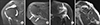 | Fig. 1Preoperative magnetic resonance images of right shoulder in 64-year-old male after trauma. (A) T2-weighted coronal image shows retracted supraspinatus tendon and no muscle edema. T2-weighted axial (B) and coronal image (C) show intramuscular high signal intensity corresponding acute muscle edema and wavy contoured torn tendon edges (white arrows). (D) T1 weighted oblique sagittal image shows fatty degeneration of cuff muscles; supraspinatus grade II, Infraspinatus and teres minor; grade I.
|
 | Fig. 2Intraoperative arthroscopic images of right shoulder in 59-year-old male show (A) smooth torn edge and relatively poor mobility of preexisting supraspinatus tear and (B) bursal hematoma on infraspinatus and (C) final construct is shown with direct repair of infraspinatus and partial repair of supraspinatus using biceps tendon.
|
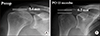 | Fig. 3Anteroposterior radiographs of right shoulder to measure the acromiohumeral interval in 57-year-old male with superimposed infraspinatus tear show (A) marked narrowing of acromiohumeral interval to 2.4 mm and upper migration of humeral head after trauma. (B) After arthroscopic repair, increased the interval to 6.2 mm in 15 months postoperative follow-up. Preop: preoperation, PO: postoperation.
|
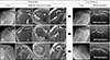 | Fig. 4Three cases of postoperative retear after complete repair of superimposed acute posterior cuff tear in pre-existing supraspinatus tear and preoperative and follow-up X-ray, magnetic resonance and ultrasonographic images. (A) Fifty-nine-year-old male with grade III fatty degeneration of teres minor (white arrow) had upper migration of humeral head and retear of infraspinatus. (B) Sixty-seven-year-old male also had grade II teres minor fatty degeneration (white arrow). Hamada III rotator cuff tear arthropathy and infraspinatus retear were revealed in final follow-up. (C) Sixty-two-year-old male with grade II infraspinatus fatty degeneration (white arrow) showed marked upper migration of humeral head and complete retear of infraspinatus on ultrasonography in final follow-up.
|
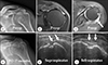 | Fig. 5A case of partial repair of posterior cuff in 58-year-old male. (A) Preoperative radiograph shows marked narrowing of acromiohumeral interval. (B) T2 weighted coronal and (C) axial image show retracted supraspinatus tear and acute superimposed infraspinatus tear (white arrow). (D) Seventeen months after partial repair with margin convergence, acromiohumeral interval is recovered and no sign of retear of (E) supraspinatus and (F) infraspinatus on ultrasonography (white arrows). Preop: preoperative, PO: postoperative.
|


References
1. Minagawa H, Yamamoto N, Abe H, et al. Prevalence of symptomatic and asymptomatic rotator cuff tears in the general population: from mass-screening in one village. J Orthop. 2013; 10:8–12.
2. Bassett RW, Cofield RH. Acute tears of the rotator cuff: the timing of surgical repair. Clin Orthop Relat Res. 1983; 18–24.
3. Petersen SA, Murphy TP. The timing of rotator cuff repair for the restoration of function. J Shoulder Elbow Surg. 2011; 20:62–68.
4. Sorensen AK, Bak K, Krarup AL, et al. Acute rotator cuff tear: do we miss the early diagnosis? A prospective study showing a high incidence of rotator cuff tears after shoulder trauma. J Shoulder Elbow Surg. 2007; 16:174–180.
5. Jeong JY, Song SY, Yoo JC, Park KM, Lee SM. Comparison of outcomes with arthroscopic repair of acute-onchronic within 6 months and chronic rotator cuff tears. J Shoulder Elbow Surg. 2017; 26:648–655.
6. Hamada K, Fukuda H, Mikasa M, Kobayashi Y. Roentgenographic findings in massive rotator cuff tears: a longterm observation. Clin Orthop Relat Res. 1990; 92–96.
7. Goutallier D, Postel JM, Bernageau J, Lavau L, Voisin MC. Fatty muscle degeneration in cuff ruptures. Pre- and postoperative evaluation by CT scan. Clin Orthop Relat Res. 1994; 78–83.
8. Beaton D, Richards RR. Assessing the reliability and responsiveness of 5 shoulder questionnaires. J Shoulder Elbow Surg. 1998; 7:565–572.
9. Tae SK, Rhee YG, Park TS, et al. The development and validation of an appraisal method for rotator cuff disorders: the Korean Shoulder Scoring System. J Shoulder Elbow Surg. 2009; 18:689–696.
10. Miller BS, Downie BK, Kohen RB, et al. When do rotator cuff repairs fail? Serial ultrasound examination after arthroscopic repair of large and massive rotator cuff tears. Am J Sports Med. 2011; 39:2064–2070.
11. Sano H, Hatta T, Yamamoto N, Itoi E. Stress distribution within rotator cuff tendons with a crescent-shaped and an L-shaped tear. Am J Sports Med. 2013; 41:2262–2269.
12. Miller RM, Thunes J, Musahl V, Maiti S, Debski RE. Effects of tear size and location on predictions of supraspinatus tear propagation. J Biomech. 2018; 68:51–57.
13. Namdari S, Henn RF 3rd, Green A. Traumatic anterosuperior rotator cuff tears: the outcome of open surgical repair. J Bone Joint Surg Am. 2008; 90:1906–1913.
14. Goutallier D, Le Guilloux P, Postel JM, Radier C, Bernageau J, Zilber S. Acromio humeral distance less than six millimeter: its meaning in full-thickness rotator cuff tear. Orthop Traumatol Surg Res. 2011; 97:246–251.
15. de Jesus JO, Parker L, Frangos AJ, Nazarian LN. Accuracy of MRI, MR arthrography, and ultrasound in the diagnosis of rotator cuff tears: a meta-analysis. AJR Am J Roentgenol. 2009; 192:1701–1707.
16. Cho YM, Kim SJ, Oh JC, Chun YM. Characteristics of magnetic resonance arthrography findings in traumatic posterosuperior rotator cuff tears. Clin Should Elbow. 2015; 18:211–216.
17. Mochizuki T, Sugaya H, Uomizu M, et al. Humeral insertion of the supraspinatus and infraspinatus: new anatomical findings regarding the footprint of the rotator cuff. J Bone Joint Surg Am. 2008; 90:962–969.
18. Duncan NS, Booker SJ, Gooding BW, Geoghegan J, Wallace WA, Manning PA. Surgery within 6 months of an acute rotator cuff tear significantly improves outcome. J Shoulder Elbow Surg. 2015; 24:1876–1880.
19. Yoo JC, Ahn JH, Koh KH, Lim KS. Rotator cuff integrity after arthroscopic repair for large tears with less-than-optimal footprint coverage. Arthroscopy. 2009; 25:1093–1100.
20. Hantes ME, Karidakis GK, Vlychou M, Varitimidis S, Dailiana Z, Malizos KN. A comparison of early versus delayed repair of traumatic rotator cuff tears. Knee Surg Sports Traumatol Arthrosc. 2011; 19:1766–1770.
21. Ohzono H, Gotoh M, Nakamura H, et al. Effect of preoperative fatty degeneration of the rotator cuff muscles on the clinical outcome of patients with intact tendons after arthroscopic rotator cuff repair of large/massive cuff tears. Am J Sports Med. 2017; 45:2975–2981.
22. Kim SJ, Lee IS, Kim SH, Lee WY, Chun YM. Arthroscopic partial repair of irreparable large to massive rotator cuff tears. Arthroscopy. 2012; 28:761–768.
23. Wellmann M, Lichtenberg S, da Silva G, Magosch P, Habermeyer P. Results of arthroscopic partial repair of large retracted rotator cuff tears. Arthroscopy. 2013; 29:1275–1282.
24. Iagulli ND, Field LD, Hobgood ER, Ramsey JR, Savoie FH 3rd. Comparison of partial versus complete arthroscopic repair of massive rotator cuff tears. Am J Sports Med. 2012; 40:1022–1026.
 XML Download