

 PDF
PDF Citation
 Print
Print


INTRODUCTION
Hair follicle nevus (HFN) is a rare, benign, follicular hamartoma that usually presents as a congenital, asymptomatic nodule on the face. Differential diagnoses of HFN include accessory tragus, cervical chondrocutaneous branchial remnants (CCBR) and trichofolliculoma, but recently there has been a debate regarding whether they are on a spectrum of the same entity1234. Here we report the case of a HFN which presented as a solitary papule on the neck. In order to investigate and compare the characteristics of hair follicles in HFN and its differential diagnoses, a pilot immunohistochemical study with cytokeratin 19 (CK19, a marker for hair follicle stem cell) was performed in the presented case and each case of accessory tragus, CCBR, and trichofolliculoma. We received consent from the patient's parents about publishing all photographic materials.
CASE REPORT
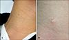
A 14-year-old boy presented with a skin tag-like lesion on his right lower neck, and a red-colored papule on his right upper neck (Fig. 1A). The lower lesion was asymptomatic and had been present since birth. The upper lesion developed 1 month ago and bled frequently. The patient had been otherwise healthy with no known prenatal or developmental abnormalities or significant family history. Physical examination revealed a soft, pedunculated, skin-colored papule, 3 mm in diameter, on the slightly elevated surface on the right lower neck (Fig. 1B), and a soft, domeshaped, red-colored papule, 2 mm in diameter, on the right upper neck (Fig. 1A). Under the clinical impressions of skin tag and pyogenic granuloma, respectively, punch excision was performed for each lesion. Histopathologically, the lower skin-colored papule demonstrated a polypoid skin segment with mild epidermal hyperplasia and many vellus hair follicles in the dermis (Fig. 2A). Several sebaceous and eccrine glands connected with the hair follicle were also seen (Fig. 2A), and perifollicular stroma showed fibrous thickening (Fig. 2B). Serial sections did not reveal any cartilage, cystic follicular structures in the stroma, or prominent connective tissue framework in the subcutaneous fat. The upper red papule showed an exophytic lesion with lobular proliferation of capillaries, consistent with pyogenic granuloma. Based on the clinical and pathologic findings, diagnoses of HFN and pyogenic granuloma were made, respectively.
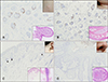
With CK19 (1:200, M0888; DAKO, Glostrup, Denmark) immunohistochemical stainining, follicular epithelium of some of the follicles in HFN and CCBR demonstrated positive expression (Fig. 3A, B). In accessory tragus, virtually all hair follicles showed negative expression except for a few follicles which showed weak expression of CK19 focally (Fig. 3C). In contrast, trichofolliculoma demonstrated no expression of CK19 in the hair follicles at all (Fig. 3D).
DISCUSSION
HFN is a rare, benign, follicular hamartoma typically presenting as a solitary, asymptomatic, skin-colored papule or nodule on the face at birth15. Histologically, HFN is characterized by proliferation of vellus hair follicles with perifollicular fibrous thickening occasionally surrounded by a cellular stroma. Sebaceous and eccrine glands and smooth muscle fibers are occasionally seen167.
The most difficult differential diagnoses of HFN include accessory tragus, CCBR, and trichofolliculoma1. They show similar clinical and histological features to HFN (Table 1)13456789. Some authors have suggested histologic differential points between HFN and accessory tragus; cartilaginous components in the stroma, prominent connective tissue framework in the subcutaneous fat, and abundant subcutaneous fat favors accessory tragus over HFN (Fig. 3B, inset)1. However, other authors have stated that HFN and accessory tragus are on the same spectrum1. Accessory tragus is considered to be of branchial arch origin. Typical preauricular lesion originates from the first branchial arch, and cervical lesion from the second branchial arch4. According to Davis and Cohen6, half of the reported HFN cases presented within the distribution of the first branchial arch. Moreover, several cases of accessory tragus without cartilage have been reported34. In particular, Asahina et al.4 observed a case of multiple accessory tragi without cartilage in the preauricular area and neck. They proposed the concept that HFN and accessory tragus are basically within the same spectrum of hamartomas, although they may differ according to component or location through the developmental process4. In line with Asahina et al.4, the present case with solitary HFN in the neck also indicates that HFN and accessory tragus are within the same spectrum of disease.
Trichofolliculoma is another lesion that resembles HFN morphologically, which is histologically characterized by a central follicular cyst connecting to the peripheral hair follicles (Fig. 3D, inset). Ackerman et al.10 insisted that HFN is actually a trichofolliculoma which includes no central cystic structures because it has been sampled from its periphery. In contrast, other authors still consider HFN as a separate entity from trichofolliculoma, and they suggest that it is necessary to exam multiple serial sections in order to exclude the presence of central cystic structures8. Recently, Karabulut et al.2 compared HFN, accessory tragus, and trichofolliculoma from a histopathologic aspect. There was no significant difference among the three diseases in the density of hair follicles, subcutaneous fat score, and presence of connective tissue framework2. In the present study, we performed a pilot immunohistochemical study with CK19. CK19, a useful marker for hair follicle stem cell, represents differential potential of hair follicle, and its expression is greater in newborn than older skin11. Misago et al.12 observed that CK19 was not expressed in trichofolliculoma, and they proposed that this finding indicates that the hair follicle in trichofolliculoma may not be normal. Consistent with the previous report, in our study hair follicles in trichofolliculoma showed no expression of CK19. In contrast, hair follicles in HFN, accessory tragus, and CCBR expressed CK19 in varying degrees. These findings imply that hair follicles in trichofolliculoma might be different from those in HFN, accessory tragus, and CCBR. Trichofolliculoma seems to result from abortive differentiation of pluripotent skin cells toward hair follicles, whereas HFN, accessory tragus, and CCBR are thought to be hamartomas, overgrowths of mature cells and tissues. However, this study has a limitation in that single cases of each diagnosis were included. Further accumulation of cases is needed to understand more fully the relationship between HFN and differential diagnoses. Varying degrees of CK19 expression in HFN, accessory tragus, and CCBR could be attributed to the different age or location of each lesion.
To our knowledge, this is the first report of HFN solely presenting as a solitary papule on the neck. Our results add to the evidence for HFN, accessory tragus, and CCBR being within the same spectrum of hamartomas and for trichofolliculoma being a separate disease entity from HFN.

 XML Download