

 PDF
PDF Citation
 Print
Print


INTRODUCTION
High-grade serous ovarian cancer (HGSOC) patients with pathogenic germline or somatic mutations in either BRCA1 or BRCA2 are most responsive to poly (ADP-ribose) polymerase (PARP) inhibitors, and PARP inhibitors combined with angiogenesis inhibition showed improved anti-cancer response [12]. The recent United States Food and Drug Administration (USFDA) approval of olaparib applies a fourth line treatment for germline BRCA-mutant ovarian cancer. Additionally, the European Medical Agency (EMA) obtained approval for olaparib maintenance in both germline and somatic BRCA-mutant platinum-sensitive ovarian cancers in 2014 [234]. Treatment of ovarian cancers in individuals with somatic mutations of BRCA genes is similar to that of these cancers with germline mutations of BRCA genes. However, currently available techniques are not sufficient to ensure identification of cancer-predisposing allelic variants in BRCA1 or BRCA2 from whole blood or tissue. Direct testing of tumor samples is needed to identify additional patients with somatic mutations who might benefit from PARP inhibitor treatment. Recently, a few studies evaluated the practicality of next-generation sequencing (NGS) based vendor-developed BRCA1/BRCA2 testing or open source testing with formalin-fixed paraffin-embedded (FFPE) tumor samples [5678]. Many diagnostic laboratories are adopting NGS technology to increase screening capacity and reduce processing time and costs. However, NGS technology is neither simple nor homogeneous in NGS workflow (type of samples, NGS platforms, enrichment methods, and analytical procedures) [9].
Here, we compare the sequencing quality and variant call data obtained from buffy coat, fresh-frozen (FF) and FFPE samples of the same individual to determine the accuracy and concordance between the sample types using an Oncomine™ BRCA1/2 NGS panel (Thermo Fisher Scientific, Carlsbad, CA, USA). We suggest acceptable quality measures of BRCA1/2 NGS test and cutoff values for variant calls to obtain reliable variant call information from different sample types in this NGS workflow.
MATERIALS AND METHODS
1. Study samples
We obtained a total of 50 ovarian cancer patients' sample sets (n=130) stored at Keimyung University Dongsan Medical Center Biobank, a member of the National Biobank of Korea. All samples derived from the National Biobank of Korea were obtained with informed consent under Institutional Review Board-approved protocols (DSMC IRB file No. 201609021). The sample size was determined to achieve a 95% confidence level in sensitivity and specificity analysis, with a less than 1% of error [1011]. Ninety of the 130 samples were placed in subset A, which consisted of 30 matched buffy coat, FF, and FFPE samples, and 40 of the 130 samples were placed in subset B, which consisted of 20 matched FF and FFPE sample sets. All samples were obtained from pretreated OC patients who underwent surgical resection between 2008 and 2016. The ages of these samples range from 0.7 to 8.6 years (median 3.7 years). A board-certified pathologist reviewed hematoxylin and eosin (H&E)-stained sections from the FFPE blocks and the FFPE slides were macro-dissected to achieve sections that had at least 50% tumor content. All methods were performed in accordance with the approved protocols.
2. NGS sample preparation, library preparation, and ion S5 XL sequencing
Genomic DNA was extracted using the QIAamp DNA Blood Mini Kit (Qiagen, Venlo, The Netherlands), the QIAamp FFPE Tissue Kit (Qiagen), and the G-DEX™ Genomic DNA Extraction Kit (iNtRON Biotechnology, Seongnam, Korea) for buffy coats, FFPE, and FF tissues, respectively. Input DNA quantitation was performed using a Qubit 3.0 Fluorometer (Thermo Fisher Scientific) with 10 ng input per sample.
NGS libraries were constructed using an Oncomine™ BRCA1/2 panel (Thermo Fisher Scientific). The Oncomine™ BRCA1/2 panel was designed for 100% amplicon coverage of all targeted coding exons and exon-intron boundaries. And, the entire length of the region of interest (ROI) of this panel was 22,404 bp. The quality of final libraries was evaluated using the 4200 TapeStation (Agilent Technologies, Santa Clara, CA, USA). The average value (range) of final library concentration (pM) was 71.6 (17.3–207). The prepared libraries were then sequenced on an Ion S5 XL Sequencer using an Ion 520 Chip and an Ion 520 kit–Chef Kit (all Thermo Fisher Scientific).
3. Sequence alignment, variant calling, and annotation
Sequence data were aligned and mapping using the Torrent Mapping Alignment Program aligner implemented in v5.2 of the Torrent Suite software (Thermo Fisher Scientific). Variant calling was performed using Torrent Variant Caller v5.2 (Thermo Fisher Scientific) with the default setting of germline low-stringency parameters or somatic low-stringency parameters according to the type of samples. After variant calling, variants were annotated using ANNOVAR (http://www.openbioinformatics.org/annovar/) and Alamut version 2.10 (Interactive Biosoftware, Rouen, France). The reference sequences used were NM_007294.3 for BRCA1 and NM_000059.3 for BRCA2. The discovered variants are classified as s pathogenic, likely pathogenic, uncertain significance, likely benign and benign according to American College of Medical Genetics and Genomics standards and guidelines [12].
4. Coverage analysis
Amplicon coverage data for targeted regions was generated from Torrent Coverage Analysis Plugin (Thermo Fisher Scientific). The details of the quality metrics of sequencing were described in Supplementary Table 1. The average read depth (>500×) and 100% of the targeted region covered with at least 20× and 100× were used to check the quality of sequencing (Fig. 1). To obtain reliable sequencing data for the detecting germline and somatic variants, we have retested the specimens which did not satisfy both 100% of the targeted region >20× and an average read depth >500×. A minimum coverage of 20× is required to ensure covering all bases of the ROI for the detection of germline heterozygote variants [1013]. Generally, candidate variants should be obtained only when a variant frequency at a given position of ≥20% and variant coverage of ≥20× [10]. In a case of somatic mutation, higher coverage is required (≥500×) since mutations are usually present at subclonal levels resulting in low percentages [1415]. Minimum 100×1 is required for detecting clonal mutations in tumor samples at sufficient power (>0.8) [16].
Fig. 1
Summary of quality metrics in the different form of specimens.
FF, fresh-frozen; FFPE, formalin-fixed paraffin-embedded; QC, quality control.
*Final results excluded the results of the 21 first replicates that were reexamined because they did not meet the criteria of quality metrics and included the re-tested results from these 21 replicates. Fail sequencing QC metrics rate included replicates that did not meet the requirements of quality metrics 1) a coverage of 100% at a minimum depth of 20× and 2) an average depth of on-target regions>500×.
5. Adaptive thresholds for variant calling in FFPE DNA
To balance the desirable sensitivity and PPV of FFPE samples, we set 2 different thresholds; 5% and 10%. To determine the values of metrics used for selecting allele frequency threshold (5% or 10%), we had adopted a threshold setting strategy for variant calls according to a previous study [17]. A threshold setting strategy consisted of 1) variants per kilobase, 2) percent annotation, and 3) transition (Ti)/transversion (Tv) ratio. The thresholds of the three metrics were calculated using receiver operating characteristic curve analysis or the non-parametric percentile method according to Clinical and Laboratory Standards Institute C28-A3. Transitional change (C:G>T:A) is the most frequent sequence artifact arising from FFPE DNA [18]. Ti/Tv ratio is used to check the risk of false positives [17].
6. Confirmatory study with Sanger sequencing and multiplex ligation-dependent probe amplification (MLPA)
Sanger sequencing was performed to verify pathogenic variants of BRCA1/2. A MLPA assay was applied for samples showing aberrant copies in copy number variation (CNV) analysis using NextGENe software (SoftGenetics, LLC, State College, PA, USA). MLPA was performed using P002-D1 BRCA1 and P045 BRCA2/CHEK2 probe mixes (MRC-Holland, Amsterdam, The Netherlands) according to the manufacturer's instructions. MLPA results were analyzed using GeneMarker software (SoftGenetics, LLC).
7. Data analysis
Evaluation of analytic sensitivity, analytic specificity, and accuracy was performed with the candidate variants in the ROI which spanned all protein-coding regions and intron-exon boundaries (±20 bp). In the analytical performance analysis, “positive” indicates the case where the variants were detected in both a buffy and tumor samples (FFPE and/or FF) or confirmed by Sanger sequencing. “Negative” means that the variants were not detected in buffy or not confirmed by Sanger sequencing.
In a previous study, we deduced parameters corresponding to CNV analysis using NextGENe software (SoftGenetics, LLC). Similarly, CNV analysis was performed using NextGENe software (SoftGenetics, LLC).
All statistical analyses were performed using MedCalc Software (https://www.medcalc.org/), and p values less than 0.05 were regarded as significant. Mann-Whitney U was used to compare differences between two independent groups that were not normally distributed.
RESULTS
1. Comparison of quality metrics in different types of specimens
Quality metrics of buffy coat, FF, and FFPE are summarized in Supplementary Table 1 and Supplementary Fig. 1. Five FFPE samples that did not have sufficient input DNA (10 ng, 0.67 ng/uL) for NGS were re-obtained from FFPE blocks. Two of the re-obtained FFPE samples were failed in DNA extraction (Fig. 1). Finally, the DNA yields from all 128 samples exceeded 0.67 ng/uL (Supplementary Fig. 1).
The mean of mapped reads for each type of specimen ranged from 318,663 to 503,020. The average depths of on-target region in buffy coat, FF, and FFPE were 1,442×, 2,243×, and 1,834×, respectively (Supplementary Table 1). A high degree of coverage uniformity (>98%) and on-target reads (>94%) were achieved on both buffy coat and FF samples. The overall values of quality metrics of FFPE including on-target (%), uniformity (%), and 20× coverage (%) were lower than those of buffy coat and FF (Supplementary Table 1). We repeated 21 tests (FF 4 samples; FFPE 17 samples) that did not satisfy 2 quality metrics simultaneously; 100% coverage of minimum read depth of 20× and more than 500× average read depth of on-target regions (Supplementary Table 2 and Supplementary Fig. 1). The rate of repeat testing was higher in FFPE (34%) than in buffy coat (0%) and FF (8%). In the initial results before 21 repeated tests, 91.5% showed a coverage 100% at a minimum depth of 20x, and only 86.9% had a sufficient average read depth of on-target regions (>500×). After 21 repeated tests, 97.7% of 130 samples showed a coverage 100% at a min depth of 20× and an average read depth of on-target regions (>500×), respectively (Supplementary Table 3 and Supplementary Fig. 1). Five samples were excluded in sequencing data analysis because they did not meet the criteria of acceptable quality metrics after repeated tests.
2. The storage period of the formalin-fixed paraffin-embedded tissues
The ages of these samples range from 0.7 years to 8.6 years (median, 3.7 years). The median storage time of FFPE (n=28) and re-tested FFPE (n=17) blocks were 5.6 and 4.0 years, respectively. The storage time of FFPE blocks has no significant effect on the quality of sequencing (p value: 0.0534, Mann-Whitney U test) (Supplementary Fig. 2 and Supplementary Table 2).
3. Pathogenic variants
Pathogenic variants in BRCA1/2 genes were found in 12 of the 50 (24%) ovarian cancers. Among them, 9 and 3 were in BRCA1 and BRCA2, respectively. We found only one somatic pathogenic variant in this study (Fig. 2). Six pathogenic germline variants (20%) and one somatic variant (3.3%) were detected in subset A. And, six pathogenic variants were detected in subset B (Table 1), whether these variants arose from germline or somatic cells could not be assessed due to the absence of the matched buffy coat in subset B. The averages of variant allele frequency (VAF) (%) of pathogenic variants were approximately 50%, 75%, and 90% in buffy coat, FFPE, and FF, respectively (Supplementary Fig. 3). The VAF (%) of germline BRCA1 pathogenic variants (c.5080G>T, p.Glu1694*) in one case (ID: 130227014) in FFPE (11.1%) were notably lower than VAF (%) in buffy coat (50%), and FF (91%) (Table 1). In subset B, a BRCA1 pathogenic variant (c.5339T>C, p.Leu1780Pro) was called in FFPE with 373 coverage depths and VAF 17%, while it was not observed in FF. Sanger sequencing was performed with DNA extracted from FFPE and FF. We could not confirm this sequence variation in FFPE due to PCR failure. However, through successfully performed sequencing with DNA from FF, the absence of variation (c.5339T>C) was confirmed in FF (data not shown). We conducted CNV analysis using NextGENe software (SoftGenetics, LLC) across all cases and carried out MLPA assay for cases that show indefinite calls. We did not observe pathogenic duplication/deletion of the BRCA gene in our cases.
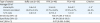
Fig. 2
Somatic mutations detected by NGS and validated by Sanger sequencing.
On the left, the results of NGS where the reads are aligned to the reference genome as provided by NextGENe software (SoftGenetics, LLC). On the right, we performed sequencing to validate the somatic nature of the mutation (c.3309dup, p.Lys1104*) by its absence in the matching buffy coat DNA. This somatic nonsense mutation was shown in FF (B and E) and FFPE (C). Wild-type was demonstrated in buffy coat (A and D).
FF, fresh-frozen; FFPE, formalin-fixed paraffin-embedded; NGS, next-generation sequencing.
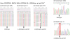
Table 1
Identified pathogenic variants* and its VAF (%) in 30 matched buffy coat, FF, and FFPE samples (subset A) and 20 matched FF & FFPE samples (subset B)

Cov., coverage; FF, fresh-frozen; FFPE, formalin-fixed paraffin-embedded; ND, not detected; NT, not tested; VAF, variant allele frequency.
*Pathogenic Variant was including pathogenic and likely pathogenic variants that were classified based on American College of Medical Genetics and Genomics standards and guidelines. GenBank accession numbers NM_007294.3 for BRCA1 and NM_000059.3 for BRCA2 were used as reference sequences.
4. False positive calls in FFPE
For reliable mutation detection across BRCA genes using FFPE samples, artifacts not of biological origin should be filtered. Of the total, 290 false positive calls were observed in subset A, and the average allele frequency of false positive calls was 8.4% (range, 5%–36%). Among the 290 false positive calls, 47.9% consisted of non-synonymous variants (41.7%, n=121) and protein-truncating variants (6.2%, n=18), while 85.8% presented as G>A or C>T Ti. Transitions (changes from A <-> G and C <-> T) occurred 35 times as frequently as Tv (Supplementary Fig. 4).
5. Filtering strategies for minimizing of sequence artifacts from FFPE DNA
The lowest limit of detection for low-frequency variants is approximately 5%–10% in tumor samples [19202122]. We used Torrent Variant Caller v5.2 after adjusting its somatic low-stringency parameters using AcroMetrix Oncology Hotspot Control at 5% allele frequency for Oncomine™ BRCA1/2 panel NGS testing. In FFPE samples, the average VAF of false positive calls was 8.6% and sequence artifacts were apparent at the range of 5%–10% VAFs. And the positive predictive value (PPV) in subset A was 51.4% using a 5% allele frequency threshold. The average calls per kilobase (kb) were 0.6, 2.3, and 0.8 in buffy coat, FFPE, and FF, respectively. The percentage of annotated variants was 96.2%, 81.7%, and 97.5% in buffy coat, FFPE and FF, respectively. The average the Ti/Tv ratio was 0.6, 6.7, and 1.0 in buffy coat, FFPE, and FF, respectively (Table 2). The three metrics consisted of 1) variants per kilobase, 2) percent annotation, and 3) Ti/Tv ratio showed significant differences between samples with or without false positive calls (p<0.001, Mann-Whitney U test). To determine the values of metrics used for selecting allele frequency threshold (5% or 10%), the cut-offs of the three metrics as a set were modulated to achieve 100% sensitivity for identifying samples with false positives (Table 2).
Table 2
The thresholds of 3 metrics for the post-filtration process in FFPE samples
Among the 125 samples, 16 (12.8%) including 2 FF and 14 FFPE, did not meet the criteria of 1) <1.2 calls/kb and >64.3% annotated, or 2) <3.8 Ti/Tv ratio. When the 14 FFPE samples were processed the post-filtration with a 10% allele frequency threshold, the PPV increased from 39.3% (95% confidence interval [CI]=36.6%–41.9%) to 71.0% (95% CI=66.3%–75.2%) and the sensitivity and negative predictive value (NPV) was 98.0% and 99.9%, respectively.
6. Sequence variants and VAF shifted heterozygous variants
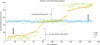
In order to create a fair comparison, among the 310 single-nucleotide variant (SNV) calls from buffy coat in subset A, the VAF (%) of 186 heterozygous variants in buffy coat were compared with those of each SNV call from FFPE and FF in subset A. Of 186 heterozygous variants, the differences of allele frequency between buffy coat and matched tumor tissue were more than 10% at 152 heterozygous variants. This phenomenon was seen across BRCA1 or/and BRCA2 genes from 19 of the 27 matched samples in subset A. Interestingly, the disproportionate allele frequencies of variants occurred both in wild type cases and BRCA1/2 mutation harboring cases (Fig. 3). Therefore, we referred to this phenomenon as ‘VAF shifted heterozygous variants.’
Fig. 3
Comparison of VAF (%) among 186 SNVs detected as “heterozygous variants” from buffy coat and other forms of samples including FFPE and FF in subset A. The variant allele frequency (%, VAF) of SNVs from buffy coat, FFPE, and FF are depicted as a square dots, triangle dots and diamond dots, respectively. The black arrow indicates the direction of change in variant allele frequency of the 186 VAF shifted heterozygous variants in FFPE and FF.
FF, fresh-frozen; FFPE, formalin-fixed paraffin-embedded; SNV, single-nucleotide variant; VAF, variant allele frequency.
7. Analytical performance
We analyzed the analytical performance using subset A. The analytical performance of BRCA1/2 NGS test was evaluated with or without VAF shifted heterozygous variants (Table 3). The sensitivity, specificity, and accuracy of BRCA1/2 NGS test were all 100% for buffy coat with or without VAF shifted heterozygous variants. The sensitivity, specificity, and accuracy for FF including heterozygous variants with VAF shift was 95.5%,100%, and 100%, respectively. When excluding heterozygous variants with VAF shift in FF, the sensitivity was increased up to 100% (Table 3). In this study, VAF shifted heterozygous variants were frequently shown in tumor tissues. When using 5% allele frequency threshold in FF, 17 VAF shifted heterozygous variants (45% to 58% VAFs in buffy coat, 7% to 22% in FFPE) were filtered out. Therefore, these VAF shifted heterozygous variants could affect the analytical performance of BRCA1/2 NGS test with tumor materials, especially the sensitivity of FF.
Table 3
Analytical performance of subset A

FF, fresh-frozen; FFPE, formalin-fixed paraffin-embedded; FP, false positive; FN, false negative; n, number of variants; NPV, negative predictive value; PPV, positive predictive value; TN, true negative TP, true positive; VAF, variant allele frequency; VAF shifted, variant allele frequency shifted heterozygous variants.
*The somatic variant was called when the variant frequency at a given position was ≥5% and variant coverage was ≥100×; †The germline variant was called when the variant frequency at a given position was ≥20% and variant coverage was ≥20×; ‡Seventeen VAF shifted heterozygous variants that detected in buffy coat with up to 50% VAFs were called with below 5% VAFs in FF.
DISCUSSION
Screening BRCA1 and BRCA2 germline variant using NGS is well established in clinical practice and is used primarily to determine hereditary breast and ovarian cancer risk [10132324]. The screening methods using buffy coat are optimized in several literatures. In our study, we compared the sequencing quality and variant call data obtained in buffy, FF and FFPE samples from all 130 samples. 100% of buffy coat and 98% of FF achieved 100% coverage at a minimum depth of 20× and mean coverage depth of 500× without re-sampling and re-testing. However, when the BRCA1/2 test was performed using FFPE, the rate of resampling and re-testing were 10% and 34% of total FFPE cases, respectively, which might be due to the damage of FFPE DNA [1725]. In a previous study, over 76% of FFPE samples were successfully analyzed, with >95% coverage of the BRCA1/2 coding regions and a mean average read depth >1,000-fold using the GeneRead DNAseq Targeted Exon Enrichment Panel [8]. In this study, re-testing increased the quality of sequencing, and the failure rate decreased from 34% to 8%.
Buffy coat, FFPE, and FF samples showed ≥99.9% of the accuracy in BRCA1/2 NGS testing. However, FFPE showed 51.4% of the positive predictive value on account of sequence artifacts. Among the 290 false positive calls in FFPE, 47.9% consisted of non-synonymous variants (41.7%, n=121) and protein-truncating variants (6.2%, n=18). The average allele frequency of false positive calls was 8.4% (range: 5%–36%). To distinguish candidate somatic mutations from sequence artifacts with more than 20% allele frequency, Sanger sequencing was applied [26]. And, from that, we confirmed the variant with maximum allele frequency (36%) in NGS was negative. Most of the false positive calls of FFPE materials in NGS were failed to confirm with Sanger sequencing. Therefore, FFPE DNA could be limited to correctly assess the somatic mutations with low allele frequency [2728]. And, tumor samples with loss of heterozygosity (LOH) [29], the allele frequency of germline BRCA pathogenic variation in tumor tissues is higher than that in buffy coat. However, one of our FFPE showed 11.1% of the allele frequency of germline BRCA1 pathogenic variation (p.Glu1694*, ID: 130227014), which is close to the 10% allele frequency threshold. In this study, the ranges of allele frequency (%) of true calls (5.3%–100.0%) and false positive calls (5.0%–36.0%) were overlapped in FFPE samples. To be used in routine BRCA NGS assay, filtering strategies for minimizing of sequence artifacts from FFPE DNA should be required to ascertain the reliability of the final report. Most artifactual changes in FFPE occur in the 1-10% allele frequency range [30]. When a 10% cut-off is applied to FFPE, clinically relevant somatic mutations with 5%–10% VAFs [31] will be missed. Therefore, we suggest applying not only a 5% cut-off strategy but also a 10% cut-off to minimize the false positive calls in FFPE. For example, when the 14 FFPE samples that did not meet the criteria (variants per kilobase, percent annotation, and Ti/Tv ratio) were proceeded using the 10% allele frequency thresholds, the PPV was increased from 39.3% to 71.0%.
In a previous study, the excellent quality of DNA extracted from FF was considered as an alternative to DNA from FFPE samples, and FF samples showed >99.7% sensitivity, specificity, and accuracy in BRCA1/2 testing [32]. However, in this study, the sensitivity of FF was 95.46% using a 5% allele frequency threshold due to VAF shifted heterozygous variants with low VAF (%). When VAF shifted heterozygous variants were excluded in evaluating analytical performance, the sensitivity would increase to 100%. In tumor tissue (FFPE and FF), VAF shifted heterozygous variants were observed in both wild-type cases and BRCA1/2 mutation harboring cases. Majority of VAF shifted heterozygous variants phenomenon in this study could be explained by chromosomal abnormalities on 13q12-q14 or 17q where BRCA2 and BRCA1 harbor, respectively. According to previous studies, more than 50% of HGSOC were showed copy number aberration on these regions [3334]. And, the allele frequency of coexisted cis-variants with BRCA1/2 pathogenic variants also contributed to shift the allele frequency based on LOH in tumor suppress gene [29]. And, this ‘VAF shifted heterozygous variants’ were observed 70% (n=19/27 pairs) cases of subset A in our study. As VAF shifted heterozygous variants showed a decreasing trend of allele frequency, the number of false negative calls increased in tumor tissue. These findings from FFPE and FF indicate that the false-positive calls and VAF shifted variants might be drawbacks of BRCA1/2 NGS assay using only tumor tissue.
In December 2014, the EMA approved the PARP inhibitor olaparib as monotherapy treatment for relapsed platinum-sensitive HGSOC patients with pathogenic germline or somatic mutations in BRCA1/2 [3]. Approximately 6%–25% of OC patients have a BRCA1/BRCA2 germline mutation [35], and somatic BRCA mutations occur in approximately 3%–7% of ovarian cancer cases [63637]. Since a simultaneous detection of germline and somatic mutations in OC is cost-effective, there is an increasing clinical need for routinely screening BRCA using tumor materials [38]. In this study, buffy coat achieved 100% coverage at 20× and 100× without re-sampling or re-analysis, and they showed 100% of the sensitivity, specificity, and accuracy. The buffy coat was not affected by shifted allele frequency of variants or false calls which are intrinsic obstacles of tumor materials. However, due to VAF shifted heterozygous variants, tumor materials (FFPE and FF) showed lower sensitivity (95.5%–99.0%) than buffy coat (100%). Our results showed that the VAF shifted heterozygous variants with low VAF (%) can be neglected during the variant calling in tumor samples when the usual 5 allele frequency threshold were applied. Furthermore, sequence artifacts could not be distinguished from pathogenic variants because the ranges of VAF (%) of true calls and false positive calls are overlapped. According to recently updated guidelines for BRCA1/2 and above findings, it is reasonable to test OC blood samples for BRCA mutations first, and then, if negative, carry testing with a tumor sample to extend patients group who might potentially benefit from PARP inhibitors therapy [3940].
We improved the performance of BRCA1/2 NGS assay through the post-filtration process using the thresholds of metrics and exclusion of VAF shifted heterozygous variants from different tissue types. In conclusion, we suggest the strategy of germline screening BRCA1/2 from buffy coat prior to testing from tumor materials to obtain clinically reliable results.
 XML Download