

 PDF
PDF ePub
ePub Citation
 Print
Print


Introduction
Amblyopia is the most common cause of preventable visual disability in children, occurring in approximately 2% to 5% of the general population [1]. Generally, amblyopia refers to the reduction of best-corrected visual acuity (BCVA) in one or both eyes that cannot be exclusively attributed to a structural abnormality of the eye. Poor vision caused by structural abnormalities of the eye and brain (such as congenital cataracts, optic nerve atrophy, retinal dystrophy, or anoxic brain damage) is regarded as organic amblyopia. Amblyopia interrupts normal visual pathway development during childhood. Amblyopia can be reversed when appropriate visual stimulation is provided within the sensitive period of visual development.
Normal Visual Development and Vision Screening
Amblyopia is caused by abnormal visual processing early in life. Visual acuity (VA) at birth is poor due to the immaturity of visual processing. Appropriate visual stimulation, including clear retinal images, equal image clarity in both eyes, and proper ocular alignment are needed for normal visual development. VA improves rapidly during the first few months of life as clear retinal images begin to stimulate neurodevelopment of visual centers [23]. Although the length of the critical period for visual development is somewhat controversial, it is generally considered to be from birth to 3 months of age [23]. Visual development continues until 7 to 8 years of age (sensitive period) and neural plasticity for visual development is progressively reduced in late childhood [234].
Children under 7 years of age who are not provided appropriate visual stimulation are vulnerable to amblyopia, and the earlier the onset of abnormal visual stimulation, the greater the visual deficit. Children should therefore be screened for vision problems and properly treated early in their life [56]. In South Korea, a nationwide infant and toddler vision screening program was initiated in 2008 as a part of the Infant Health Screening Project of the Korean National Health Insurance Corporation [7]. The program screens children aged 4 months to 6 years for the presence of frequent and important ocular problems. Unilateral or bilateral vision problems, strabismus, and refractive errors are important target diseases for vision screening in childhood.
Diagnosis and Classification of Amblyopia
Abnormal visual stimulation by a unilateral or bilateral blurred retinal image or a misalignment of two eyes can lead to permanent damage to the visual processing to the visual center in the brain. The diagnosis of amblyopia requires detection of decreased VA and identification of the cause associated with the visual problem. Amblyopia rarely develops in the absence of strabismus, unequal refractive error, media opacity, or other ocular and neural abnormalities. When the cause for the visual deficit is not obvious, clinicians should search for a possible alternative diagnosis that predisposed the child to decreased vision. Bilateral amblyopia is usually diagnosed when BCVA is worse than 20 / 50 in children aged ≤4 years, <20 / 40 in children aged 4 to ≤5 years, or <20 / 30 in children aged >5 years [1]. Unilateral amblyopia is defined clinically as a between-eye difference in BCVA of 2 or more lines [1]. Anisometropia and strabismus are the two main amblyogenic factors, and some children have combined anisometropic-strabismic amblyopia. Amblyopia is generally classified as follows: strabismic (misalignment of the visual axis), refractive, anisometropia (difference in refraction of the two eyes), isoametropic (bilateral high refractive error), visual deprivation (media opacity, ptosis, etc.), and occlusion.
Treatment of Amblyopia
The basic strategy to treat amblyopia is to provide a clear retinal image to each eye and correct ocular dominance. Treatment modalities include correcting any underlying organic disease, prescribing appropriate optical correction, and providing occlusion/penalization therapy for the dominant eye within the sensitive period of visual development. Amblyopia treatment can effectively restore normal or near-normal visual function by eliminating eccentric fixation and/or developing more extensive synaptic input to the visual cortex [89]. Detection and management of amblyopia should be initiated as early as possible during the sensitive period for visual development because the success of amblyopia treatment decreases with increasing age [5671011].
The Pediatric Eye Disease Investigator Group (PEDIG) is a clinical network of pediatric ophthalmologists funded by the US National Eye Institute to conduct clinical research studies related to pediatric eye disorders. The Amblyopia Treatment Studies conducted by PEDIG have focused on evaluating the effectiveness and duration of different amblyopia treatment modalities for children and adolescents. Their results have provided an evidence-based treatment approach and changed clinical practice guidelines for children with amblyopia. Many pediatric ophthalmologists now follow the current recommendations suggested by the PEDIG. This review is mainly based on the results from PEDIG studies. We also searched for other clinical studies for amblyopia and describe the results in this review
Optical correction
Optical correction of refractive error is the initial step to provide a clear retinal image to each eye for children with amblyopia. A guideline for consideration of refractive correction for children is described in the Preferred Practice Pattern guidelines published by the American Academy of Ophthalmology in 2018 [12]. Optical correction should be tailored to the individual child together with consideration of the findings of the clinical examination, visual symptoms, and patient history.
A PEDIG trial has investigated the effect of spectacle correction on bilateral refractive amblyopia in children aged 3 to less than 10 years with BCVA of 20 / 40 to 20 / 400 without previous treatment [13]. Spectacle correction resulted in improvement of binocular BCVA: 73% of children achieved VA of 20 / 25 or better within one year of starting treatment; binocular VA improved by an average of 3.9 lines and children with worse baseline VA had greater improvement in terms of the number of lines; however, the probability of achieving 20 / 25 or better binocular VA was greater in children who had better baseline VA [13]. Some studies have suggested that spectacle correction alone can be used as a treatment for anisometropic amblyopia [1415]. Refractive correction alone improved VA by ≥2 lines in 77% of patients and resulted in resolution of amblyopia in at least one third of children aged 3 to 7 years who had untreated anisometropic amblyopia [15]. Most cases where resolution was reported with refractive correction alone had moderate (20 / 40 to 20 / 100) amblyopia. For children with greater degrees of amblyopia, initial treatment with spectacles resulted in a 3-line improvement in VA on average, which may have reduced the burden of subsequent amblyopia therapy. In addition, a study of children aged 7 to 17 years found that amblyopia improved by 2 or more lines with optical correction alone in about one fourth of children enrolled [16]. The effects of spectacle correction on strabismic amblyopia have also been investigated [1718]. Refractive correction with spectacles alone resulted in a clinically meaningful improvement in VA in the amblyopic eye in the majority of previously untreated strabismic children. Overall, a mean improvement of 2.6 lines occurred in the amblyopic eye, with 75% of children showing improvement of ≥2 lines and 54% of children showing improvement of ≥3 lines. Resolution of amblyopia occurred in 32% of patients [17].
Patching
Patching or occlusion therapy involves covering the sound eye to stimulate the amblyopic eye. It has been the mainstay of amblyopia treatment for the past 250 years. Patching is currently the preferred treatment option among ophthalmologists, although other methods such as atropine penalization have been shown to provide equivalent remediation for moderate and severe amblyopia [11011].
Over the last decade, several randomized controlled clinical trials have provided evidence as to how to tailor amblyopia therapy more precisely to achieve the best visual outcome while minimizing negative impacts on children and their family. There have been many studies on the number of hours of patching per day [1920212223]. In a PEDIG trial, 6 hours per day occlusion treatment produced an improvement of VA that was of similar magnitude to that obtained by full-time occlusion in children 3 to less than 7 years of age with severe amblyopia (20 / 100 to 20 / 400) [19]. In children who had moderate amblyopia (20 / 40 to 20 / 80), initial occlusion therapy for 2 hours daily resulted in an improvement in VA that was similar in magnitude to the improvement produced by prescribing 6 hours of daily patching [20]. After 17 weeks of treatment, children with severe amblyopia showed a mean improvement of 3.6 lines with 2 hours of daily patching [21]. For children with residual amblyopia after treatment with refractive correction and 2-hour patching, increasing the daily patching time to 6 hours resulted in an average of 1.2 lines of statistically significant additional improvement at 10 weeks, compared to an average 0.5 line additional improvement when continuing with just 2 hours of patching [22]. Improvement of ≥2 lines occurred in 40% of participants after patching for 6 hours versus 18% of those who continued 2-hour patching. When vision in the amblyopic eye stops improving with two hours of daily patching, increasing the daily patching duration to 6 hours is recommended to obtain greater improvement in VA [2223]. Patching should be considered even in older children and teenagers, particularly if they have not been previously treated [101624]. For patients aged 7 to 17 years, prescribing 2 to 6 hours per day of patching together with near visual activities can improve VA even if the amblyopia has been previously treated [16].
Clinicians should be aware that children treated with patching may develop occlusion amblyopia or strabismus. Amblyopia patients who had previously been treated with patching were found to have deterioration and improvement of ocular alignment [2526]. Parents should be informed that change in ocular alignment may occur in some amblyopia patients after patching.
Atropine penalization
Penalization using atropine produces cycloplegia in the non-amblyopic eye. It is an effective choice for treating children who do not show improvement with spectacles alone. Atropine (1%) eye drops lead to loss of accommodation and optical defocusing in the non-amblyopic eye and forced use of the amblyopic eye. Atropine is most effective when the non-amblyopic eye is hyperopic. The importance of a fixation switch to the amblyopic eye has been considered an important predictor of success with atropine penalization [2728].
PEDIG trials have compared patching and atropine as treatments for moderate amblyopia [27282930]. Both atropine penalization and patching were effective treatments for children with moderate amblyopia. VA improved by 3.16 lines in the patching group and 2.84 lines in the atropine group after 6 months of treatment in children younger than 7 [29]. Although improvement was initially more rapid in the patching group, the between-group difference in VA was small and statistically non-significant after 6 months [29]. A PEDIG trial that enrolled children from 3 to 12 years of age with severe amblyopia (20 / 125 to 20 / 400) found that their vision also improved with weekend atropine [31].
Atropine is initially prescribed daily for amblyopia treatment. The cycloplegic effect of topical atropine usually lasts for several days. Less frequent dosing of atropine may enhance compliance with treatment. Several studies have investigated daily use and weekend use of atropine for amblyopia treatment [3233]. A study by Simons et al. [32] has reported that intermittent atropine therapy (one or two days a week) is as successful as daily atropine therapy. The first prospective clinical trial by PEDIG found that the magnitude of improvement after 4 months of treatment was 2.3 lines with both daily and weekend atropine regimens [27]. Prescribing weekend atropine appeared to be as effective as daily atropine in treating children with moderate amblyopia [27]. Weekend atropine seems to be equally effective and produces greater compliance. It should be noted that atropine had less pronounced dilating and cycloplegic effects in children with dark iris pigmentation [3334]. When initiated before age 7, improvement achieved with patching or atropine treatment for moderate amblyopia due to strabismus, anisometropia, or both was maintained until 10 to 15 years of age in children with a mean VA in their amblyopic eye of about 20 / 25 [35].
Amblyopia treatment in older children
Although it is true that amblyopia can be treated more effectively in younger children, several studies have shown that older children and adults with amblyopia are also able to respond to amblyopia treatment [41016243637383940]. A randomized PEDIG trial assessing treatment of amblyopia in children aged 7 to 17 years [16] revealed the following results: (1) amblyopia improved with optical correction alone in about one fourth of patients aged 7 to 17 years, (2) for patients aged 7 to 12 years, 2 to 6 hours per day of patching together with near visual activities and atropine improved VA even if the amblyopia had been treated previously, (3) even for patients 13 to 17 years of age, patching for 2 to 6 hours per day together with near visual activities improved VA when amblyopia had not been treated previously. A recent analysis of the Preferred Practice Pattern for amblyopia by the American Academy of Ophthalmology recommended treatment for children up to 10 years of age [1].
Compliance with prescribed amblyopia treatment
Compliance is defined as the extent to which a patient's behavior matches the prescribed treatment. Many studies have demonstrated that treatment compliance is the most critical factor for predicting a successful outcome in amblyopic children [4142]. Reported rates of compliance with prescribed treatment range widely. Compliance with amblyopia therapy may be improved by educational programs or more effective communication with children and their parents. Poor parental knowledge has been reported to be associated with poor compliance [424344]. A study by Newsham [44] reported that a large proportion of patients could benefit by increasing parental knowledge in several key areas, such as critical period, importance of amblyopia therapy, and potential negative consequences of not treating amblyopia. Written information may be a simple, inexpensive, and effective method to improve parental understanding of amblyopia and subsequent compliance of their children with prescribed treatment/s. Increasing the frequency of physician check-up and a more direct communication between physician and child can also contribute to improved compliance with treatment and better final visual outcome [41]. Beginning therapy with a less intensive patching regime can also help improve patient compliance [20212223]. Splitting part-time patching hours into 2 or more smaller sessions has been reported to provide visual improvement comparable to equivalent numbers of continuous hours of part-time patching for children with anisometropic amblyopia while creating less emotional stress [45]. Patching is usually administered by applying an opaque adhesive patch directly to the skin surrounding the non-amblyopic eye. Periocular skin irritation due to adhesive skin patches during amblyopia treatment can cause considerable distress and reduce compliance with treatment. A recent study has shown that treatment with either adhesive skin patching or over-glasses patching produced similar degrees of vision improvement of the amblyopic eye [46].
New Treatments for Amblyopia
Alternative therapies for amblyopia treatment have long been a topic of interest among researchers and clinicians. New technologies have been incorporated into standard therapy for amblyopia as our understanding of the pathophysiological basis of amblyopia has increased.
Intermittent occlusion glasses
With regard to advances in occlusion therapy for amblyopia, an electronic device, Amblyz liquid crystal intermittent occlusion glasses (XPAND 3D Group, Ljubljana, Slovenia), has been introduced. Intermittent occlusion glasses are programmed to unilaterally alternate between opaque and transparent phases at 30-second intervals, providing effective occlusion of the fellow eye 50% of the time they are worn. Because these glasses are more child friendly and do not produce the side effects seen with adhesive skin patches, they may potentially improve compliance with occlusion therapy. However, a recent study to monitor objective compliance with intermittent occlusion glasses using a microsensor affixed to the glasses found that general compliance was not as high as was anticipated for this new technology (averaging 51.6%) and varied greatly from patient to patient (range, 10% to 97%), and in addition, daily compliance decreased slightly over time [47]. There have been several studies assessing the effectiveness of the intermittent occlusion glasses [484950]; however, there has been only one randomized clinical trial comparing the effectiveness of liquid crystal occlusion glasses and adhesive occlusion patches [50]. After 12 weeks of treatment, 4 hours' daily intermittent occlusion therapy with liquid crystal glasses was not inferior to 2 hours' daily patching when treating children 3 to 8 years of age with moderate, unilateral amblyopia [50]. However, there have been no further studies to confirm the effectiveness of intermittent occlusion therapy glasses, in terms of age, severity and subtype of amblyopia.
Perceptual learning
Perceptual learning refers to any relatively permanent and consistent change in the perception of a sensory task following repeated practice [5152]. Visual performance may be improved with repetitive practice of specific controlled visual tasks. Persistence of binocular cortical communication in subjects with amblyopia is the basis for the hypothesis that activation of these persistent binocular neural circuits might awaken an amblyopic eye. Since the Cambridge Stimulator treatment described in the 1970s, which was the first application of perceptual learning theory to amblyopia [53], perceptual learning to various visual tasks has resulted in improvement in orientation discrimination, stereoacuity and contrast sensitivity, even in adults with amblyopia [545556]. Perceptual learning seems to be a promising method; however small numbers of participants in the previously published studies and lack of long-term follow-up currently limit widespread use of perceptual learning as a therapeutic option for amblyopia.
Dichoptic training
It has been suggested that patching may further disrupt binocularity and theoretically may not be an ideal method to restore binocular cortical function in amblyopic patients [5758596061626364]. Binocular visual stimulation using computer games played on a smartphone or computer tablet has been suggested as a means to improve not only VA, but also binocular function [5758596061626364]. Dichoptic presentation refers to presenting different images to each eye. Given the amblyopic eye has lower contrast sensitivity compared to the sound eye, when employing dichoptic presentation as a treatment method, children with amblyopia are trained on tasks in which reduced contrast images are presented to the sound eye while higher contrast images are shown to the amblyopic eye in order to balance cortical input and overcome inter-ocular suppression. Early non-randomized studies of binocular visual stimulation have demonstrated promising results [596061]. Recently, several clinical trials employing different testing protocols and different age groups with or without prior amblyopia treatment have been completed to investigate the effectiveness of binocular computer tablet treatment for amblyopia [6061626364]. A PEDIG trial has compared VA improvement in children with amblyopia treated with a binocular iPad game for 1 hour a day vs. part-time patching for 2 hours a day in 385 children aged 5 to 12 years with amblyopia [60]. At 16 weeks, mean amblyopic-eye VA improved 1.05 lines in the binocular group and 1.35 lines in the patching group, with an adjusted treatment group difference of 0.31 lines (favoring patching). Binocular treatment of amblyopia using videogames (BRAVO) has been performed to compare the effectiveness of a binocular video game with a placebo video game for improving visual functions [64]. The results indicated that the binocular video game did not improve visual outcomes more than the placebo video game in older children and adults [64]. In children aged 13 to 16 years, improvement in amblyopic eye VA with a binocular iPad game was not better than patching [62]. There was no benefit with respect to VA or stereoacuity after 4 or 8 weeks of treatment with the dichoptic binocular Dig Rush iPad game in children aged 7 to 12 years who had received no previous treatment for amblyopia other than spectacles [63]. Although research has been ongoing, to date, evidence supporting the inclusion of binocular treatment for amblyopic patients remains insufficient.
Transcranial magnetic stimulation
Transcranial magnetic stimulation (TMS) is an established, safe, and noninvasive technique for stimulating the human brain. The technique is based on the principle of electromagnetic induction, whereby a brief magnetic field is generated within a plastic-coated coil of wire that is placed on the head above the cortical area to be stimulated. The magnetic field passes painlessly through the skull and induces a weak electrical current within the underlying region of cortex. As a result, the neural excitability of the stimulated region may be temporarily altered. The first study to assess the effect of repetitive TMS on visual function in adult with amblyopia demonstrated a transient improvement in contrast sensitivity [65]. TMS is expected to enhance the effects of traditional amblyopia treatments [656667]. The effects of repeated applications of TMS as a therapeutic option in amblyopia are currently being investigated.
Pharmacologic therapy
Dopamine concentrations have been found to be decreased in the retina with deprivation amblyopia [68]. Increasing retinal dopamine concentrations may have a beneficial effect in amblyopia. In the f irst study using levodopa, the immediate metabolic precursor of dopamine, as a possible treatment in adult amblyopia, a single administration of levodopa temporarily improved contrast sensitivity and decreased scotoma size in the amblyopic eye [69]. A PEDIG randomized trial of levodopa as a treatment for residual amblyopia in children aged 7 to 12 years showed that treatment with oral levodopa while continuing to patch for 2 hours daily did not produce a clinically or statistically meaningful improvement in VA compared to placebo and patching [70]. Cytidine diphosphatecholine is a complex organic molecule that acts as an intermediate in the biosynthesis of cell membrane phospholipids and has been hypothesized to protect the anatomic and structural integrity of cell membranes, thereby preventing nerve cell damage [717273]. A study with adult amblyopic patients demonstrated improvement in VA with citicoline augmentation of patching that was not sustained following cessation of the medication [7172]. Early studies in amblyopic children were promising, showing treatment effects with citicoline both alone and in addition to patching [73]. Potential long-term effects of using levodopa and other medications with psychoactive and extrapyramidal effects in an immature nervous system of children are of concern [74]. Restoring cortical plasticity and reducing interocular suppression have received attention as novel therapeutic strategies for amblyopia [7576777879].
Follow-up and Cessation of Amblyopia Treatment
The response to amblyopia treatment should be monitored and the treatment plan should be adjusted as necessary: VA in the amblyopic eye and fellow eye, refraction in both eyes, adherence to the prescribed treatment, and treatment-related side effects should be included in follow-up examinations. Follow-up assessments are usually arranged at six week intervals after initiating amblyopia treatment. Based on the results of previous studies, amblyopia treatment is typically initiated using spectacles alone, followed by the addition of low-dose patching or weekend atropine in a stepwise manner. If VA in the amblyopic eye has been improving with stable VA in the fellow eye, the current treatment may be continued. If no further improvement in VA is found and residual amblyopia persists in spite of good adherence to treatment, increasing treatment intensity (for example, increasing patching from 2 to 6 hours daily) or changing/adding a treatment modality (for example, switching to topical atropine administration) should be considered. An alternative diagnosis compatible with decreased vision should be considered for children who fail to show any improvement in VA after amblyopia treatment. Thorough ophthalmologic re-examinations should be performed to rule out any organic disorders, such as inherited retinal dystrophy or optic nerve/macular abnormalities.
Although amblyopia can be successfully treated by optical correction, patching, and atropine penalization, recurrence of amblyopia has been reported, with incidences ranging from 6% to 67% [80818283]. In a PEDIG prospective observational study of successfully treated amblyopic children less than 8 years of age for whom treatment had been discontinued, the overall amblyopia incidence of recurrence was 24% over 1 year of follow-up [80]. Recurrence was similar among those patients who discontinued patching (25%) and those who stopped atropine (21%). Recurrence occurred throughout the 52-week follow-up period, although recurrences occurred more frequently during the first 13 weeks after cessation of treatment. In patients treated with moderately intense patching (6 to 8 hours per day), recurrence was more common (42%) when treatment was not reduced prior to cessation than when treatment was reduced to 2 hours per day prior to cessation (14%).
When maximum VA is achieved, treatment should be tapered and then discontinued with monitoring for amblyopia recurrence. De Weger et al. [84] have recommended the follow-up period be longer than 2 years in the presence of risk factors such as increasing anisometropia. Clinicians should be aware of the possibility of vision deterioration after cessation of amblyopia treatment. Careful and prolonged follow-up is needed for all children who have been successfully completed previous treatment for amblyopia [80838485].
Conclusion
Amblyopia may be reversible with appropriate visual stimulation. The detection and management of amblyopia should begin as early as possible. Several studies have shown that older children and adults with amblyopia also respond to amblyopia treatment. Thus, treatment should be attempted in older children.
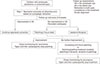
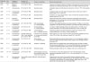
Amblyopia should be treated in a stepwise manner (Fig. 1). The results from various PEDIG studies are summarized in Table 1 [1315161718192021222730313980]. Prescribing an appropriate refractive correction is an initial step when treating amblyopic patients. Patching for 2 hours each day or pharmacologic penalization may be considered as an additional treatment option for amblyopia. If there is no improvement between two consecutive visits, increasing the prescribed patching duration to 6 hours per day could be a reasonable option. Good compliance with prescribed amblyopia treatment is critical for a successful visual outcome. Compliance with amblyopia therapy may be improved by educational programs, direct communication with children and their parents, and increasing the frequency of follow-up visits. Treatment should be tapered and then discontinued with monitoring for amblyopia recurrence. Follow-up visits are required after cessation of treatment to detect any deterioration of vision and provide prompt re-treatment, if necessary.
 XML Download