

 PDF
PDF ePub
ePub Citation
 Print
Print


INTRODUCTION
The simultaneous presentation of a mediastinal cyst with an accompanying vertebral abnormality is the hallmark of a neurenteric cyst (1). Neurenteric cysts arise during early embryogenesis when the foregut and notochord are in close proximity. Adhesion between the two may cause the foregut to invaginate and pinch off, forming an enteric cyst that may demonstrate intraspinal extension. Therefore, the coexistence of vertebral anomalies is strongly suggestive of neurenteric cysts. Neurenteric cysts are typically located in the posterior mediastinum (2). The patient in our case had a posterior mediastinal cystic mass and a butterfly vertebra, whose later pathological examination proved to be a bronchogenic cyst.
CASE REPORT
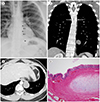
A 27-year-old man presented with chest X-ray abnormalities. He had no clinical symptoms and no medical history. Physical examination and routine laboratory test results were normal. Chest radiography showed an approximately 5 cm, sharply-defined round mass in the left paravertebral area, obscuring left paraaortic interface, and a paravertebral stipe. These findings indicated that the mass was located in the posterior mediastinum (Fig. 1A). On CT scan, the mass was a well-circumscribed, hypodense cystic lesion without contrast enhancement (28 Hounsfield unit) in the posterior mediastinum, near the descending thoracic aorta and 11th thoracic vertebra. The cyst had no definable wall or calcification (Fig. 1B). Coronal reformatted CT images revealed that the thoracolumbar curvature was “S”-shaped and the 6th thoracic vertebral body had a sagittal cleft through the body with the appearance of a butterfly. There was no communication between the cystic mass and the cleft (Fig. 1C). Our preoperative diagnosis was a neurenteric cyst associated with a butterfly vertebra. The patient underwent video-assisted thoracoscopic surgery for excision of the mass. Examination of the pathologic specimen revealed that the cyst wall was lined by ciliated pseudostratified columnar epithelium, including mature cartilage and submucosal glands (Fig. 1D).
DISCUSSION
Foregut cysts probably result from aberrant development of the primitive foregut and are classified into three types: bronchogenic, esophageal duplication, and neurenteric cysts. Bronchogenic cysts are thought to arise from the abnormal budding of the ventral foregut, which forms the tracheobronchial tree. Histologic findings of respiratory epithelium provide the definitive diagnosis of a bronchogenic cyst. Esophageal duplication cysts are thought to arise from the dorsal foregut, destined to be the alimentary tract. Histologic findings of a submucosal or muscular layer of the gastrointestinal tract provide the definitive diagnosis of an esophageal duplication cyst (34). Neurenteric cysts result from the failure of the alimentary tract to separate from the primitive neural crest, leaving a persistent communication between the foregut and the spinal cord (1). Histologically, both neural elements and gastrointestinal epithelium are typically seen (3).
Each type of foregut cyst has similar CT findings, with characteristic locations in the chest. On CT, all three types of foregut cysts usually appear as solitary well-circumscribed round or oval cystic masses with fluid attenuation (35). Bronchogenic cysts are most frequently found in the middle mediastinum, near the carina, and less frequently in any part of mediastinum or chest (67). Esophageal duplication cysts are usually located along the esophagus in the lower posterior mediastinum. Most neurenteric cysts are located in the posterior mediastinum (68). In cases where these cysts share anatomic locations, differentiation by CT becomes very difficult (6).
Foregut cysts may be accompanied by other congenital anomalies. Bronchogenic cysts are occasionally associated with congenital pulmonary malformations and 12% of patients with esophageal duplication cysts also have malformations of the gastrointestinal tract (1). Half of all neurenteric cysts are associated with cervical and upper thoracic vertebral anomalies, such as hemivertebrae, butterfly vertebrae, anterior spina bifida, and scoliosis. Intravertebral extension of the foregut can disrupt vertebral body development and induce a sagittal cleft defect. Because of the cephalic growth of the notochord and caudal growth of the foregut, any associated vertebral defects are typically superior to the mediastinal cyst (4). CT can demonstrate a connection of the cyst to the spinal defect. However, they can also manifest as an isolated mediastinal cyst with no spinal connection or they may have a fibrous tract to the attached spine. Therefore, the combination of a posterior mediastinal cyst with vertebral defect anomalies is virtually diagnostic of a neurenteric cyst, with or without a spinal connection (1).
In our case, a typical mediastinal cyst was located in the posterior mediastinum, ventral to the 11th thoracic vertebral body. There were two vertebral anomalies, a butterfly vertebra of the 6th thoracic vertebral body and scoliosis. The vertebral anomaly was superior to the cyst. All these findings strongly identified the cyst as a neurenteric cyst. However, examination of the pathologic specimen revealed respiratory epithelium lining in the cyst and identified it as a bronchogenic cyst. We could not find any case of a bronchogenic cyst associated with the presence of a butterfly vertebra and scoliosis using Google and Pubmed. A bronchogenic cyst can be present in a patient with vertebral anomalies. If the communication between the cyst and the vertebral defect is not clearly identified, a bronchogenic cyst, as well as a neurenteric cyst, should be included in the differential diagnosis.
 XML Download