

 PDF
PDF ePub
ePub Citation
 Print
Print


INTRODUCTION
The rapid development of imaging modalities for identification and evaluation of the heart has revealed a variety of normal cardiac anatomical variations. The coumadin ridge, one such normal variation, often appears as a pseudotumor between the left atrial appendage and left upper pulmonary vein. This ridge is called the “coumadin ridge,” because, in the past, it was mistaken for a thrombus and treated unnecessarily with anticoagulants such as coumadin or warfarin (1). In this report, we describe the findings for a patient with a past history of stroke who underwent echocardiography and cardiac CT and was then surgically confirmed as having a coumadin ridge.
CASE REPORT
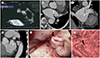
A 66-year-old woman visited our hospital with a complaint of nonspecific chest pain. The patient had experienced a cerebral infarction 2 years ago and had a history of hypertension and diabetes. Transesophageal echocardiography was performed in the cardiology department, and an echogenic mobile mass was found at the Q-tip (between the left atrial appendage and left upper pulmonary vein). The size of this mass was 0.6 × 0.7 cm, and the mass undulated with cardiac motion (Fig. 1A). The cardiologist considered this lesion to be a cardiac tumor or thrombus.
A prospective cardiac CT scan was then performed and a low-attenuation lesion approximately 7 mm in size with a stalk-like structure at the junction of the left atrial appendage orifice and left upper pulmonary vein was found on CT (Fig. 1B–D). Considering the patient's previous history of stroke and the results of transesophageal echocardiography, the lesion on the cardiac CT scan was thought to be a benign tumor, such as myxoma, or an intracardiac thrombus. Since the patient's cerebral infarction history was likely to be due to cardioembolism, the neurologist and cardiologist concluded that it would be better to remove the lesion by surgery to prevent a secondary stroke, without having a radiologist interpret the cardiac CT findings.
The patient underwent surgery at the cardiac surgical department for removal of the lesion. During the operation, the operator could not observe any abnormal finding, including any mass. The mass seen on the CT image was a prominent left atrial appendage tissue in the left atrium (Fig. 1E).
Thus, the findings noted on transesophageal echocardiography and cardiac CT were surgically confirmed to be a pseudotumor coumadin ridge, which was a fold of the left atrial wall protruding to the left atrial surface. Subsequently, we retrospectively reconstructed the CT image and compared it with the operative findings. The findings on the reconstructed image were consistent with the operative findings (Fig. 1F). The patient's chest pain symptoms improved, and she was then discharged. Although the cause could not be determined, the patient's initial chest pain did not seem to be due to a cardiac lesion.
DISCUSSION
A variety of normal anatomical variants involving the heart have been identified, and their locations and properties have been elucidated with the development of various imaging modalities such as cardiac CT (12). Radiologists should be aware of the normal variants and should be able to distinguish these from lesions mimicking a mass or abnormal lesion in cardiac imaging.
The coumadin ridge is a linear or nodular mass protruding into the left atrium. It can be mistaken as a tumor or thrombus owing to similarities in its shape and location. Moreover, the distal tip of the coumadin ridge may be large and can protrude into the left atrium and left atrial appendage, making it look like a clot or a small tumor (34). In the past, patients with this ridge were prescribed anticoagulants such as coumadin, and thus, it was named “coumadin ridge” (1). However, because the coumadin ridge is a normal variant, unlike myxoma or thrombus, it does not cause embolism and should be differentiated from other lesions. Therefore, CT as well as cardiac MRI or echocardiography may be helpful for this purpose.
The unique location of the coumadin ridge (i.e., between the left atrial appendage and left upper pulmonary vein) is an important clue to correct identification of the ridge. In addition, because of its round shape, the coumadin ridge is often referred to as a “Q-tip sign” on echocardiography (2). Moreover, the intensity or density of the coumadin ridge on cardiac MRI or CT is similar to those of a thrombus and some cardiac tumors (4).
According to previous studies, the characteristics that could differentiate a thrombus from a coumadin ridge are that the thrombus has different echogenicity in comparison with the surrounding tissues and that it moves with adjacent tissues (3). In addition, the most common location of the left atrial thrombus is the left atrial appendage. Moreover, atrial myxoma can be sessile or pedunculated, and about 75% of myxoma cases are found in the left atrium (34). However, most myxomas can be distinguished from a coumadin ridge because they are located on the interatrial septum, near the fossa ovalis in the left atrium.
In the present case, the echogenic mobile mass was found between the left atrial appendage and left pulmonary vein on transesophageal echocardiography and cardiac CT in a patient who presented with nonspecific chest pain. While the mass showed similar echogenicity with the surrounding tissues, a thrombus could not be ruled out, since the mass undulated with cardiac motion. In addition, while it was found in the typical location of the coumadin ridge, the possibility of myxoma was also considered, because it looked like a nodular mass with a stalk.
Therefore, considering the patient's previous history of stroke and the results of transesophageal echocardiography, the lesion on cardiac CT was thought to be a benign tumor such as myxoma or an intracardiac thrombus. The patient underwent surgery for removal of the lesion to prevent the recurrence of cerebral infarction. However, the surgical examination revealed no abnormal finding other than the left atrial appendage tissue at the site of the preoperatively identified mass-like lesion in the left atrium. The chest pain improved by itself, and the patient was discharged.
In this case, we misidentified the case as a myxoma, and we were able to confirm the operative findings for a coumadin ridge for the first time. Subsequently, we reconstructed the CT image; the findings on the reconstructed image were consistent with the operative findings (Fig. 1F). Therefore, cardiac CT reconstruction allows more accurate diagnosis by identifying the typical position of the coumadin ridge, i.e., the junction of the left atrial appendage orifice and left upper pulmonary vein.
In conclusion, herein, we present a surgically confirmed case of coumadin ridge. This case emphasizes the need for radiologists to be aware of the typical location and features of the coumadin ridge and distinguish it from lesions mimicking a mass or abnormal lesion in cardiac imaging.
 XML Download