

 PDF
PDF ePub
ePub Citation
 Print
Print


INTRODUCTION
Necrotizing fasciitis (NF) is a life-threatening infection characterized by extensive necrosis of subcutaneous tissue and the fascia (1). NF is relatively rare, although its prevalence is increasing due to an increase in the number of immunocompromised patients with human immunodeficiency virus infection, diabetes mellitus, cancer, alcoholism, vascular insufficiencies, and organ transplants. NF can be idiopathic, and it occurs after trauma or around foreign bodies in surgical wounds (234). Although NF can occur anywhere in the body, it commonly affects the extremities, followed by the trunk and perineum. Only a few cases of NF in the breast have been reported, with the first reported case by Shah et al. (5); however, specific imaging findings of primary breast NF have not been described in the literature. Due to its rarity, diagnosis of primary breast NF can be delayed. Early diagnosis is crucial because delays can lead to fatal sepsis or requirement for inevitable total mastectomy. It has been reported that mortality rates can reach 73% but can be reduced with early diagnosis and prompt institution of appropriate management strategies (67).
Herein, we present a rare case of primary NF of the breast in a patient with untreated diabetes, who was diagnosed using breast enhanced CT and successfully managed with local debridement.
CASE REPORT
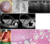
A 53-year-old female presented to the emergency department with a five-day history of breast pain and abrupt swelling of the right breast. The patient had undergone an operation for mastitis 10 years previously. On inspection, diffuse skin erythema and swelling were observed in the right breast, and the patient reported severe pain and abrupt onset of breast swelling (Fig. 1A).
The results of initial laboratory tests revealed leukocytosis (31620/µL), increased erythrocyte sedimentation rate (100 mm/h), and increased C-reactive protein level (164.9 mg/L). Her body mass index was 33 kg/m2, with no other identified underlying medical history; however, her initial serum glucose level was 470 g/dL and hemoglobin A1c was 12.3%, suggesting untreated diabetes. Chest radiography revealed diffuse subcutaneous emphysema in the right chest wall and neck (Fig. 1B). Emergency ultrasonography (US) was performed and revealed diffuse posterior shadowing with limited evaluation due to an air collection (Fig. 1C). Due to suspected soft tissue necrosis, contrast-enhanced breast CT was promptly performed for further evaluation. Enhanced breast CT scans were acquired using a dual-source 128-slice CT scanner (Somatom Definition FLASH, Siemens Healthcare, Erlangen, Germany). Scanning was performed from the lower part of the breast to the level of the upper neck. Scanning was performed before and after intravenous administration of contrast medium (120 mL Iohexol, Bonorex 350, Central Medical Service, Seoul, Korea) with a power injector (Dual-shot GX7, Nemoto Kyorindo Co., Ltd., Tokyo, Japan) at an injection rate of 2.5 mL/sec. The scanning parameters were as follows: power, 120 kVp; current, 35–257 mA; tube rotation time, 0.28 seconds; and pitch 0.6.
Enhanced CT scan of the breast revealed a cystic mass, 23 × 20 cm in size, with an air-fluid level and a thickened deep fascia without remarkable enhancement. Ancillary findings revealed extensive subcutaneous emphysema along the right chest wall, upper arm, and neck space, with subcutaneous fat infiltrations (Fig. 1D). These CT findings suggested a massive abscess with NF in the right breast, with spread to the right chest wall and right neck.
Intravenous antibiotic therapy was started, and an emergency operation was performed to debride the necrotic skin and soft tissue; the wound was left open (Fig. 1E). During the operation, approximately 5000 cc of pus was aspirated. The intraoperative findings were highly suggestive of NF given that the deep fat layer exhibited necrosis and loss of normal fascia resistance to finger dissection.
Microscopically, the excised soft tissue exhibited breast tissue with extensive acute inflammatory reaction and abscess formation. Higher power examination demonstrated gas inclusion with impressive suppuration and extensive fascial and fat necrosis. There was associated intravascular thrombosis. These findings were compatible with NF (Fig. 1F). The specimen was negative for malignancy. Both blood and necrotic tissue cultures showed growth of Escherichia coli only.
Eight days following the emergency operation, a culture of the operation site grew Pseudomonas aeruginosa. The patient underwent 17 incision and drainage procedures and antibiotic treatment; insulin therapy was started for the untreated diabetes mellitus diagnosed in the emergency department. On postoperative day 15, the operation site cultures were negative for Pseudomonas aeruginosa and the patient remained stable. She was discharged without additional complications after one month and was scheduled to be examined again at a regular outpatient follow-up after wound healing was confirmed 15 days after discharge.
DISCUSSION
NF is a life-threatening infection characterized by spreading necrosis of subcutaneous tissue and fascia. Comorbidities associated with NF include diabetes mellitus, peripheral vascular disease, alcoholic liver disease, immunosuppression, and obesity (234). It most commonly affects the extremities followed by the trunk and perineum. The breasts are rarely affected, with most cases presenting after trauma or surgical intervention. In our case, the patient had no other infection site or history of recent trauma or surgical intervention but had untreated diabetes mellitus and obesity. Therefore, we believed it was likely idiopathic primary NF of the breast. Unfortunately, NF can be difficult to recognize in its early stages and progresses rapidly (2). The skin may have a normal appearance in the early stages of disease because the infection spreads along the subcutaneous tissue. Skin changes will be seen only with resulting skin ischemia, usually a late feature of the disease (4).
The overall morbidity and mortality rates are 70–80%, and one of the most important predictors of mortality is a delay in the diagnosis of necrosis (2). Therefore, prompt diagnosis is critical for survival and mitigating the extent of surgery.
NF remains a clinical diagnosis. However, clinical suspicion may not be sufficient because the equivocal signs can initially mimic simple cellulitis or mastitis. Imaging studies (US, MRI, and CT) can be useful adjunct(s) in establishing the diagnosis, mapping the extent of disease, supporting plans for the surgical approach, identifying necrotic margins, and excluding other processes. The imaging findings in NF are similar to those in cellulitis but are more severe and reveal involvement of deeper structures. It reveals skin thickening and increased attenuation of the subcutaneous fat, with both subcutaneous and intermuscular stranding. One specific distinguishing sign of NF is the presence of gas in the subcutaneous tissues, although gas is not observed in all cases. Other features include thickening of the affected fascia, fluid collections along the deep fascial sheaths, and extension of edema into the intermuscular septa and the muscles. On contrast-enhanced CT/MRI, there is no demonstrable enhancement of the fascia, a finding that confirms the presence of necrosis (2). US may demonstrate posterior shadowing due to gas tracking along the fascial planes but has limitations in revealing the extent of disease or characteristics of soft tissue due to artifact. Additionally, radiography, such as chest radiography or mammography, may show subcutaneous emphysema, but detailed delineation can be limited, and mammography may induce severe pain. In this case, emergency chest radiography revealed subcutaneous emphysema and US revealed posterior shadowing suggesting air collection. Although enhanced breast CT had not been considered the primary method to evaluate breast lesions, in this case, CT played an important role in the assessment of soft-tissue infection with necrosis. Enhanced breast CT is superior to US in that it can not only detect air collection but can also be used to assess the extent of the lesion and demonstrate fascial necrosis. Therefore, the presence of air collection or tissue ischemia detected in the emergency department should not delay examination using enhanced CT. Additionally, CT has wide availability, scanning speed, high spatial resolution, and multi-planar reformatting capabilities. Although MRI may provide superior analysis of the breast soft tissue compared with CT, MRI is more time consuming and more expensive than CT in the diagnosis of NF (8).
In summary, we present a case of primary breast NF with untreated diabetes mellitus diagnosed using enhanced breast CT. Although enhanced breast CT had not been considered the primary method to evaluate breast lesions, in cases of clinical suspicion for primary breast NF, CT can play an important role in the assessment and diagnosis of NF in the emergency department and should not be delayed. Nevertheless, differential diagnosis of rapidly enlarging and painful breast masses should include abscess, cellulitis, and inflammation with comorbid neoplasm, such as inflammatory cancer.
 XML Download