

 PDF
PDF ePub
ePub Citation
 Print
Print


INTRODUCTION
Paraganglioma is a tumor that originates in the chromaffin cells, which are located in the sympathetic and parasympathetic ganglia of the entire body. Reportedly, in 75% of the cases, abdominal paraganglioma occurs in the posterior abdominal cavity. However, it can occur along the entire distribution of the sympathetic nervous system, i.e. from the bladder to the cranium (12). Abdominal paraganglioma reportedly arises in the bladder, liver, and mesentery and rarely in the stomach (3456789). Herein, we report the computed tomography (CT) and magnetic resonance imaging (MRI) findings of a nonfunctioning gastric paraganglioma with liver metastasis that was detected on follow-up and review the relevant literature.
CASE REPORT
A 61-year-old man visited our hospital with a palpable and painless mass located in the upper right abdominal region. There were no other presenting symptoms or relevant medical history. Physical examination revealed no abdominal tenderness, and the results of laboratory examination were non-specific. Endoscopy showed mild erythematous change in the mucosa and external compression in the gastric antrum, which indicated peritoneal mass, gastric subepithelial mass, or a mass in another organ.
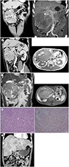
Contrast-enhanced abdominal CT in the portal venous phase showed a mass measuring 15 × 12 × 14 cm, with clear boundaries and good contrast-enhancement, in the right subhepatic space. Enlarged blood vessels were found around and inside the mass and seemed to be connected to the gastroduodenal, right gastric, and gastroepiploic arteries and some parts of the superior mesenteric artery (Fig. 1A). The mass was large, and its origin could not be detected, but the liver, gallbladder, and stomach were seen in the superior portion of the mass, while the duodenum and pancreas were seen in the middle portion of the mass, where it was connected to the gastric antrum (Fig. 1B). Heterogeneous contrast-enhancement and calcification were observed inside the mass. As the boundary between the vessel's origin and the gastric antrum was unclear, the exophytic mass was assumed to be originating from the stomach. Therefore, the differential diagnosis from CT findings included gastrointestinal stromal tumor and schwannoma. For the definitive diagnosis, MRI was performed.
The mass showed heterogeneous signal intensity on T1-weighted images, with mostly low signal intensity and some high signal intensity inside the mass (Fig. 1B). In addition, the mass showed heterogeneous signal intensity on T2-weighted images, including low signal intensity and signal void in some parts inside the mass. Similar to CT, the mass showed heterogeneous enhancement on MRI (Fig. 1C). The central non-enhancing area showed high signal intensities on both T1- and T2-weighted images, suggesting hemorrhage and necrosis. Based on the signal void on MRI, gastric paraganglioma was included in the differential diagnosis along with gastrointestinal stromal tumor and schwannoma.
Positron emission tomography (PET)/CT revealed no metastases. Mass excision, distal gastrectomy, and gastrojejunostomy were performed. The surgical findings revealed a hypervascular mass measuring approximately 15 × 15 cm, located in the greater curvature of the gastric antrum, connected to the antrum, and adhered to the surrounding mesentery and omentum. The mass was encapsulated, which distinguished it from other anatomical structures. The feeding arteries anastomosed with the right gastric, gastroduodenal, gastroepiploic, and superior mesenteric arteries and drained into the gastroepiploic and superior mesenteric veins.
On microscopy, a pathologic specimen of the tumor showed epithelial and mesenchymal cells, with eosinophilic granular cytoplasm and presence of nested patterns. On immunohistochemistry, the mass tested positive for neuron-specific enolase (Fig. 1D) but negative for PanCK, CD31, CD34, C-kit, hepatocytes, chromogranin A, and synaptophysin. Considering the laboratory, radiologic, surgical, and pathologic findings, the patient was diagnosed with nonfunctioning gastric paraganglioma. As the pathologic examination revealed a mitotic count of the tumor cells of over 5/50 high-power field (HPF), a follow-up test was needed to confirm malignancy.
In the 1-year follow-up, CT did not reveal any new masses in the remaining gastric wall but showed enhancement of multiple hepatic masses in the arterial phase and washout in the portal and venous phases (Fig. 1E). Furthermore, multiple enhancing masses were observed along the region of the transverse mesocolon. As the mass was identified as a metastasis from paraganglioma on liver biopsy, chemotherapy with adriamycin, ifosfamide, and mesna was performed for nine cycles and was discontinued as tests in the 1-year follow-up did not show any improvement in the diagnosed condition.
DISCUSSION
Paraganglioma is a tumor arising from the sympathetic or parasympathetic nerve branches where cells with high chromatin are located outside the adrenal glands (12). Abdominal paraganglioma mainly arises in the retroperitoneum along the distribution of the paraganglion but rarely in the gallbladder, bladder, prostate, and duodenum (210).
Similar to pheochromocytoma, paraganglioma can be divided into functioning and nonfunctioning. Paraganglioma can be diagnosed as a malignant mass or tumor when metastatic lesions are found, and the metastatic sites include the lymph nodes, bones, liver, and lungs (12310). A study on tumors arising from chromaffin cells reported that pheochromocytoma is characterized by a diameter of 5 cm or more, weight of over 80 g, high level of cell division, invasion of blood vessels and membranes, DNA abnormalities, and Ki67 of 6% or higher (1). In the present case, based on the size, weight, and cell-division level of over 5/50 HPF, we considered the possibility of malignancy, which required a detailed follow-up examination of the patient. In the 1-year follow-up, liver metastasis was detected, and the patient was diagnosed with metastatic paraganglioma on histologic examination. Paraganglioma is malignant in approximately 30–40% of the cases, and the overall 5-year survival rate is 50% (8). Because of the poor prognosis, there is no specific therapy for malignant pheochromocytoma, and individualized therapy is used for palliation (2).
In the literature, only a few cases of gastric paraganglioma have been reported, which included only a description of the condition and no CT or MRI images. Therefore, the literature review was performed only for the description of the condition (3456789). The length of gastric paraganglioma varies from 3 to 15 cm, and different occurrence sites were reported, with 1, 3, 1, 1, and 1 cases in the fundus, lesser curvature, posterior wall, greater curvature, and antrum, respectively. The number of cases of nonfunctioning and functioning paragangliomas were 5 and 1, respectively. Metastasis to the patient's spine, abdominal cavity, and liver occurred in one case each (Table 1). In the present case, a hypervascular mass measuring 15 cm was located in the greater curvature of the gastric antrum, and similar to most previous cases, it was non-functional. As liver metastasis was observed in the follow-up, it was diagnosed as malignant gastric paraganglioma. In the present case, the mass was big and showed exophytic growth. Differentiation was required to confirm primary gastric paraganglioma or pheochromocytoma invasion to the gastric wall. However, since the mass showed connection only to the antrum and a clear capsule separating it from the duodenum, gallbladder, etc., and the CT image and surgical findings showed that it anastomosed with the right gastric and superior mesenteric arteries, it was diagnosed as primary paraganglioma arising in the stomach.
In this case, there was a hypervascular solid mass with good contrast enhancement, and in the non-enhancing parts, necrosis with high signal intensity on T2-weighted images, hemorrhage with high signal intensity on T1-weighted images, and calcification were observed on CT. These findings are typical of paraganglioma involving various parts of the body (110). The preoperative differential diagnosis included gastrointestinal stromal tumor, schwannoma, and other tumors that show exophytic growth. Although the masses in previous studies were diagnosed as a gastrointestinal stromal tumor preoperatively, it was difficult to find out differential points based only on the results of imaging in this case (67). Paraganglioma rarely originates in the stomach, which makes its differentiation from other similar conditions difficult; however, a hypervascular mass and presence of signal void on MRI, as seen in this case, can be used to diagnose gastric paraganglioma.
Clinically, a functioning paraganglioma can be differentiated from a gastrointestinal stromal tumor based on the clinical symptoms, laboratory findings, and vital signs of the patient at the time of surgery. However, since many previous cases and the present case showed a nonfunctioning paraganglioma, it was difficult to diagnose the condition clinically before the surgery (6). There have been no cases of a submucosal tumor with exophytic growth detected in the preoperative endoscopic biopsy. In this case, the endoscopic findings showed no gastric ulcers, but previous case reports have described the presence of ulceration in the gastric wall. As the gastrointestinal stromal tumor is often accompanied by additional gastric ulcers, differentiating a paraganglioma from a gastrointestinal stromal tumor is more difficult (3).
In conclusion, this was a case of malignant nonfunctioning gastric paraganglioma with exophytic growth and a strongly enhanced vascular internal structure, accompanied by liver metastasis, which was detected in the postoperative follow-up.
 XML Download