

 PDF
PDF ePub
ePub Citation
 Print
Print


INTRODUCTION
Spinal meningiomas, which account for 25% of all intraspinal tumors, are typically intradural extramedullary lesions in the thoracic spine of a middle-aged woman. The diagnosis of the tumor is usually not difficult based on radiological findings and typical location. However, spinal meningiomas rarely occur in epidural location, which account for only 2.5% to 3.5% of all spinal meningiomas. Spinal epidural meningiomas may show different growth patterns; en plaque, dumbbell-shaped, or ovoid. Among them, en plaque epidural meningioma may easily be mistaken for a lymphoma due to same growth pattern and signal intensity on MRI. We report a case of pathologically confirmed spinal epidural meningioma, which was misdiagnosed as a lymphoma on preoperative imaging study.
CASE REPORT
A 79-year-old woman presented with progressive numbness of both fingertips and weakness of both arms for seven months. Neurologic examination revealed paresthesia in both first to third fingertips and normal power of motor Grade V in both upper and lower limbs.
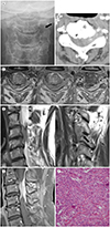
On anteroposterior radiograph of C-spine, the left neural foraminal widening of C3–4 level was suspected (Fig. 1A). Enhanced CT showed subtly enhanced spinal and extraspinal mass at that level (Fig. 1B). A spine MRI demonstrated an en plaque mass, approximately 2.4 cm × 1.6 cm in size, in the left lateral and posterior epidural space of the third and fourth cervical vertebral level, surrounding and compressing the spinal cord (Fig. 1C). The mass showed left neural foraminal and extraforaminal extension of the C3–4 with an encasement of left C4 nerve root (Fig. 1D). The neural foramen was enlarged but no bony destruction was observed. On T1-weighted images with contrast enhancement, the mass displayed intense and homogeneous enhancement (Fig. 1E). Presumptive diagnosis before surgery was a lymphoma.
The patient underwent tumor resection with partial laminectomy of the left C3–4. The tumor was located epidurally and hardly adhered to the adjacent dura mater, which made complete resection difficult to perform. The final pathologic diagnosis was a transitional (WHO Grade I) meningioma. Histologically, the tumor was typical of transitional type meningioma having features of transitional features between those of meningothelial meningioma and fibrous meningioma (Fig. 1F).
DISCUSSION
Spinal meningiomas are the second most common intraspinal tumors, which accounts for 25% of all intraspinal tumors (1). Meningiomas arise from meningothelial cells in the arachnoid villi, most of whom are intradural (2). Spinal epidural meningiomas are rare and account for only 2.5% to 3.5% of all spinal meningiomas (3). Their origin is still unclear but previous studies suggest that they may be originated from aberrant arachonid islets in the extradural space, islands of arachnoidal tissue that migrated into the extradural space, or arachnoid villi that separated from the main arachnoid layer and invaded into the dura (4).
Spinal epidural meningiomas are not essentially different from the common intradural types. They show the same histology, the same frequent location in the thoracic spine, and the same sex predilection of female patients (5). However, recent studies found the cervical spine to be the most common site of these tumors (14). Similar to those of common intraspinal tumors, the clinical symptoms of extradural meningiomas are sensory, motor, or sphincter dysfunctions, which eventually appear in the late stages of disease progression (4).
MRI is the modality of choice for the diagnosis of spinal meningioma because it clearly defines the mass and its relation to the spinal cord. Mostly, the lesion shows iso to hypointensity on T1-weighted images, variable signal intensity on T2-weighted images, with homogeneous enhancement after Gadolinium injection (16). Epidural meningiomas may be en plaque, dumbbell-shaped, or fusiform/ovoid (1). The term “en plaque” was used by Cushing to describe the tumor, which grows like a flat and spread carpet (7). This kind of meningioma grows in a sheetlike form along the dura mater, which commonly occurs in the sphenoid ridge of the skull base (4). Although the exact incidence is uncertain due to its rarity, Zhang and Yuan. (1) reported that nine of fourteen cases (64%) were en plaque form. Neural foraminal extension and foraminal widening were commonly found in this kind of meningioma (4), mimicking neurogenic tumor such as schwannoma. Neurogenic tumors usually show high signal intensity on T2-weighted images with heterogeneous enhancement, whereas meningiomas are characterized by homogeneous contrast enhancement (8). The dural tail sign, linear thickening and contrast enhancement of the meninges adjacent to the mass, is commonly seen in meningiomas, but it is not specific finding, which is also seen in metastatic tumors or lymphomas (1).
The differential diagnosis of spinal epidural meningiomas includes schwannoma, metastatic tumors, lymphoma, and tuberculoma. They are easily mistaken preoperatively for malignant metastatic epidural tumors, which can alter the surgical approach and increase morbidity (9). Lymphoma is the other mimicker of meningioma, especially en plaque type, because of the same growth pattern and signal intensity on T2-weighted images with intense and homogeneous enhancement (4). They are characterized by formation of a paravertebral soft tissue mass and infiltration of adjacent bone (1), but discrimination would be difficult without these findings. Preoperative differential diagnosis is important when planning surgical strategies and determining the extent of the required resection. Particularly, as lymphomas are very chemo- and radiosensitive tumors, surgery for lymphomas has most commonly been limited to spinal decompression and biopsy. In our case, the mass showed infiltrative growth encircling the spinal cord and extending to neural foramen, and intermediate high signal intensity on T2-weighted images with homogeneous and intense enhancement, making confusion with a lymphoma.
The recurrence rate after surgery for epidural meningioma is four times higher than that of intradural meningiomas (10). The recurrence may be due to close adherence of tumor to the dura or nerve roots, which makes complete resection difficult to perform (1). In our case, the tumor was also severely adherent to the dura and totally encased the nerve root, which resulted in incomplete resection. The long-term prognosis of epidural meningiomas remains uncertain because of contradicting reports (9). However, if complete resection is possible, there may be no difference in patients' prognoses.
In conclusion, we describe here a rare case of spinal epidural en plaque meningioma in the cervical spine. They are easily mistaken for metastatic tumors or lymphomas, which affects the extent of surgery and patients' outcome so the diagnosis needs to be carefully done. Epidural meningioma should be considered in the differential diagnosis of a middle-aged patient with an epidural spinal tumor involving the cervical or thoracic regions.
 XML Download