

 PDF
PDF ePub
ePub Citation
 Print
Print


INTRODUCTION
Compartment syndrome (CS) is defined as the increased pressure within a compartment that limits blood supply and results in ischemia. CS mainly occurs in the lower legs but can also occur in the hands, feet, forearms, buttocks, thighs, and even, the paraspinal muscles. CS in the paraspinal muscles has recently received more attention, possibly due to an increasing number of people engaging in weight-lifting exercises, as well as improved means of detecting and recognizing CS (12). Here, we present a case of a characteristic MRI finding from an incidence of acute lumbar CS.
CASE REPORT
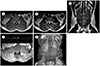
A 26-year-old male visited the hospital for severe low back pain. He had been doing a deadlift exercise for a short time and had begun to feel pain at about 12 hours after having finished the exercise. The pain increased in intensity. He had no significant medical history and denied any form of recent medication. His vital signs were stable. Physical examination revealed severe swelling and intense rigidity of lumbar paraspinal muscles with direct tenderness. Hypoactive bowel sounds on auscultation and a tingling sensation with mild numbness in the lumbar area were also noted. The range of motion was limited due to the pain and the straight leg raising test was about 60 degrees for both sides. Initial laboratory tests showed the serum myoglobin was more than 3000 ng/mL (normal range: 28–72 ng/mL), serum creatine kinase (CK) was 74280 IU/L (normal range: 26–200 IU/L), lactate dehydrogenase (LDH) was 2000 IU/L (normal range: 140–271 IU/L), aspartate aminotransferase (AST) was 643 IU/L (normal range: 0–37 IU/L), alanine aminotransferase (ALT) was 171 IU/L (normal range: 0–41 IU/L), and the white blood cell count was slightly high at 10350/µL (normal range: 4000–10,000/µL). A urine test revealed that the urine erythrocytes were four-positive that turned out to be myoglobinuria of 1626 ng/mL (normal range: 0–9.9 ng/mL) after two days. A plain radiography of the lumbosacral spine showed no bony abnormalities. A lumbosacral spine MRI was conducted and a T1-weighted image demonstrated diffuse swelling of the bilateral lumbar paraspinal muscles with no definite abnormal signal intensity (Fig. 1A). Heterogeneous and diffuse slight hyperintensity was noted on a T2-weighted image (Fig. 1B, C). A contrast-enhanced T1-weighted spectral presaturation with an inversion recovery image showed a craniocaudally-oriented area of hypoperfusion along the bilateral multifidus, longissimus, and iliocostalis lumborum muscles, as well as the non-visualization of the vasculature, within this area. The region of hyperperfusion was also noted bilaterally, making a heterogeneous appearance on contrast-enhanced scans (Fig. 1D, E).
The patient was diagnosed as lumbar paraspinal rhabdomyolysis with secondary CS and admitted to the neurosurgery department under a nephrology consultation. By the administration of a massive intravenous fluid (80 cc/hour) supply, diuretics, and pain relievers, his symptoms and laboratory findings soon improved, so conservative treatment was continued for a week. No surgical intervention or hyperbaric oxygen therapy was administered. One day before discharge, abnormal laboratory results dropped to 235 ng/mL for serum myoglobin, 6990 IU/L for CK, 563 IU/L for LDH, and 173 IU/L for AST. In an outpatient follow-up study two weeks later, serum myoglobin was less than 21 ng/mL, CK was 527 IU/L, LDH was 400 IU/L, ALT was 47 IU/L, and AST was normal. Although he still complained of mild numbness in the lumbar region and stiffness during flexion, he had no pain or functional impairment when using his paraspinal muscles.
DISCUSSION
CS, defined as increased pressure within a fascial compartment, causes impaired circulation that leads to ischemia and congestion, which in turn leads to edema and worsens high pressure. This vicious cycle is well reflected in its classical symptoms, known as the 6P's: pain, pallor, pulselessness, paresthesia, paralysis, and poikothermia. Fractures are the most common cause while other variable injuries can also trigger CS. A diagnosis of a typical CS is made clinically on the basis of these clinical clues. An intracompartmental pressure measurement can confirm a diagnosis (3).
CS can develop in any body part that has fascial compartments. Middle and posterior layers of thoracolumbar fascia encase ipsilateral compartment muscles anteroposteriorly and spinous processes cover the compartment medially, making a separate space that is susceptible to increased compartmental pressure. With less than 20 reports extant in English on paraspinal CS, three types of presentations have been recognized so far: following intense physical activity, surgery-related complications, or drug overuse (2).
The diagnosis of paraspinal CS can be made when detailed history-taking, physical exam, laboratory finding along with radiologic study are comprehensively considered to avoid misdiagnosing as many other pathologic conditions causing low back pain. Radiologic study also can detect other differential diagnoses. It may also evaluate the extent of the involved region and detect complications. Cross-sectional imaging best depicts findings related to paraspinal CS. Both CT and MRI show bulging of bulky paraspinal musculature, which would suggest an increased volume in a limited space. A diffuse increased signal intensity in a T2-weighted image would imply an edematous condition within a compartment.
With the administration of a contrast medium, uneven enhancements can be seen. Only a few reports (2) describe MRI findings on a gadolinium-enhanced image of paraspinal CS and show the variable extent of hypo- and hyperenhancement. Rominger et al. (4) stated that strong enhancement of the affected compartment is a reflection of disturbed cellular membrane permeability and the non-enhancing portion results from liquefactive myonecrosis in a pathological examination. The area of hyperenhancement can also be explained by venous congestion caused by disrupted venous outflow. A contrast-enhanced scan better visualizes the area of impaired blood circulation than does a T2-weighted image and is useful when only subtle changes are seen in the T2-weighted image. Vivid localization of affected region and affective selective fasciotomy is possible.
The differential diagnosis of CS with similar image findings includes other causes of muscle edema, such as denervation, inflammation or infection, ischemia, iatrogenic (e.g. radiotherapy or postoperative), systemic diseases (e.g. sickle cell anemia or diabetes), and rhabdomyolysis (5).
Rhabdomyolysis can be both the cause and result of CS. Our patient were diagnosed as rhabdomyolysis and CS when he visited our hospital 12 hours after deadlift. Since there was no cause for CS or rhabdomyolysis other than extensive exercise, exercise-induced rhabdomyolysis and secondary CS was the reasonable diagnosis. Rhabdomyolysis differs from CS in that increased compartmental pressure is not mandatory and it may extend beyond the fascial space. The presented case showed altered signal intensity confined to paraspinal muscle compartment. Decreased enhancement at ischemic area also favored CS rather than rhabdomyolysis alone.
Diagnostic criteria for CS in the paraspinal region has been proposed by Nathan et al. (6) that include acute characteristic pain, unique physical examination, and other investigations. Our subject fulfilled all above criteria except elevated intramuscular pressure, not performed in our case for its invasiveness. Instead, tense configuration and marked rigidity of lumbar paraspinal muscles group were considered as sign of increased compartmental pressure. A recent study supports T2-weighted imaging as being able to show diagnostic outcomes similar to those of a needle manometry (7). Compressed and absent intramuscular vasculature in contrast-enhanced image also contributed to the diagnosis of increased compartmental pressure. This finding is rarely described in literature but correlates well with pathophysiology of CS.
Complications of CS can be detected by radiologic studies. An intramuscular hemorrhage can be detected as a hyperintensity in a T1-weighted image. Fibrosis, calcification, and muscular atrophy can be seen as a sequel in the chronic phase. Some authors consider the decreased uptake of gadolinium as a sign of necrosis, which suggests irreversible damage and demands emergent surgical intervention (8), whereas other authors have suggested observing if the rhabdomyolysis is in control (9). The MRI findings of our case showed no definite complications of infarction, hemorrhages, or liquefactive necrosis that may be seen as bright signal intensities on a T2- and high or low signal intensity on a T1-weighted image. Hence, the patient was treated conservatively. The early improvements in the symptoms and laboratory results also contributed to maintaining this decision.
In conclusion, we report a rare case of lumbar paraspinal CS following an intense weight-lifting exercise. MRI revealed diffuse edematous swelling and uneven gadolinium uptake of the bilateral lumbar paraspinal muscles. In addition, diminished vascular space by increased compartmental pressure may result as loss of intramuscular vasculature.
 XML Download