

 PDF
PDF ePub
ePub Citation
 Print
Print


INTRODUCTION
The incidence of ductal carcinoma in situ (DCIS) has increased since the introduction of breast cancer screening. Approximately 20–25% of all screening-detected breast cancers consist of DCIS (12). DCIS is often diagnosed by image-guided biopsy techniques, which are known to have an underestimation rate for invasive cancer (3). The rate of underestimation was found to be greater than 20% for 14-G core biopsy and up to 11–19% for 11-G vacuum biopsy (3456). Because the high rate of underestimation of invasive cancer leads to secondary surgical procedures for axillary sampling, the role of sentinel lymph node biopsy (SLNB) at the primary surgical procedure in all patients with pure DCIS is disputed. However, as the incidence of axillary lymph node metastases in pure DCIS is low, SLNB in all patients with DCIS may lead to overtreatment (12). Several studies therefore defined subgroups of patients at a high risk of invasion indicated for SLNB, e.g. patients undergoing mastectomy, patients with high grade lesions, presence of a palpable mass, a mass on mammography, large tumor size or patients with DCIS diagnosed on core biopsy (78910). Preoperative breast MRI has been widely used because of its high sensitivity for the detection of invasive breast cancer. The sensitivity of MRI has been reported to vary between 90% and 94% in larger studies and meta-analyses (1112). Dynamic contrast-enhanced MRI of breasts helps the surgical planning, the preoperative staging, and predicting the surgical outcome based on morphological information, tissue perfusion and enhancement kinetics as well (13). Furthermore, MRI reflects the biology of breast lesions (14), and the technique may be useful in distinguishing between patients at a high risk of invasive cancer and those at a low risk. Previous studies addressing this issue were, however, not focused on MRI, and reported the imaging findings that showed increased risk of invasive lesion. The potential value of MRI as an adjunct to conventional imaging is largely unknown. The aim of this study was to evaluate the imaging criteria for high diagnostic performance of preoperative breast MRI in biopsyproven DCIS. Ultimately, these findings may improve the selection of patients indicated for initial axillary sampling at the time of primary surgery.
MATERIALS AND METHODS
PATIENTS
This retrospective study was approved by our Institutional Review Board, which waived the need for informed consent (IRB No. 2010-01-023). Our study population consisted of 77 consecutive patients carrying 80 lesions of DCIS based on preoperative percutaneous biopsy from January to December 2014. One patient had bilateral lesions and two patients had two separated lesions in different quadrants of the same breast. Patients age ranged from 26 to 72 years (mean age, 50 years). Preoperative diagnosis was established by stereotactic vacuum-assisted biopsy (VAB) (15/80), ultrasound (US)-guided VAB (10/80), and US-guided 14-gauge core needle biopsy (55/80). All patients underwent preoperative MRI for evaluating the extent of primary tumor, multiplicity, and contralateral breast cancer.
MR IMAGING
Breast MRI was performed using a 1.5 Tesla (T) Philips Intra Achieva in 6 cases, using a 3.0 T GE Signa in one case, and a 3.0 T Philips Intra Achieva in 73 cases. All patients were imaged in a prone position using a dedicated double breast surface coil. Both breasts were imaged in all cases. An axial T1-weighted spin echo sequence and fat-suppressed T2-weighted fast spin echo sequence were performed. For dynamic contrast enhanced images, a three-dimensional, axial/sagittal, fat-suppressed T1-weighted fast gradient echo sequence was obtained before, and every min (until 7 min) after a bolus injection of 0.1 mmoL/kg of gadolinium diethylenetriamine pentaacetic acid (Magnevist; Berlex Laboratories Inc., Wayne, NJ, USA). The parameters were as follows: repetition time and echotime (8.7/4.3 ms for the axial dynamic images; 16/4.1 ms for the sagittal dynamic images), a 20° flip angle, a 27-cm field of view, 1.5-mm sections with no gap, and a 512 × 512 matrix. Two signals were averaged and spectrally selected for inversion recovery-prepared fat suppression. After examination, two subtraction images were generated automatically on a pixel-by-pixel basis: the unenhanced images were subtracted from the early post-contrast images (standard subtraction), and the last post-contrast images were subtracted from the early post-contrast images (reverse subtraction). Reformatted images (maximum-intensity projection) in a cranio-caudal, medio-lateral, or anteriorposterior projection were created from the standard and reverse subtraction images.
ANALYSIS OF BREAST MRI FINDINGS
The morphological and dynamic image parameters of breast MR images were retrospectively analyzed by two radiologists who were blinded to the final pathological results of the lesions. The morphological type was analyzed with distribution of non-mass enhancement, as well as the extent, and enhancement kinetics of the lesions.
Based on the morphologic type, the lesions were classified into mass and non-mass enhancement according to the categories of the American College of Radiology (ACR) Breast Imaging Reporting and Data System (BIRADS) for MRI (15). A mass is a space-occupying tumor that has three dimensions, with a visible correlate on pre-contrast T1- or T2-weighted images. A non-mass enhancement occurs in an area of the fibroglandular tissue that otherwise appears normal on pre-contrast images. There is no space-occupying effect.
The enhancing area usually not correlated with the fat-suppressed or non-fat-suppressed T2-weighted images. Cases, which showed both areas of mass and non-mass enhancement concurrently, were classified as the mass lesions. In the non-mass enhancement, the distribution of enhancement was assessed by the orientation: linear or segmental (along the milk ducts) and diffuse or regional and focal. The enhancement kinetics was analyzed by assessing the early enhancement and washout, based on the visual assessment using standard subtraction and reverse subtraction images and measured signal intensity within the region of interest on pre-, and post-contrast (early and delayed) enhanced images. To determine the extent of lesions, we measured the longest diameter of the contrast-enhanced lesion at the early enhancement time (2 min) and the cut-off value that predicted invasive lesion was determined using the receiver operating characteristic (ROC). In case of lesions without early enhancement, we measured the extent on the delayed images.
After evaluating significantly different MRI findings between pure DCIS and DCIS with invasive lesions, we applied multiple criteria using a combination of significant MRI findings and assessed their diagnostic performance.
STATISTICAL ANALYSIS
We compared the MR findings between pure DCIS and DCIS with invasive lesions. We used the chi-square test to analyze the morphology and enhancement, and the t-test to analyze the extent of lesion. p-value ≤ 0.05 was considered statistically significant. A cut-off value was obtained for the extent that predicted invasive lesion using the ROC analysis. MR findings that showed statistical significance in differentiating pure DCIS from DCIS with invasive lesions were evaluated. The sensitivity, specificity, positive predictive value (PPV) and negative predictive value (NPV) of significant MR findings in predicting co-existing invasive lesion were calculated.
RESULTS
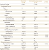
All surgical specimens were carefully assessed for concomitant invasive lesion. Four cases showed no residual lesions on the pathology after surgery. Among the 76 lesions, 49 lesions were confirmed as pure DCIS and 27 lesions were confirmed as DCIS with invasive lesions after surgery. Among the 27 DCIS with invasive lesions, 19 lesions were invasive ductal carcinoma (IDC) in the background of DCIS which included 9 cases of micro IDC; one lesion was invasive lobular carcinoma with surrounding DCIS; and two were mucinous carcinoma with surrounding DCIS; five cases were confirmed as pure IDC (extensive intraductal component less than 25%) after surgery. The overall MR features of pure DCIS and DCIS with invasive lesion are listed in Table 1. The morphologic types were classified into mass and non-mass enhancement according to the ACR BIRAD for MRI (15). All DCIS with invasive lesions and 42/49 (86%) of pure DCIS showed positive MR findings.
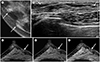
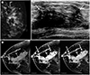
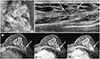
The lesions that showed negative MR findings were all pure DCIS (7/49). Four lesions (4/49) showed very subtle findings that were hard to differentiate from normal breast parenchyma without knowing that the patient had a biopsy-proven DCIS lesion. Lesions with subtle or imperceptible contrast enhancement on the preoperative MRI were pure DCIS without a concurrent invasive lesion (Fig. 1). Regarding the morphologic type, the mass or non-mass enhancement and their distribution were not statistically significant in both pure DCIS and DCIS with invasive lesions. In the kinetics of contrast enhancement, DCIS with invasive lesions exhibited higher rates of early enhancement (92.6%), and washout pattern (55.6%) than pure DCIS (71.4% and 24.5%), which were statistically significant (p = 0.030 and p = 0.048) (Fig. 2). In pure DCIS lesions, 14.3% of the lesions showed no abnormal enhancement in early phase. Among the 35 lesions with early enhancement, 23/35 (65.7%) did not show washout kinetics. The extent of DCIS with invasive lesions was significantly larger than that of pure DCIS lesions (5.01 ± 2.2 cm vs. 2.82 ± 1.9 cm) (p < 0.001) (Fig. 3).
Table 2 shows the diagnostic performance of significant MR findings that differentiate pure DCIS and DCIS with invasive lesions. After the ROC analysis, we determined a size of 4 cm as the cut-off value for differentiating pure DCIS and DCIS with invasive lesion. Among the three MR findings that showed statistical significance, a cut-off extent of at least 4 cm showed the best diagnostic performance. When we applied the combination criteria based on extent of at least 4 cm or early enhancement and washout kinetics, the sensitivity, specificity, PPV and NPV were 92.6%, 61.2%, 56.8 % and 93.8%, respectively.
DISCUSSION
DCIS is a non-invasive breast lesion without metastatic potential. DCIS can, however, be associated with the presence of invasive cancer. Because of the underestimation rate of invasive disease using image-guided biopsy, preoperative selection of patients at a relatively high risk of invasion is essential to avoid a second surgical procedure for axillary nodal staging. Although MRI has a high sensitivity for the detection of invasive cancer, the role of MRI in assessing the presence of invasive disease is as yet unclear. There have been only few reports about preoperative predictive MR findings for invasive cancer and despite recent advances in the diagnosis, evaluation, and treatment of breast cancers, surgical treatment of DCIS remains unreliable (16). According to Morakkabati-Spitz et al. (17), segmental or linear enhancement patterns on dynamic breast MRI are hallmarks of DCIS. However, invasive carcinomas that coexist with a background of diffuse DCIS also frequently show segmental non-mass enhancement. In this study, non-mass enhancement with ductal or segmental distribution was seen most frequently in both groups of pure DCIS and DCIS with invasive lesions. The biological explanation of enhancement on MRI of a malignant tumor is believed to be the presence of tumor-induced angiogenesis. The increased density of microvessels will increase blood flow, thereby causing contrast enhancement. Furthermore, these tumor-induced microvessels demonstrate structural abnormalities that give rise to leakage of contrast medium. This causes characteristic contrast enhancement kinetics (plateau and washout phenomenon) (1819). Angiogenesis-induced vessel sprouting and alteration of vessel wall permeability are thought to lead to the strong and fast contrast enhancement observed in dynamic MRI study of invasive breast cancers (20). However, DCIS has been shown to exhibit variable angiogenetic activity, yielding variable contrast enhancement values. Even though Van Goethem et al. (21) reported that enhancement kinetics such as washout and moment of maximal enhancement could not aid the differential diagnosis between pure DCIS and invasive carcinoma, in our study, a washout pattern was observed significantly more in DCIS with invasive lesions than in pure DCIS (p < 0.05). In the extent of lesions, DCIS with invasive lesion showed significantly larger extent than pure DCIS (5.01 ± 2.2 cm vs. 2.82 ± 1.9 cm) (p < 0.001). Based on our results, a larger lesion size is most frequently used as a predictive factor for invasive cancer on mammography, US or MRI. The larger lesion size on mammography was reported to be an independent predictive factor for invasion with the cut-off ranging from 20 to 50 mm (6, 22, 23). It was reported that sonographic lesion size of 20 mm or larger was a significant factor for invasion (24). In an MRI study, lesions of 60 mm or larger showed the likelihood of having invasive component (25). These could be a reflection of the assumption that the larger the lesion, the more likely DCIS and invasive cancer coexist in the same lesion. Some investigators stated that the incidence of microinvasion or invasion was directly related with tumor size (2627). In addition, a larger target lesion increased the target area for sampling, and a greater probability of missing invasive component (2628).
We compared the performance of three statistically significant MR findings in differentiating pure DCIS and DCIS with invasive lesions and in predicting co-existing invasive lesions. Among the three features, the extent of 4 cm showed the best diagnostic performance in predicting co-existent invasive lesions, although the sensitivity was 74.1%. The combined criteria of either extent ≥ 4 cm or early enhancement and washout kinetics for predicting invasive lesion, the sensitivity and NPV increased to greater than 90%. Even though the specificity and PPV of combination criteria were around 60%, we expected that their predictive ability for co-existing invasive lesions may help preventing a two-step operation by selectively using SLNB. In cases of subtle or imperceptible contrast enhancement on preoperative MRI, we could predict that the lesion would be pure DCIS and that lumpectomy alone would be adequate treatment.
Our study has a few limitations. First, the radiologists interpreted MR images with a knowledge of previous histological diagnosis of DCIS, and mammographic and US findings, which may increase the percentage of positive MR findings for all lesions. Second, inter-observer variation is possible with regards to mass and non-mass enhancement. Third, our study focused on MR features. We did not include US and mammographic findings in our analysis. Therefore, further studies, which include all imaging findings and clinical aspects, are needed to evaluate the most reliable predictors of co-existent invasive lesions in patients with biopsy-proven DCIS.
In conclusion, DCIS with invasive lesions showed larger extent and more frequent washout kinetics compared with pure DCIS. The combined criteria of either extent ≥ 4.0 cm or early enhancement and washout kinetics of the lesions are useful in predicting co-existent invasive lesions in biopsy-proven DCIS patients, and SLNB could be recommended for such selected patients.

 XML Download