

 PDF
PDF ePub
ePub Citation
 Print
Print


INTRODUCTION
Vesicoureteral reflux (VUR) is relatively common in children with febrile urinary tract infection (UTI). VUR occurs in 35–40% of children with a history of febrile UTI; this UTI develops in approximately 1–2% of children (1). VUR has been associated with hypertension, chronic renal disease, and end-stage renal disease; hence, it is an important consideration when treating children with UTI (234).
The key change between the 1999 American Academy of Pediatrics (AAP) guideline and the 2011 revised AAP guideline for children aged 2–24 months regarding the first febrile UTI relates to imaging modalities, as follows (56): voiding cystourethrography (VCUG) should not be performed routinely after the first febrile UTI; VCUG is indicated if renal and bladder ultrasonography (US) reveals hydronephrosis, scarring, or other findings that would suggest either high-grade VUR or obstructive uropathy, as well as in other atypical or complex clinical circumstances (5).
Infants younger than 2 months were excluded from the AAP guideline because of their special age-related considerations (5). In practice, these recommendations are often extended to younger or older children (7). Notably, the updated guideline recommends that only children with high-grade VUR should receive antibiotic prophylaxis and/or undergo surgery, as high-grade VUR is less likely to resolve and more likely to result in renal scarring. Conversely, over half of the cases of all-grade VUR are expected to resolve within 2 years (89). Further, understanding the implications of high-grade VUR in renal US is important for pediatric radiologists. Few studies have compared the diagnostic accuracy of renal US criteria for VUR with the accuracy of VCUG. According to a study by Leroy et al. (3), evaluation of ureteral dilation by using renal US provides the best accuracy for suspected high-grade VUR in comparison with other predictors in infants and children. Wallace et al. (10) reported that normal renal US findings suggest little possibility of grade 4 and 5 VUR, but do not exclude low-grade VUR in infants less than 2 months of age. However, most studies have not clearly defined the sonographic findings for high-grade VUR (5).
Therefore, our goal was to compare the diagnostic accuracy of the renal US criteria for VUR with the accuracy of VCUG to ultimately facilitate clinical management of children experiencing their first febrile UTI who are recommended to undergo VCUG.
MATERIALS AND METHODS
STUDY DESIGN AND POPULATION
Our Institutional Review Board approved this retrospective study, and the need for informed consent was waived (IRB No. HPIRB2017-06-013). We included infants and children under 24 months of age diagnosed with UTI who underwent both renal US and VCUG at our hospital between March 2011 and September 2017. Patients with UTI were defined as having a fever of at least 37.5℃, positive findings of pyuria (white cell counts ≥ 5 per high-powered field, or approximately 25 cells/µL), and positive urine culture findings (growth of only one microorganism at ≥ 100000 colony-forming units/mL). Urine was obtained using a sterile urinary bag or aseptic catheter before initial antibiotic treatment was administered to patients. Patients' clinical information was collected through electronic medical records. We conducted a retrospective study of the included patients and collected data using standardized data formats.
Among the patients with UTI who underwent both renal US and VCUG, we excluded patients who had been diagnosed with other genitourinary abnormalities in which altered renal size was a risk factor, patients who had already received antibiotic treatment for > 3 days due to UTI, and those who had undergone follow-up assessments for VUR that was already diagnosed.
During the study period, a total of 179 patients with diagnosed UTI underwent renal US and VCUG; 41 were excluded from this retrospective study for the following reasons: 7 patients had been diagnosed with other genitourinary abnormalities (Herlyn-Werner-Wunderlich syndrome, crossed fused ectopic kidney, and multicystic dysplastic kidney) and the remaining 34 patients had already received antibiotic treatment or had been followed-up for previously diagnosed VUR. A total of 138 patients were included in the study, with a greater number of male patients (109/138; 79.0%); the mean age of the patients was 3 months.
PERFORMANCE AND REVIEW OF RENAL US AND VCUG
Renal US and VCUG were performed by a single pediatric radiologist with 9 years of experience in pediatric radiology. Renal US with gray-scale and color Doppler was performed by using an 8–5 MHz sector and 12–5 MHz linear transducer (iU22; Philips Medical Systems, Bothell, WA, USA). Patients underwent renal US after filling the bladder by feeding or drinking water. The patients first underwent examination of the urinary bladder and kidney in the supine position, and then underwent reexamination of the kidney in the prone position. Measurements of renal length were always obtained in the prone position. Abnormal renal size was defined as kidneys outside of the 95% confidence interval (CI) for age by Han and Babcock (11), or kidneys with a length difference of > 1 cm (12). In cases with a normal renal length range, a globular kidney shape was considered to indicate increased renal size. Right and left renal echogenicity were compared with the echogenicity of the adjacent liver and spleen, respectively. Normal renal echogenicity in patients older than 6 months was lower than the echogenicity of adjacent organs; normal patients younger than 6 months exhibited similar renal echogenicity with that of adjacent organs (1113). However, increased renal parenchymal echogenicity is a subjective finding; this was determined at the discretion of the reviewer. To overcome this limitation, increased renal parenchymal echogenicity was considered to indicate an inflammatory lesion in the kidney; color power Doppler US was used as an adjunct to detect this type of lesion (14). Ureteral dilation was defined by the presence of a hypoechoic tube connected with the bladder of any diameter (3). Renal pelvic and ureteral thickening were noted when a measurable wall appeared on renal US. Renal pelvic dilation was divided into subcategories, namely hydronephrosis and prominent renal pelvis. Hydronephrosis and prominent renal pelvis were defined by anteroposterior (AP) renal pelvis diameters ≥ 10 mm and ≥ 5 mm in the supine position, respectively (1516). Further, accentuated pelvic dilation was defined as a new hydronephrosis or prominent renal pelvis in the prone position compared to the normal renal pelvis AP diameter in the supine position. Two attending radiologists (one was the pediatric radiologist mentioned above; the other was a radiologist with 2 years of experience in general radiology who was trained by the pediatric radiologist), who were blinded to VCUG results, each analyzed the renal US results independently.
To perform the VCUG examination, the bladder was catheterized using a 5- to 8-French soft plastic catheter. The inserted catheter was fixed to the external genitalia and thigh by medical tape. Sodium ioxitalamate (Telebrix; Guerbet, Aulnay-sous-Bois, France) was instilled using gravity from a height of approximately 1 m above the patient, who was in the supine position. Intermittent fluoroscopic and spot images were obtained while the bladder was filled with contrast material and the patient was voiding. The male patients provided spot images in the oblique position, in order to detect urethral abnormalities. A postvoid image was obtained to access reflux and postvoid residual urine volume. Three cycles of filling and voiding were performed, unless the patient was incontinent or reflux was detected.
VUR was classified into five grades (according to the International Reflux Study in Children): grade 1 indicates that VUR is present only in the ureter and does not reach the renal pelvis; grade 2 indicates that VUR reaches the renal pelvis without definite pelvic dilation; grade 3 indicates mild-to-moderate renal pelvic or ureteric dilation, without obliteration of the forniceal angle; grade 4 indicates moderate renal pelvic or ureteric dilation, with obliteration of the forniceal angle; and grade 5 indicates gross renal pelvic or ureteric dilation with tortuosity (17). Grades 3 and higher were considered high-grade VUR; grades 2 and 1 were considered low-grade VUR (18).
STATISTICAL ANALYSIS
Sensitivity, specificity, positive predictive value (PPV), and negative predictive value (NPV) of renal US abnormalities were calculated for all-grade and high-grade VUR by using VCUG findings as the reference standard. We used a multivariate logistic regression model to analyze the data, and then calculated the odds ratio (OR) of each renal US finding for all-grade and high-grade VUR. With the exception of interobserver agreement, only the results obtained by the pediatric radiologist with 9 years of experience in pediatric radiology were used in the statistics. The interobserver agreements were analyzed for each renal US finding by using Cohen's kappa statistics. A kappa value of 0.81 to 0.99 was considered to indicate almost perfect agreement; 0.61 to 0.80, substantial agreement; 0.41 to 0.60, moderate agreement; 0.21 to 0.40, fair agreement; 0.01 to 0.20, slight agreement; and less than 0.01, poor or less than chance agreement. Data analyses were performed using a commercially available statistical software (SPSS 23.0 for Windows, IBM Corp., Armonk, NY, USA). A p value < 0.05 was considered statistically significant.
RESULTS
Among 138 patients, 53 (38.4%) patients were diagnosed with unilateral VUR, 17 (12.3%) patients exhibited bilateral VUR, and 43 (31.2%) patients exhibited high-grade VUR in one or both kidneys. Both kidneys of each patient were analyzed separately; thus, a total of 276 kidneys were analyzed.
The incidence of each abnormal finding on renal US was as follows: decreased renal size, 5.4% (n = 15); increased renal size, 12.7% ((n = 35); increased renal parenchymal echogenicity, 24.3% ((n = 67); ureteral dilation, 29.7% ((n = 82); ureteral thickening, 16.3% ((n = 45); prominent renal pelvis, 29.7% ((n = 82); hydronephrosis, 8.7% ((n = 24); renal pelvic thickening, 36.6% ((n = 101); and accentuated pelvic dilation, 10.9% ((n = 30).
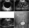
A total of 109 kidneys showed normal US findings (apart from prominent renal pelvis). Six of these exhibited VUR; five showed low-grade VUR, and one showed high-grade VUR (grade 3) (Fig. 1). Sensitivities and NPVs for negative results in VCUG within normal US findings (apart from prominent renal pelvis) were 91.9% (95% CI: 83.2–97.0%), and 94.5% (95% CI: 88.7–97.4%), respectively.
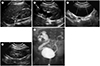
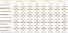
The sensitivity, specificity, PPV, and NPV of each US factor for all-grade VUR and high-grade VUR are described in Table 1. The OR was obtained using a multivariate logistic regression model; the significance values are summarized in Table 2. Decreased renal size was significantly related to all-grade VUR (OR, 16.6; 95% CI: 3.4–81.3; p = 0.001) and high-grade VUR (OR, 29.7; 95% CI: 5.7–155.3; p < 0.001) (Fig. 2). The values of each decreased renal size parameter were all smaller than the mean of the reference values and were more than 1 cm smaller than the opposite (19,20). Accentuated pelvic dilation was also significantly related to all-grade VUR (OR, 8.0; 95% CI: 3.2–20.4; p < 0.001) and high-grade VUR (OR, 11.9; 95% CI: 4.5–31.9; p < 0.001) (Fig. 1). Increased renal parenchymal echogenicity and ureteral dilation were significantly related to all-grade VUR, with ORs of 2.7 and 2.4, and to high-grade VUR, with ORs of 4.4 and 3.4, respectively. A significant relationship between renal US factors and high-grade VUR compared with all-grade VUR was detected.
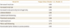
The interobserver agreement for each renal US finding showed almost perfect agreement, except for ureteral thickening, hydronephrosis, and pelvic wall thickening, for which it showed moderate agreement (Table 3).
DISCUSSION
Children with low-grade VUR are more likely to show spontaneous resolution of VUR and are less likely to develop renal scarring than those with high-grade VUR. Additionally, whereas children with high-grade VUR require antibiotic prophylaxis, this measure is typically unnecessary in patients with low-grade VUR. Therefore, it is important to distinguish between low-grade and high-grade VUR using a renal screening tool (89).
There are concerns regarding missed or delayed diagnoses of clinically important VUR in children with UTI. After the revised AAP guideline was published, several studies have reported that renal US is an inadequate screening test for VUR (2122). Nelson et al. (21) recommended that renal US be used as a complementary tool for the diagnosis of VUR in children with UTI because renal US showed lower sensitivity and lower PPV than VCUG for VUR. Despite this controversy, the current trend within the radiological community represents a shift toward the reduction or removal of radiation exposure; notably, renal US is a first-line imaging tool in pediatric radiology that does not involve radiation exposure, invasiveness, sedation, or discomfort. Furthermore, it can be used for screening purposes prior to the use of other tools (23). Our study analyzed the detection of VUR using a larger variety of US findings than that used in previous studies, such as the five US findings in 117 patients reported by Leroy et al. (3); thus, more diverse statistical results were obtained to overcome the inherent limitations of the procedure.
In our results, 5.7% of patients with UTI exhibited decreased renal size; decreased renal size observed on renal US was the most significant factor for the diagnosis of all-grade VUR and high-grade VUR, with a higher OR (29.7) for high-grade VUR compared with the OR (16.6) for all-grade VUR. Lee et al. (18) noted that a small kidney or thinned renal cortex is the most reliable renal US factor for predicting high-grade VUR, and our result supports their findings. This may result from the increased frequency with which repeated severe reflux, a precursor to decreased renal size, occurs in high-grade VUR relative to low-grade VUR (7). The low sensitivity (18.9%) of decreased renal size for VUR suggests that decreased renal size can be masked when the kidney exhibits acute inflammation and undergoes parenchymal edematous changes. However, as the kidney suffers repeated, severe reflux injuries, there is a progression to renal atrophy with an associated decrease in renal size. Therefore, even in cases in which the kidney undergoes parenchymal edematous changes, decreased renal size may be observed on renal US. We believe this accounts for the high specificity (99.5%) of decreased renal size for VUR in our study.
Despite some discussion over the changing diameter of the pelvicalyceal system as an indirect sonographic sign of VUR, accentuated pelvic dilation measured in the prone position was not examined in previous studies (24). Positional changes lead to alterations in the degrees of filling of the renal pelvocalyx and ureter (19). Since VUR inherently indicates an abnormality in the ureter or ureteral valve, we suspect that it will have a greater effect on the flow upstream of the ureter and pelvocalyx, thus causing a change in the diameter of the pelvicalyceal system upon position change. We believe that these factors contributed to our findings indicating that accentuated pelvic dilation may be an indicator of VUR.
In several previous studies, ureteral dilation has been associated with VUR (325); our results also showed significant associations with the diagnosis of all-grade VUR and high-grade VUR, with ORs of 2.4 and 3.4, respectively. We believe the rationale underlying these findings will be similar to those for accentuated pelvic dilation in the prone position.
Acute pyelonephritis (APN) is diagnosed when a patient has fever, pyuria, and increased renal parenchymal echogenicity and decreased flow in color Doppler images on renal US (24). Renal infection including APN is the most common acute disorder causing increased renal parenchymal echogenicity in children (26). According to Lee et al. (18), APN is a risk factor for high-grade VUR; further, VUR increases the incidence of UTIs, including APN.
A prominent renal pelvis (AP diameters of the renal pelvis between 5 and 10 mm) is occasionally observed on renal US as an incidental finding and is not associated with VUR in children (15). Based on our calculated NPV for renal US, normal US findings (apart from a prominent renal pelvis) can predict a negative result in VCUG. Normal US findings (aside from a prominent renal pelvis) can exclude high-grade VUR, as has been previously reported (27). Only one kidney with high-grade VUR (grade 3) underwent VCUG due to suspicion of high-grade VUR in the opposite kidney because of the presence of increased parenchymal echogenicity, ureteral dilation, ureteral wall thickening, and accentuated pelvic dilation in prone position in the opposite kidney (grade 5). When evaluating bilateral VUR, the kidney with lower-grade VUR may be inappropriately assigned a lower grade than clinically indicated because image findings are compared with those seen in the high-grade VUR in the opposite kidney.
Interobserver agreement was rather high, as the general radiologist with 2 years of experience in our study was a direct disciple (learning via “man-to-man” training) of the pediatric radiologist with 9 years of experience also involved in our research. Although the subspecialty was different, a previous study showed that man-to-man training is critical for the resident or general radiologist to show improvement in diagnostic performance and interobserver variabilities (28). Similar studies should examine interobserver reliability specifically within pediatric radiology, and more particularly for US; the minimization of interobserver variability may serve to further increase the utility of US in the detection of VUR.
There were several limitations in our study. First, it was a retrospective study. Second, other studies have used different modalities, such as technetium 99m-dimercaptosuccinic acid scan, whereas our study was conducted solely using renal US and VCUG. However, these two modalities are the most used tools in practice; therefore, we believe it is important to determine the diagnostic accuracy of renal US in predicting VUR in VCUG. Third, the renal US findings were subjective, and there were biases in interobserver agreement.
We demonstrated that decreased renal size may have the highest overall diagnostic accuracy for the US-based diagnosis of both all-grade and high-grade VUR. Accentuated pelvic dilation in the prone position, increased renal parenchymal echogenicity, and ureteral dilation may aid in the diagnosis of high-grade VUR. Although the sensitivity of each renal US factor is relatively low, radiologists can obtain findings that indicate high-grade VUR when all factors are considered together. VCUG should be recommended for patients with a high suspicion of VUR on renal US.

 XML Download