

 PDF
PDF ePub
ePub Citation
 Print
Print


Abstract
Purpose
Fluorodeoxyglucose (FDG) uptake of bone marrow (BM) and adipose tissue is known to reflect systemic inflammatory response to cancer cell. The objective of this study was to evaluate the prognostic value of F-18 FDG uptake of BM and determine characteristics of visceral adipose tissue (VAT) and subcutaneous adipose tissue (SAT) on PET/CT images in malignant melanoma.
Materials and Methods
We retrospectively reviewed 33 patients histopathologically diagnosed with malignant melanoma via FDG PET/CT staging. BM-to-liver uptake ratio (BLR), volume of VAT and SAT, CT Hounsfield unit (HU), and mean of standardized uptake value (SU-Vmean) of VAT and SAT on PET/CT were measured and prognostic values of these parameters for prediction of disease progression-free survival (DPFS) were evaluated.
Results
Patients with stage III–IV melanoma had higher CT HU and SUVmean for SAT and VAT but lower volume of VAT compared with patients at stage I–II (p < 0.05). Survival analysis, patients with high CT HU of VAT and SAT, high SUVmean of VAT and SAT, and high BLR showed worse DPFS (allp < 0.05), indicating significant association. However, volume of SAT or VAT failed to show significant association with DPFS (p > 0.05).
Go to : 
References
1. Bandarchi B, Jabbari CA, Vedadi A, Navab R. Molecular biology of normal melanocytes and melanoma cells. J Cl/iin Pathol. 2013; 66:644–648.

2. McCourt C, Dolan O, Gormley G. Malignant melanoma: a pictorial review. Ulster Med J. 2014; 83:103–110.
3. Mohr P, Eggermont AM, Hauschild A, Buzaid A. Staging of cutaneous melanoma.Ann Oncol. 2009; 20(Suppl 6):vi14–vi21.
4. Ghuman S, Van Hemelrijck M, Garmo H, Holmberg L, Malmström H, Lambe M, et al. Serum inflammatory markers and colorectal cancer risk and survival. Br J Cancer. 2017; 116:1358–1365.
5. Aino H, Sumie S, Niizeki T, Kuromatsu R, Tajiri N, Nakano M, et al. The systemic inflammatory response as a prognostic factor for advanced hepatocellular carcinoma with extrahepatic metastasis.Mol Clin Oncol. 2016; 5:83–88.
6. Stotz M, Gerger A, Eisner F, Szkandera J, Loibner H, Ress AL, et al. Increased neutrophil-lymphocyte ratio is a poor prognostic factor in patients with primary operable and inoperable pancreatic cancer.Br J Cancer. 2013; 109:416–421.
7. Nieman KM, Romero IL, Van Houten B, Lengyel E. Adipose tissue and adipocytes support tumorigenesis and metastasis.Biochim Biophys Acta. 2013; 1831:1533–1541.
8. Parekh N, Chandran U, Bandera EV. Obesity in cancer survival.Annu Rev Nutr. 2012; 32:311–342.
9. Demark-Wahnefried W, Platz EA, Ligibel JA, Blair CK, Courneya KS, Meyerhardt JA, et al. The role of obesity in cancer survival and recurrence.Cancer Epidemiol Biomarkers Prev. 2012; 21:1244–1259.
10. Renehan AG, Tyson M, Egger M, Heller RF, Zwahlen M. Body-mass index and incidence of cancer: a systematic review and metaanalysis of prospective observational studies. Lancet. 2008; 371:569–578.
11. Lee JW, Na JO, Kang DY, Lee SY, Lee SM. Prognostic significance of FDG uptake of bone marrow on PET/CT in patients with non-small-cell lung cancer after curative surgical resection.Clin Lung Cancer. 2017; 18:198–206.
12. Lee JW, Jeon S, Mun ST, Lee SM. Prognostic value of fluorine-18 fluorodeoxyglucose uptake of bone marrow on positron emission tomography/computed tomography for prediction of disease progression in cervical cancer.Int J Gynecol Cancer. 2017; 27:776–783.
13. Lee JW, Lee MS, Chung IK, Son MW, Cho YS, Lee SM. Clinical implication of FDG uptake of bone marrow on PET/CT in gastric cancer patients with surgical resection.World J Gastroenterol. 2017; 23:2385–2395.
14. Van de Wiele C, Van Vlaenderen M, D'Hulst L, Delcourt A, Copin D, De Spiegeleer B, et al. Metabolic and morphological measurements of subcutaneous and visceral fat and their relationship with disease stage and overall survival in newly diagnosed pancreatic adenocarcinoma: Metabolic and morphological fat measurements in pancreatic adenocarcinoma. Eur J Nucl Med Mol Imaging. 2017; 44:110–116.
15. Neagu M, Constantin C, Dumitrascu GR, Lupu AR, Caruntu C, Boda D, et al. Inflammation markers in cutaneous melanoma-edgy biomarkers for prognosis.Discoveries. 2015; 3:e38.
16. Fang S, Wang Y, Sui D, Liu H, Ross MI, Gershenwald JE, et al. C-reactive protein as a marker of melanoma progression.J Clin Oncol. 2015; 33:1389–1396.
17. Hayes AJ, Larkin J. BMI and outcomes in melanoma: more evidence for the obesity paradox.Lancet Oncol. 2018; 19:269–270.
18. Fang S, Wang Y, Dang Y, Gagel A, Ross MI, Gershenwald JE, et al. Association between body mass index, C-reactive protein Levels, and melanoma patient outcomes.J Invest Dermatol. 2017; 137:1792–1795.
19. Sergentanis TN, Antoniadis AG, Gogas HJ, Antonopoulos CN, Adami HO, Ekbom A, et al. Obesity and risk of malignant melanoma: a metaanalysis of cohort and case-control studies.Eur J Cancer. 2013; 49:642–657.
20. Zer A, Domachevsky L, Rapson Y, Nidam M, Flex D, Allen AM, et al. The role of 18F-FDG PET/CT on staging and prognosis in patients with small cell lung cancer.Eur Radiol. 2016; 26:3155–3161.
21. Perng P, Marcus C, Subramaniam RM. 18F-FDG PET/CT and melanoma: staging, immune modulation and.
22. Tan TH, Boey CY, Lee BN. Role of pre-therapeutic 18F-FDG PET/CT in guiding the treatment strategy and predicting prognosis in patients with esophageal carcinoma.Asia Ocean J Nucl Med Biol. 2016; 4:59–65.
23. Kwon HW, Lee SM, Lee JW, Oh JE, Lee SW, Kim SY. Association between volume and glucose metabolism of abdominal adipose tissue in healthy population. Obes Res Clin Pract. 2017; 11:133–143.
24. Murata Y, Kubota K, Yukihiro M, Ito K, Watanabe H, Shibuya H. Correlations between 18F-FDG uptake by bone marrow and hematological parameters: measurements by PET/CT.Nucl Med Biol. 2006; 33:999–1004.
25. Inoue K, Goto R, Okada K, Kinomura S, Fukuda H. A bone marrow F-18 FDG uptake exceeding the liver uptake may indicate bone marrow hyperactivity. Ann Nucl Med. 2009; 23:643–649.
26. Bural GG, Torigian DA, Chen W, Houseni M, Basu S, Alavi A. Increased 18F-FDG uptake within the reticuloendothelial system in patients with active lung cancer on PET imaging may indicate activation of the systemic immune response. Hell J Nucl Med. 2010; 13:23–25.
27. Van Kruijsdijk RC, Van der Wall E, Visseren FL. Obesity and cancer: the role of dysfunctional adipose tissue.
28. Okumura T, Ohuchida K, Sada M, Abe T, Endo S, Koikawa K, et al. Extra-pancreatic invasion induces lipolytic and fibrotic changes in the adipose microenvironment, with released fatty acids enhancing the invasiveness of pancreatic cancer cells. Oncotarget. 2017; 8:18280–18295.
29. Lee JW, Lee SM, Chung YA. Prognostic value of CT attenuation and FDG uptake of adipose tissue in patients with pancreatic adenocarcinoma.Clin Radiol. 2018; 73:1056.e1–1056. .e10.
30. Murphy RA, Register TC, Shively CA, Carr JJ, Ge Y, Heilbrun ME, et al. Adipose tissue density, a novel biomarker predicting mortality risk in older adults.J Gerontol A Biol Sci Med Sci. 2014; 69:109–117.
31. Veld J, Vossen JA, De Amorim Bernstein K, Halpern EF, Torriani M, Bredella MA. Adipose tissue and muscle attenuation as novel biomarkers predicting mortality in patients with extremity sarcomas. Eur Radiol. 2016; 26:4649–4655.
32. Yoo ID, Lee SM, Lee JW, Baek MJ, Ahn TS. Usefulness of metabolic activity of adipose tissue in FDG PET/CT of colorectal cancer. Abdom Radiol (NY). 2018; 43:2052–2059.
33. Amjadi F, Javanmard SH, Zarkesh-Esfahani H, Khazaei M, Narimani M. Leptin promotes melanoma tumor growth in mice related to increasing circulating endothelial progenitor cells numbers and plasma NO production. J Exp Clin Cancer Res. 2011; 30:21.
34. Oba J, Wei W, Gershenwald JE, Johnson MM, Wyatt CM, Ellerhorst JA, et al. Elevated serum leptin levels are associated with an increased risk of sentinel lymph node metastasis in cutaneous melanoma. .Medicine.
35. Lavie CJ, De Schutter A, Patel DA, Milani RV. Body composition and fitness in the obesity paradox–body mass index alone does not tell the whole story.Prev Med. 2013; 57:1–2.
36. Ma J, Kuzman J, Ray A, Lawson BO, Khong B, Xuan S, et al. Neutrophil-to-lymphocyte Ratio (NLR) as a predictor for recurrence in patients with stage III melanoma.Sci Rep. 2018; 8:4044.
Go to :
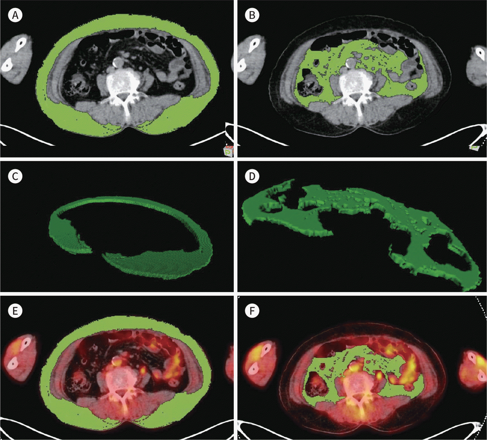 | Fig. 1.Example of measurement of adipose tissue. A, B. On consecutive transaxial CT images at L3/L4 level. The area of adipose tissue was automatically computed using a CT attenuation range from −50 to −200 HU. C, D. Within the areas of adipose tissue on the 3 images, the volume and mean HU were automatically calculated. E, F. The areas of adipose tissue on the 3 CT images were then exported to the corresponding fluorodeoxy-glucose PET/CT images. HU = Hounsfield unit |
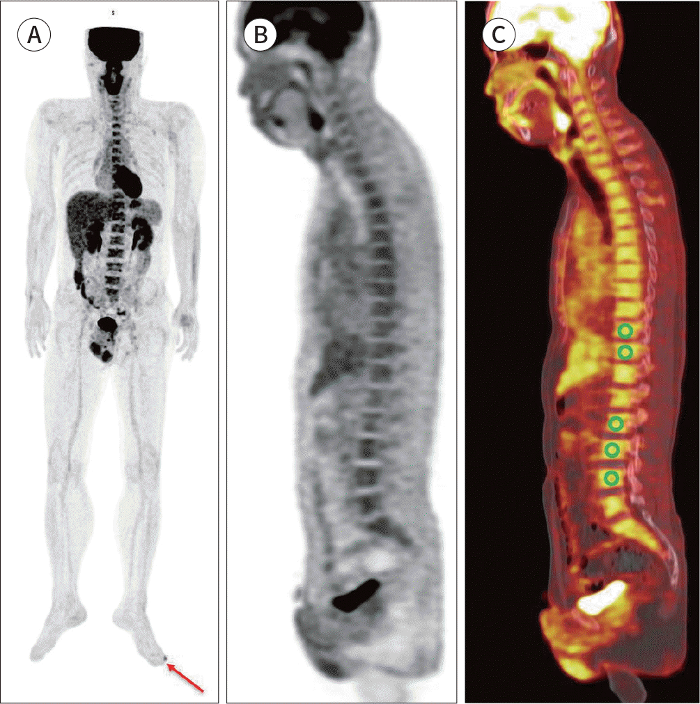 | Fig. 2.Example of measurement of bone marrow. Maximum intensity projection (A), sagittal PET (B), and sagittal fused PET/CT images of FDG PET/ CT (C). Spheroid-shaped five VOIs were drawn in vertebral bodies of thoracic and lumbar spines, and the average SUV of those VOIs was calculated. Primary malignant lesion was located left toe (arrow). FDG = fluorodeoxyglucose, SUV = standardized uptake value, VOI = volume of interest |
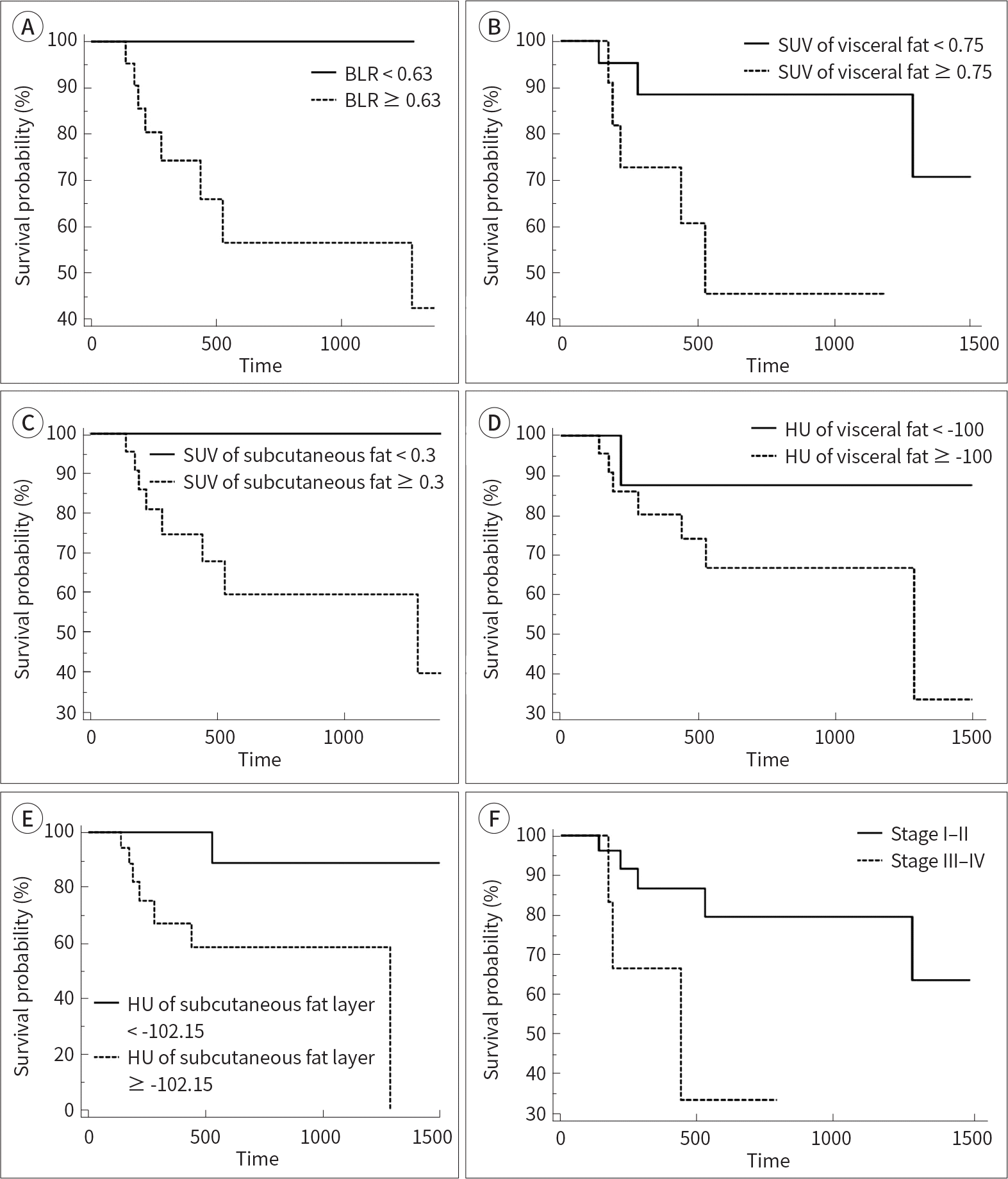 | Fig. 3.Kaplan-Meier curves for distant progression-free survival according to different parameters: BLR (A), SUV of VAT (B), SUV of SAT (C), HU of VAT (D), HU of SAT (E), and stage (F). BLR = bone marrow-to-liver uptake ratio, SAT = subcutaneous adipose tissue, SUV = standardized uptake value, HU = Hounsfield unit, VAT = visceral adipose tissue |
Table 1.
Clinical Characteristics of Enrolled Patients (n = 33)
∗Measured in 17 patients. BLR = BM-to-liver uptake ratio, BM = bone marrow, BMI = body mass index, CRP = C-reactive protein, HU = Hounsfield unit, LDH = lactate dehydrogenase, NLR = neutrophil-to-lymphocyte ratio, SAT = subcutaneous adipose tissue, SUVmean = mean of standardized uptake value, SUVtumor = maximum fluorodeoxyglucose uptake of primary tumor, VAT = visceral adipose tissue, WBC = white blood cell
Table 2.
Univariate Analysis of Disease Progression-Free Survival
Table 3.
Recurrence Rates According to Staging and Fluorodeoxyglucose Uptake of Adipose Tissue
 XML Download